Lion's Mane and Cancer
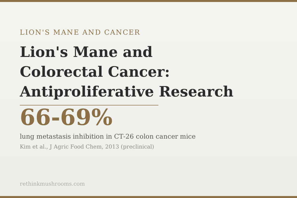
66-69% lung metastasis inhibition in CT-26 colon cancer mice treated with lion's mane extract
Kim and colleagues (2013) tested H. erinaceus mushroom extract in CT-26 colon cancer-transplanted mice and found 66-69% inhibition of cancer metastasis to the lung. This is the most clinically meaningful preclinical result in lion's mane colorectal cancer research, and it comes from an animal model, not a human trial.
Kim et al., Journal of Agricultural and Food Chemistry, 2013
3 mechanisms
three separate lion's mane compound classes target colorectal cancer through distinct pathways: erinacine A via extrinsic apoptosis (TNFR/Fas/JNK), HEFP polysaccharides via intrinsic apoptosis (ROS/caspase-9), and HEFP-2b via S-phase cell cycle arrest
IBD link
Ren et al. (2018) showed lion's mane polysaccharides reduced colitis severity via gut inflammation and microbiome modulation, addressing the IBD-to-colorectal cancer progression pathway upstream of tumor formation
0 human trials
no randomized controlled trial has tested lion's mane against colorectal cancer in humans; all antiproliferative evidence is from cell line studies and mouse models
Kim et al., Journal of Agricultural and Food Chemistry, 2013
 View as image ↗
Colorectal cancer has the richest lion's mane-specific research of any cancer type. That is not because colorectal cancer is special to mushroom researchers. It is because the CT-26 colon cancer mouse model is a widely used and well-validated experimental system, and Hericium erinaceus researchers used it to generate the most compelling preclinical metastasis data in the lion's mane cancer literature.
The 2013 Kim study showing 66-69% metastasis inhibition is the headline. But the colorectal evidence runs deeper than a single study: three structurally distinct compound classes from different parts of lion's mane inhibit colorectal cancer cells through three separate molecular mechanisms. Erinacine A from the mycelium induces extrinsic apoptosis via death receptor pathways. HEFP polysaccharides from the fruiting body induce intrinsic apoptosis via reactive oxygen species. HEFP-2b, a specific fruiting body polysaccharide, arrests cancer cells in the S-phase of the cell cycle. For the broader lion's mane cancer overview, see lion's mane and cancer: what the research shows.
Key Takeaways
- Kim and colleagues (2013) found 66-69% inhibition of lung metastasis in CT-26 colon cancer-transplanted mice treated with H. erinaceus extract, the most clinically meaningful preclinical result in lion's mane colorectal cancer research
- Lee and colleagues (2019) identified the specific mechanism of erinacine A's apoptosis induction in HCT-116 colorectal cancer cells: TNFR (tumor necrosis factor receptor), Fas, and FasL upregulation via JNK/p300/p50 signaling with histone acetylation
- Hou and colleagues (2020) showed that HEFP polysaccharides from the fruiting body induce apoptosis in colorectal cancer cells through a completely different mechanism: reactive oxygen species (ROS) generation mediating caspase-9-dependent intrinsic apoptosis pathway
- Liu and colleagues (2020) isolated and characterized HEFP-2b, a novel polysaccharide from H. erinaceus fruiting body, finding it arrests colorectal cancer cells in S-phase of the cell cycle, a third distinct anti-cancer mechanism
- Ren and colleagues (2018) showed that H. erinaceus polysaccharides attenuate colitis in mice via oxidative stress reduction and gut microbiome modulation, addressing the IBD-to-colorectal cancer progression pathway through prevention of chronic inflammation
- All evidence is preclinical; no randomized controlled trial has tested lion's mane against colorectal cancer in human patients
Why Colorectal Cancer Has the Richest Lion's Mane Evidence
The depth of colorectal cancer evidence in lion's mane research reflects the availability of validated experimental tools. The CT-26 and HCT-116 cell lines are standard, well-characterized colorectal cancer models used across the oncology research literature. When researchers want to test a compound's anti-cancer activity, these are routine choices. H. erinaceus researchers made the same choices, producing a body of work more concentrated in colorectal than in most other cancer types.
The result is that three distinct compound classes from lion's mane have been independently tested against colorectal cancer models, producing three distinct mechanistic pictures. This is unusual. Most natural compound cancer research tests one compound in one model once. The colorectal lion's mane evidence shows different research groups, testing different compounds, finding different mechanisms, in the same cancer type.
Gravina and colleagues (2023) provided the clinical review context for H. erinaceus in gastrointestinal diseases broadly, including the historical use patterns that made GI cancers a natural starting focus for the research.[8]
The Metastasis Data: Kim 2013's CT-26 Mouse Study
Kim and colleagues (2013) tested H. erinaceus mushroom extracts in mice transplanted with CT-26 colon cancer cells, a model designed to test not just primary tumor growth but metastatic spread to other organs.[5]
The experimental design specifically tested whether lion's mane treatment could reduce the number of CT-26 colon cancer metastases to the lung, one of the primary target organs in colorectal cancer dissemination. The result: 66-69% inhibition of lung metastasis in treated animals compared to untreated controls.
Metastasis data is more clinically meaningful than primary tumor cell line data because metastatic spread, not the primary tumor, is the primary cause of death in colorectal cancer. A compound that reduces primary cell proliferation in a dish addresses a limited part of the clinical problem. A compound that reduces metastatic spread in a live animal with an established tumor addresses the mechanism that kills patients.
The 66-69% figure is striking by preclinical standards. It does not establish that lion's mane prevents colorectal cancer metastasis in humans; it establishes that something in lion's mane interacts with the biological processes of metastasis in this model. Li and colleagues (2014) further documented antiproliferative activity of H. erinaceus extracts against HT-29 human colon cancer cells alongside other GI cancer lines, extending the in vitro evidence.[6]
Erinacine A: Extrinsic Apoptosis via TNFR and JNK Signaling
Lee and colleagues (2019) investigated the specific molecular mechanism by which erinacine A, the primary terpenoid compound from lion's mane mycelium, induces apoptosis in HCT-116 human colorectal cancer cells.[1]
The mechanism is extrinsic apoptosis: death receptor-mediated cell death signaling from outside the cell. Lee's study found erinacine A treatment upregulated TNFR (tumor necrosis factor receptor), Fas, and FasL (Fas ligand) expression in the cancer cells. These are the molecular components of the extrinsic apoptosis pathway: when cancer cells express more of these death receptors, they become more susceptible to immune-mediated killing (through Fas/FasL signaling) and inflammatory cytokine killing (through TNFR).
The downstream signaling was traced through the JNK/p300/p50 pathway, with histone acetylation identified as part of the mechanism. Histone acetylation changes gene expression by opening up DNA for transcription; erinacine A's effect on histone acetylation patterns in HCT-116 cells alters which apoptosis-related genes are expressed.
Importantly, the Lee 2019 findings included xenograft confirmation: the in vitro HCT-116 cell line results were validated in a mouse xenograft model, showing tumor growth inhibition in animals. This moves the evidence beyond cell line data into a live animal model.
A 2017 proteomics study by Lee and colleagues complemented this mechanistic work by identifying novel protein targets involved in erinacine A's inhibition of colorectal cancer cell aggressiveness.[2] The proteomics approach revealed changes in proteins involved in cell migration, invasion, and adhesion, consistent with the metastasis-inhibiting effects observed in the Kim 2013 mouse data.
HEFP Polysaccharides: Intrinsic Apoptosis via Reactive Oxygen Species
The extrinsic apoptosis mechanism from erinacines is mycelium-derived. The fruiting body produces a different set of compounds with a different cancer cell death mechanism.
Hou and colleagues (2020) tested HEFP (Hericium erinaceus Fruiting body Polysaccharide) against HCT-116 and DLD-1 human colorectal cancer cell lines.[3] The mechanism was intrinsic apoptosis: cell death signaling initiated from within the cell through the mitochondrial pathway.
HEFP treatment increased reactive oxygen species (ROS) inside the cancer cells. ROS at elevated levels cause oxidative stress that damages cellular structures; cancer cells under oxidative stress trigger caspase-9-dependent apoptosis through the intrinsic mitochondrial pathway. This is a fundamentally different mechanism from erinacine A's extrinsic death receptor pathway.
He and colleagues (2017) reviewed the broader biological activities of H. erinaceus polysaccharides, providing the chemical context for why HEFP compounds have cancer-relevant activity: their beta-glucan structure activates pattern recognition receptors, but specific structural features of the colorectal-active fractions also have direct antiproliferative effects through the ROS mechanism.[9]
HEFP-2b: S-Phase Cell Cycle Arrest as a Third Mechanism
Liu and colleagues (2020) isolated and structurally characterized a novel polysaccharide fraction from H. erinaceus fruiting bodies, designated HEFP-2b.[4]
Testing HEFP-2b against HCT-116 colorectal cancer cells revealed a third mechanism: cell cycle arrest at S-phase. Cancer cells must progress through the cell cycle (G1, S, G2, M phases) to divide. S-phase is when DNA is replicated. Arresting cells at S-phase prevents them from completing replication and dividing, effectively halting tumor growth without necessarily killing the arrested cells.
This is mechanistically distinct from both apoptosis pathways. Erinacine A and HEFP cause cancer cells to die. HEFP-2b causes them to stop dividing. Both outcomes arrest tumor growth, but through fundamentally different processes.
The existence of three distinct anti-cancer mechanisms from different structural classes of lion's mane compounds means that the anti-tumor activity is not a single trick. Multiple parts of the mushroom, with different chemical structures, affect colorectal cancer cells through different biological routes. This breadth of mechanism is the scientific basis for calling colorectal cancer lion's mane's best-studied cancer indication.
The IBD-to-Cancer Connection: Chronic Inflammation and Prevention
Colorectal cancer does not arise solely from isolated mutations. In a significant proportion of cases, particularly those in people with inflammatory bowel disease, chronic colonic inflammation drives the sequential progression from normal tissue to dysplasia to carcinoma. Reducing chronic colonic inflammation is therefore a legitimate cancer prevention target.
Ren and colleagues (2018) showed that H. erinaceus polysaccharides attenuate colitis in C57BL/6 mice via regulation of oxidative stress and inflammation-related signaling pathways, with associated positive changes in gut microbiome composition.[7] The study tested a mouse model of DSS-induced colitis (a standard IBD model) and found that lion's mane polysaccharide treatment reduced intestinal inflammation, protected the colonic epithelium, and shifted the gut microbiome toward healthier composition.
This creates a plausible prevention pathway: if lion's mane polysaccharides reduce the chronic colonic inflammation that drives IBD-associated colorectal cancer, then regular consumption might reduce cancer risk upstream of tumor formation. This is the weakest evidence link in the colorectal cancer story because it connects mouse colitis data to human cancer risk through multiple inferential steps. But it is mechanistically coherent, and it adds a prevention dimension to the direct antiproliferative evidence.
The Bottom Line
Colorectal cancer has the most mechanistically detailed lion's mane preclinical evidence of any cancer type. Kim 2013 found 66-69% metastasis inhibition in CT-26 colon cancer mice. Three compound classes operate through three distinct mechanisms: erinacine A (Lee 2019) induces extrinsic apoptosis via TNFR/Fas/JNK signaling in HCT-116 cells, confirmed in xenograft; HEFP polysaccharides (Hou 2020) induce intrinsic apoptosis via ROS-mediated caspase-9 activation; HEFP-2b (Liu 2020) arrests cell cycle at S-phase. Ren 2018 adds an upstream prevention mechanism via colitis reduction. All evidence is preclinical; no human colorectal cancer trial has been conducted.
Frequently Asked Questions
Sources & References
- 1. Source details for this reference are not yet available.
- 2. Source details for this reference are not yet available.
- 3. Source details for this reference are not yet available.
- 4. Source details for this reference are not yet available.
- 5. Source details for this reference are not yet available.
- 6. Source details for this reference are not yet available.
- 7. Source details for this reference are not yet available.
- 8. Source details for this reference are not yet available.
- 9. Source details for this reference are not yet available.