Sanghuang Research
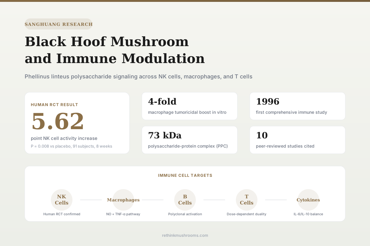
5.62 point NK cell increase
A 91-person randomized controlled trial found that 1,000 mg/day of Phellinus linteus extract significantly improved natural killer cell activity at the 12.5:1 effector-to-target ratio after eight weeks.
Ku & Kang, Medicine, 2022
1996
first immune study published
91
subjects in the largest human trial
4-fold
macrophage tumoricidal boost in vitro
Ku & Kang, Medicine, 2022
 View as image ↗
Black hoof mushroom (Phellinus linteus), known as sanghuang in Korea and China, has been used in East Asian traditional medicine for centuries. It grows on mulberry trees and produces a dark, hoof-shaped fruiting body. The mushroom entered modern immunology research in 1996, when Kim et al. isolated a polysaccharide from its mycelial culture and demonstrated that it stimulated both humoral and cell-mediated immunity in mice, across a wider range of immune parameters than beta-glucans from other Basidiomycetes.[1] Three decades of research have followed, producing two human randomized controlled trials and dozens of mechanistic studies in cell and animal models. For an overview of sanghuang's broader research profile, see what is sanghuang? Korea's cancer-fighting mushroom.
The immune modulation story of Phellinus linteus centers on its polysaccharides and polysaccharide-protein complexes. These molecules interact with immune cells at multiple levels: activating macrophages to produce nitric oxide and TNF-alpha, enhancing natural killer cell cytotoxicity, modulating T cell responses, and stimulating B cell proliferation. The evidence is strongest for macrophage activation (multiple independent labs, consistent findings) and NK cell enhancement (confirmed in a human trial). T cell and B cell effects are documented but less extensively replicated.
Key Takeaways
- A polysaccharide from Phellinus linteus stimulated T cell proliferation, cytotoxic T cell activity, NK cell activity, macrophage function, and both T-dependent and T-independent antibody responses in mice (Kim et al., 1996)
- Acidic polysaccharide from P. linteus enhanced macrophage tumoricidal activity against B16 melanoma cells 4-fold at 200 micrograms/mL, through nitric oxide and TNF-alpha upregulation (Kim et al., 2004)
- A 73 kDa polysaccharide-protein complex (PPC) from P. linteus activated macrophages, upregulated NK cell killing of YAC-1 lymphoma cells, and selectively stimulated B cell but not T cell proliferation (Kim et al., 2006)
- In a 91-person RCT, 1,000 mg/day of P. linteus extract for 8 weeks significantly improved NK cell activity at the 12.5:1 ratio (P = 0.008) compared to placebo; secondary cytokine markers did not reach significance (Ku & Kang, 2022)
- P. linteus polysaccharides shifted the IL-6/IL-10 cytokine balance in RAW 264.7 macrophages, with structural differences between P. linteus and P. igniarius polysaccharides producing distinct immune profiles (Suabjakyong et al., 2015)
- Fermented broth from P. linteus dose-dependently inhibited mitogen-induced IL-2 production and IFN-gamma/IL-4 ratio in T cells, suggesting immunosuppressive capacity at certain concentrations (Lin et al., 2016)
The 1996 Foundation: Broad-Spectrum Immune Activation
Kim et al. (1996) published the first comprehensive study of Phellinus linteus polysaccharide (PL) effects on the immune system. They purified a polysaccharide from mycelial culture and tested it across multiple immune parameters in mouse splenocytes, both in vitro and after intraperitoneal injection.[1]
The results showed activation across the full spectrum of immune responses. PL stimulated T lymphocyte proliferation in mixed lymphocyte reactions and enhanced the cytotoxicity of cytotoxic T cells responding to alloantigens. NK cell activity and macrophage function both increased with PL treatment in vivo and in vitro. On the humoral side, PL boosted both T-dependent and T-independent primary antibody responses and acted as a polyclonal B cell activator.
What distinguished PL from other mushroom polysaccharides tested at the time was its breadth. Beta-glucans and other polysaccharides from Basidiomycetes typically raised only T lymphocyte-mediated immunity. PL elevated both T cell and B cell arms, plus innate immune components. This wider range of immunostimulation suggested that PL contained structural features absent from standard beta-glucans, or that it engaged additional receptor pathways on immune cells.
Macrophage Activation: The NO and TNF-Alpha Pathway
Two studies from Kim's group at Pusan National University established the macrophage activation mechanism in detail. In 2003, Kim et al. showed that acidic polysaccharide from P. linteus induced nitric oxide (NO)-mediated tumoricidal activity in peritoneal macrophages through protein tyrosine kinase (PTK) and protein kinase C (PKC) signaling.[2] When PTK was inhibited with genistein or PKC with staurosporine, the macrophage tumoricidal activity dropped, confirming these kinases as required mediators.
The 2004 follow-up study quantified the dose-response relationship. Acidic polysaccharide at 200 micrograms/mL enhanced macrophage killing of B16 melanoma cells 4-fold compared to untreated controls. The mechanism involved simultaneous upregulation of NO and TNF-alpha production from peritoneal macrophages.[3] Both in vitro treatment of isolated macrophages and in vivo injection produced the same pattern: polysaccharide exposure primed macrophages into a tumoricidal state through NO synthesis.
These findings are consistent with a Dectin-1/TLR2 activation model, where beta-glucan-type structures in the polysaccharide engage pattern recognition receptors on macrophages, triggering downstream signaling through NF-kB and MAPK pathways. The PKC and PTK dependence documented by Kim et al. maps to known Dectin-1 signaling intermediates. For a detailed analysis of how beta-glucan structure determines immune receptor binding, see beta-glucan recognition by the innate immune system.
The Polysaccharide-Protein Complex: Cell-Type Specificity
Kim et al. (2006) isolated and characterized a novel polysaccharide-protein complex (PPC) from P. linteus with a molecular weight of approximately 73 kDa. PPC contained five monosaccharides, predominantly D-glucose and D-mannose in a 3:2 molar ratio, with aspartic acid as the main amino acid component.[4]
PPC showed a distinctive pattern of cell-type specificity. It markedly increased B cell proliferation but did not stimulate T cell proliferation. This distinguished PPC from lipopolysaccharide (LPS), which has a similar B cell activating profile; the key difference was that PPC activity was not blocked by polymyxin B (a specific LPS inhibitor), confirming PPC operates through a distinct receptor mechanism.
On macrophages, PPC increased cytokine production and NO output, enhancing tumoricidal activity against B16 melanoma through NO-dependent lysis. PPC also upregulated NK cell killing of YAC-1 lymphoma cells in vitro. The combined profile (B cell activation, macrophage priming, NK cell enhancement, but no T cell stimulation) suggests PPC engages a subset of pattern recognition receptors different from the whole polysaccharide fraction tested in the 1996 study, which activated T cells. This implies P. linteus contains multiple immunoactive fractions with complementary but distinct immune targets.
Anti-Inflammatory Effects in Macrophages
While the studies above focused on immune activation, a parallel line of research demonstrated that P. linteus also has anti-inflammatory capacity. Kim et al. (2007) showed that P. linteus extract inhibited inflammatory mediators in LPS-stimulated RAW 264.7 macrophages by suppressing redox-based NF-kB and MAPK activation.[5] The extract reduced NO production, prostaglandin E2, TNF-alpha, and IL-1beta in the inflammatory context.
Xie et al. (2019) confirmed and extended this finding. P. linteus polysaccharides inhibited NF-kB translocation from cytoplasm to nucleus in LPS-stimulated RAW 264.7 macrophages, blocking the transcription of inflammatory cytokines at the level of nuclear signaling.[8]
This creates an apparent contradiction: P. linteus polysaccharides activate macrophages in unstimulated conditions (producing NO and TNF-alpha to kill tumor cells) but suppress macrophage inflammatory output when cells are already stimulated by LPS. The resolution likely involves context-dependent signaling. In resting macrophages, polysaccharide engagement of Dectin-1 and TLR2 triggers a controlled activation response. In already-inflamed macrophages where NF-kB is hyperactive, the same polysaccharides may stabilize the signaling cascade rather than amplify it. This bidirectional capacity is sometimes described as "immunomodulation" rather than simple "immunostimulation," and it is consistent with beta-glucan biology observed in other medicinal mushroom species.
Cytokine Balance and Species Differences
Suabjakyong et al. (2015) compared polysaccharides from Phellinus linteus and Phellinus igniarius (a closely related species sometimes confused with P. linteus in commercial products) for their effects on IL-6 and IL-10 production in RAW 264.7 macrophages.[6]
Both species' polysaccharides stimulated IL-6 (pro-inflammatory) and IL-10 (anti-inflammatory) production, but the ratios differed. The structural characterization revealed differences in monosaccharide composition, molecular weight, and branching patterns between the two species' polysaccharides. These structural differences translated into different cytokine profiles, which is relevant for understanding why studies using different Phellinus species or different extract preparations may report conflicting immune outcomes. For more on how species confusion affects research interpretation, see sanghuang vs. meshima: different species, different research.
This finding has practical implications. Commercial "sanghuang" products may contain P. linteus, P. igniarius, or other Phellinus species, and the immune effects are not interchangeable. Extract preparation method, growth substrate, and species identity all influence which polysaccharide fractions dominate and what immune profile they produce.
T Cell Modulation: Suppression at Higher Concentrations
Lin et al. (2016) examined how P. linteus fermented broths and isolated polysaccharides affected T cell responses. Their findings introduced complexity to the immunostimulation narrative.[7]
PL3 broth, which contained high quantities of polysaccharides, dose-dependently inhibited mitogen-induced IL-2 production in T cells. It also decreased the IFN-gamma to IL-4 ratio, indicating a shift from Th1 (pro-inflammatory) toward Th2 (anti-inflammatory) T cell responses. Cell cycle analysis showed that PL3 and a methanol-dichloromethane fraction arrested T cells in S phase, preventing progression through division.
This immunosuppressive effect on T cells appears to contradict Kim et al.'s 1996 finding of T cell stimulation. The difference may be concentration-dependent, extract-specific, or related to the stimulation context (mitogen-induced vs. antigen-driven). At lower concentrations, P. linteus polysaccharides may prime T cells for activation; at higher concentrations, they may dampen overactivation. This dose-dependent duality mirrors the macrophage data, reinforcing the immunomodulatory (rather than purely immunostimulatory) characterization of P. linteus.
Human Evidence: Two Randomized Controlled Trials
The preclinical mechanistic work led to two human RCTs, both conducted by Ku and Kang in South Korea and published in 2022.
The pilot trial enrolled 30 healthy adults randomized to three groups: PL extract at 1,000 mg/day (n=10), PL extract at 2,000 mg/day (n=10), or placebo (n=10) for 8 weeks. NK cell activity and IL-6 levels tended to improve in the 1,000 mg group compared to placebo. Immunoglobulin levels (IgG1, IgG2, IgM) showed non-significant trends toward improvement in both PL groups.[9]
The main trial expanded to 91 participants (45 PL, 46 placebo) receiving 1,000 mg/day of PL extract or dextrin placebo for 8 weeks. The primary outcome, NK cell activity measured by lactate dehydrogenase cytotoxicity assay, showed significant improvement in the PL group at the 12.5:1 effector-to-target cell ratio: a mean change of 5.62 ± 8.24 versus 0.95 ± 8.05 in the placebo group (P = 0.008). At higher ratios (25:1 and 50:1), the differences were not statistically significant.[10]
Secondary outcomes (WBC count, TNF-alpha, IFN-gamma, IL-1beta, IL-2, IL-6, IL-12, IgG1) did not demonstrate significant improvement. Safety evaluation confirmed no hepatotoxicity or renal toxicity from 8 weeks of oral PL extract.
The pattern (significant NK cell effect at the lowest effector ratio, non-significant cytokine changes) is consistent with a modest immunomodulatory effect that primarily enhances NK cell priming rather than triggering systemic cytokine elevation. The effect size was small. These trials provide proof-of-concept that oral P. linteus extract reaches the immune system and produces a measurable change, but they do not establish clinical benefit for any disease state. For context on sanghuang's broader anti-cancer research profile, see sanghuang and cancer: research on Phellinus igniarius.
Limitations Across the Evidence Base
The preclinical studies share several constraints. Most macrophage and NK cell work comes from a single research group (Kim et al. at Pusan National University), raising the question of independent replication. The cell models use murine peritoneal macrophages and splenocytes; human immune cell responses may differ in magnitude or pathway. Polysaccharide fractions tested in different studies were extracted using different methods, making direct comparison difficult. No study has identified the specific receptor binding site on immune cells that initiates the signaling cascade, leaving the molecular mechanism partially speculative.
The human trials, while randomized and placebo-controlled, were small (30 and 91 participants), short (8 weeks), and conducted in healthy adults. They measured surrogate markers (NK cell activity in vitro) rather than clinical outcomes (infection rates, disease incidence). The significant result appeared at only one of three effector-to-target ratios tested, and secondary cytokine markers were uniformly non-significant. Both trials were conducted in South Korea, and the extract was sourced from Korean P. linteus mycelium; generalizability to other populations and extract preparations is unknown.
Species identification remains a persistent problem in the field. Commercial products labeled as sanghuang, black hoof, Phellinus linteus, or meshima may contain different species within the Phellinus/Sanghuangporus complex, and as Suabjakyong et al. demonstrated, different species produce different immune profiles. For more on this taxonomic challenge, see sanghuang: Korea and China's prized anti-cancer mushroom and its hispidin content.
The Bottom Line
Phellinus linteus polysaccharides activate macrophages, NK cells, and B cells through multiple signaling pathways, with the strongest preclinical evidence for NO-mediated macrophage tumoricidal activity. A 91-person RCT confirmed modest NK cell enhancement in healthy humans at 1,000 mg/day over 8 weeks. The mushroom shows bidirectional immune modulation: activating resting immune cells while suppressing inflammatory responses in already-stimulated cells. Evidence quality is limited by reliance on a small number of research groups, short-term human trials with surrogate endpoints, and species identification challenges across the Phellinus genus.
Frequently Asked Questions
Sources & References
- 1RTHM-00020·Kim, H M et al. (1996). “Stimulation of humoral and cell mediated immunity by polysaccharide from mushroom Phellinus linteus..” International journal of immunopharmacology.Study breakdown →PubMed →↩
- 2RTHM-00057·Kim, Gi-Young et al. (2003). “Acidic polysaccharide isolated from Phellinus linteus induces nitric oxide-mediated tumoricidal activity of macrophages through protein tyrosine kinase and protein kinase C..” Biochemical and biophysical research communications.Study breakdown →PubMed →↩
- 3RTHM-00072·Kim, Gi-Young et al. (2004). “Acidic polysaccharide isolated from Phellinus linteus enhances through the up-regulation of nitric oxide and tumor necrosis factor-alpha from peritoneal macrophages..” Journal of ethnopharmacology.Study breakdown →PubMed →↩
- 4RTHM-00102·Kim, Gi-Young et al. (2006). “Partial characterization and immunostimulatory effect of a novel polysaccharide-protein complex extracted from Phellinus linteus..” Bioscience.Study breakdown →PubMed →↩
- 5RTHM-00116·Kim, Ho Gyoung et al. (2007). “Phellinus linteus inhibits inflammatory mediators by suppressing redox-based NF-kappaB and MAPKs activation in lipopolysaccharide-induced RAW 264.7 macrophage..” Journal of ethnopharmacology.Study breakdown →PubMed →↩
- 6RTHM-00402·Suabjakyong, Papawee et al. (2015). “Structural characterization and immunomodulatory effects of polysaccharides from Phellinus linteus and Phellinus igniarius on the IL-6/IL-10 cytokine balance of the mouse macrophage cell lines (RAW 264.7)..” Food & function.Study breakdown →PubMed →↩
- 7RTHM-00434·Lin, Chun-Jung et al. (2016). “Modulation of T cell response by Phellinus linteus..” Journal of bioscience and bioengineering.Study breakdown →PubMed →↩
- 8RTHM-00599·Xie, Zhenglu et al. (2019). “Anti-inflammatory activity of polysaccharides from Phellinus linteus by regulating the NF-κB translocation in LPS-stimulated RAW264.7 macrophages..” International journal of biological macromolecules.Study breakdown →PubMed →↩
- 9RTHM-01555·Ku, Yong Ho et al. (2022). “Efficacy of Phellinus linteus extract on immunity enhancement: A CONSORT-randomized, double-blind, placebo-controlled pilot trial..” Medicine.Study breakdown →PubMed →↩
- 10RTHM-01556·Ku, Yong Ho et al. (2022). “Effects of Phellinus linteus extract on immunity improvement: A CONSORT-randomized, double-blinded, placebo-controlled trial..” Medicine.Study breakdown →PubMed →↩