Chaga for Gut Health
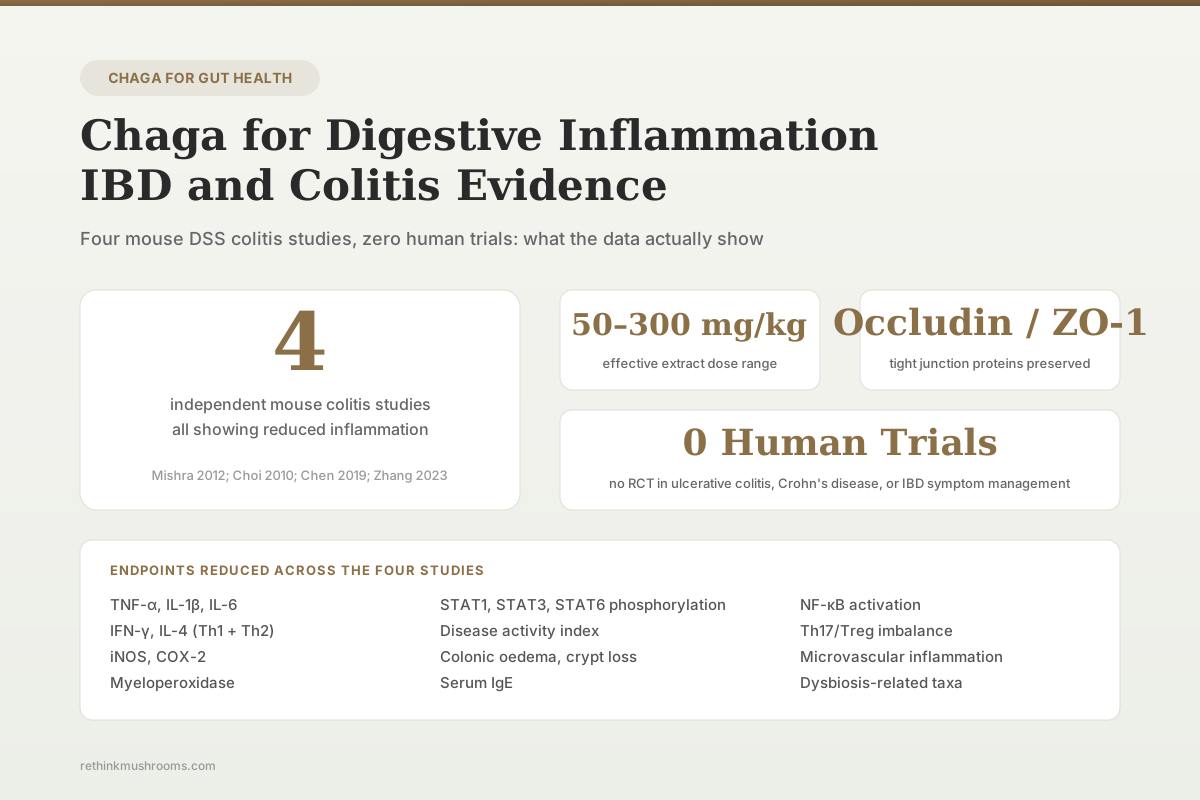
4 Mouse Studies No Human Trial
Four published mouse colitis experiments show aqueous, polysaccharide, and fermented mycelium fractions of Inonotus obliquus reduce colonic pro-inflammatory cytokines, myeloperoxidase, iNOS, and tight junction disruption. No randomised clinical trial has tested chaga in people with ulcerative colitis or Crohn's disease.
Mishra et al., J Ethnopharmacol, 2012
IOAE 50–100 mg/kg
aqueous extract doses that reduced DSS colitis severity in C57BL/6 mice (Mishra et al., 2012)
Th1/Th2 + Th17/Treg
CD4+ T-cell subsets rebalanced by IOP at 100–300 mg/kg (Chen et al., 2019)
0 human trials
no RCT of chaga in ulcerative colitis, Crohn's, or IBD symptom management
Mishra et al., J Ethnopharmacol, 2012
 View as image ↗
Digestive inflammation sits on a spectrum from transient gastroenteritis to chronic inflammatory bowel disease (IBD). IBD encompasses ulcerative colitis (UC) and Crohn's disease (CD), immune-mediated conditions affecting around 7 million people globally, with rising incidence in newly industrialising regions. Current treatment relies on aminosalicylates, corticosteroids, thiopurines, anti-TNF biologics (infliximab, adalimumab), anti-integrin agents (vedolizumab), and anti-IL-23 therapies (ustekinumab, risankizumab). Against this clinical backdrop, chaga (Inonotus obliquus) has four mouse colitis experiments and a scattering of mechanistic support. Zero human trials. For the closely related sibling covering upstream gastric inflammation, see chaga and ulcers: can it protect the stomach lining? and for the broader anti-inflammatory framework see chaga and inflammatory bowel disease: preclinical evidence.[1][2][3][4]
Key Takeaways
- Mishra et al. (2012) administered aqueous I. obliquus extract (IOAE) at 50 and 100 mg/kg to C57BL/6 mice exposed to 2% DSS; IOAE reduced colonic oedema, mucosal damage, crypt loss, iNOS, myeloperoxidase, and mRNA expression of TNF-α, IL-1β, IL-6, and IFN-γ
- Choi et al. (2010) reported that I. obliquus feeding decreased colonic TNF-α, STAT1, IL-4, and STAT6 expression and reduced serum IgE in BALB/c mice with DSS colitis; the same extract inhibited LPS-induced TNF-α and STAT1/STAT6 activation in RAW 264.7 macrophages
- Chen et al. (2019) showed that I. obliquus polysaccharide (IOP) at 100, 200, and 300 mg/kg reduced disease activity index, preserved tight junction proteins Occludin and ZO-1, rebalanced Th1/Th2 and Th17/Treg ratios, and modulated p-STAT1, p-STAT3, and p-STAT6 in chronic DSS colitis
- Zhang et al. (2023) demonstrated that polysaccharide from fermented mycelium of I. obliquus attenuated DSS ulcerative colitis and adjusted gut microbiota composition in mice, extending the evidence to fermentation-derived polysaccharide fractions
- Kim et al. (2007) documented that ethanol extract of I. obliquus inhibited LPS-induced NF-κB activation in RAW 264.7 macrophages and reduced iNOS, COX-2, TNF-α, IL-1β, and IL-6 expression, providing the mechanistic substrate for the in vivo colitis findings
- Review-level synthesis (Fordjour 2023; Ern 2024) catalogues chaga's anti-inflammatory gut activity as preclinical only; no randomised controlled trial in humans with ulcerative colitis, Crohn's disease, or related digestive inflammation has been registered or published
What Digestive Inflammation Is at the Tissue Level
Digestive inflammation in IBD involves breakdown of the intestinal barrier and dysregulated mucosal immunity. The epithelial barrier is a single cell layer sealed by tight junctions (claudins, occludin, ZO-1) that normally restrict paracellular passage of luminal antigens. Barrier breakdown allows microbial products to engage underlying immune cells, activating pattern recognition receptors (TLR4, NOD2) and driving NF-κB-mediated transcription of TNF-α, IL-1β, IL-6, and other pro-inflammatory cytokines.
CD4+ T helper cell differentiation contributes to chronicity. Crohn's disease classically associates with Th1 and Th17 responses (IFN-γ, IL-17); ulcerative colitis shows a Th2-like profile with IL-13 contribution; regulatory T cells (Tregs, FoxP3+) normally suppress effector responses. Imbalance in Th1/Th2 and especially Th17/Treg ratios drives persistent tissue damage. Cytokine-driven activation of STAT transcription factors (STAT1 for Th1/IFN-γ; STAT6 for Th2/IL-4; STAT3 for IL-6/Th17) translates these signals into tissue-level inflammation.
Neutrophil infiltration, measured by myeloperoxidase activity in tissue, is a reliable marker of acute colitis severity. Inducible nitric oxide synthase (iNOS) expression in colonic epithelium and lamina propria immune cells produces high-output NO that combines with superoxide to form peroxynitrite, extending tissue damage. A candidate agent for digestive inflammation should plausibly reduce some combination of these endpoints: pro-inflammatory cytokine transcription, NF-κB activation, Th-subset imbalance, neutrophil infiltration, iNOS induction, or barrier disruption.
The Mishra 2012 Study: Aqueous Chaga Extract in DSS Colitis
Mishra et al. (2012) randomised 5-week-old female C57BL/6 mice to prevention and treatment protocols with aqueous I. obliquus extract (IOAE) at 50 and 100 mg/kg body weight.[1] Colitis was induced by 2% dextran sulphate sodium (DSS) in drinking water for 7 days. Endpoints included histological scoring (hematoxylin and eosin), iNOS by immunohistochemistry, neutrophil infiltration by myeloperoxidase assay, and RT-PCR quantification of colonic TNF-α, IL-1β, IL-6, and IFN-γ mRNA.
Histology showed that IOAE suppressed oedema, mucosal damage, and crypt loss induced by DSS. IOAE markedly attenuated DSS-induced iNOS levels and myeloperoxidase accumulation in colon tissues, indicating reduced immune cell infiltration and reduced inflammatory enzyme activity at the tissue level. IOAE significantly inhibited mRNA expression of all four pro-inflammatory cytokines assayed. The authors framed the findings as suggesting that IOAE might be a useful supplement in the setting of inflammatory bowel disease, while noting that preclinical demonstration does not establish human efficacy.
Limitations: single species (mouse), single colitis model (DSS), short-duration acute protocol rather than chronic, two doses tested, no chronic maintenance phase, and no paired assessment of barrier function or microbiome modulation.
The Choi 2010 Study: Chaga in BALB/c DSS Colitis with STAT Pathway Detail
Choi et al. (2010) divided 28 male BALB/c mice (average weight 20.7 ± 1.6 g) into four treatment groups: commercial diet control, DSS-induced colitis, I. obliquus feeding alone, and IO feeding + DSS colitis.[2] Endpoints included colonic TNF-α, STAT1, IL-4, and STAT6 expression and serum IgE levels.
IO treatment decreased TNF-α and STAT1 expression in colon tissues compared with the DSS-only colitis group. IL-4 and STAT6 expression were reduced in the IO + DSS group compared with DSS alone. Serum IgE was lower in both IO-treated groups. In the mechanistic arm, IO extract inhibited LPS-induced TNF-α, STAT1 phosphorylation, STAT6 phosphorylation, and their unphosphorylated forms in RAW 264.7 macrophages, providing macrophage-level evidence for the in vivo cytokine and transcription factor reductions observed.
The BALB/c background is more Th2-biased than C57BL/6, making the STAT6 reduction particularly relevant to ulcerative colitis pathology. The weak cytotoxic effect of chaga extract on RAW 264.7 cells noted by the authors is a safety signal that warrants dose-response characterisation in any further macrophage work.
The Chen 2019 Study: IOP, Tight Junctions, and CD4+ T-cell Balance in Chronic Colitis
Chen et al. (2019) moved the evidence from acute to chronic DSS colitis and introduced barrier-protein and T-cell-subset endpoints.[3] I. obliquus polysaccharide (IOP) was administered orally at 100, 200, and 300 mg/kg to mice with chronic DSS-induced colitis. Primary endpoints were disease activity index, histological pathology, tight junction protein expression, CD4+ T-cell subset ratios, and STAT phosphorylation.
IOP reduced the disease activity index and alleviated colon pathology at all three tested doses. Critically for barrier biology, the tight junction proteins Occludin and ZO-1, which are characteristically degraded in colitis, were preserved in IOP-treated mice. Flow cytometry and RT-PCR on colon tissue, mesenteric lymph nodes, and spleen showed that IOP rebalanced Th1/Th2 and Th17/Treg ratios. Immunohistochemistry and western blot confirmed IOP modulation of p-STAT1 (Th1 axis), p-STAT6 (Th2 axis), and p-STAT3 (Th17 axis), linking the T-cell subset rebalancing to upstream cytokine signalling.
This study is the most mechanistically detailed of the four. The barrier protein preservation and T-cell rebalancing address two hallmarks of IBD pathology that the earlier acute-phase studies did not directly measure. The study remains preclinical: a mouse chronic DSS model is a reasonable but imperfect surrogate for human ulcerative colitis.
The Zhang 2023 Study: Fermented Mycelium Polysaccharide and Gut Microbiota
Zhang et al. (2023) extended the evidence to fermentation-derived fractions.[4] Polysaccharide isolated from fermented mycelium of I. obliquus (as distinct from wild sclerotium extract) was tested in DSS-induced ulcerative colitis in mice. The polysaccharide attenuated colitis severity and shifted gut microbiota composition.
The microbiome angle is important because IBD pathology interacts with dysbiosis. Reduced microbial diversity, depletion of short-chain fatty acid producers (Faecalibacterium prausnitzii, Roseburia), and expansion of pro-inflammatory taxa (adherent-invasive E. coli) are characteristic of IBD. A colitis-reducing intervention that also moves microbiota composition toward a healthier profile has plausibly additive mechanism. Detailed microbial shifts and durability of effects require further characterisation in longer studies. For the dedicated microbiome sibling in this cluster, see chaga and gut microbiome modulation: research review.
The Mechanistic Substrate: NF-κB, Cytokines, and Macrophage Signalling
Kim et al. (2007) established the macrophage-level mechanism that the colitis studies operate on top of.[5] Ethanol extract of I. obliquus suppressed LPS-induced NF-κB activation in RAW 264.7 cells and reduced iNOS, COX-2, TNF-α, IL-1β, and IL-6 expression. NF-κB sits at the centre of intestinal inflammation biology: TLR4-mediated recognition of microbial ligands that cross a compromised barrier drives IKK-mediated phosphorylation of IκB, freeing NF-κB for nuclear translocation and transcription of the exact cytokine panel that the colitis studies measured in vivo.
Javed et al. (2019) extended the anti-inflammatory signal to microvascular endothelium, showing I. obliquus attenuation of histamine-induced microvascular inflammation.[6] Colonic microvascular permeability contributes to oedema in IBD; attenuating endothelial inflammation is mechanistically relevant to reduced tissue swelling observed histologically in the colitis studies.
The chemical basis for these effects is partly explained by chaga's antioxidant polyphenols. Lee et al. (2007) isolated inonoblins A, B, C and phelligridins D, E, G from chaga, all with scavenging activity against ABTS, DPPH, and superoxide radicals.[7] ROS amplify NF-κB signalling and sustain iNOS activity; direct ROS scavenging is a plausible proximal mechanism for the observed tissue-level reductions.
What the Reviews Synthesise
Fordjour et al. (2023) and Ern et al. (2024) both include anti-inflammatory gut activity among chaga's documented preclinical properties, with the colitis studies cited as the central evidence base.[8][9] Both reviews frame the evidence as entirely preclinical. Neither recommends chaga for clinical use in IBD. Both flag the complete absence of randomised controlled trials in patients with ulcerative colitis, Crohn's disease, or lower-grade inflammatory digestive conditions.
A phase 2a trial of chaga polysaccharide as an adjunct to standard aminosalicylate therapy in mild-to-moderate ulcerative colitis would be scientifically reasonable given the preclinical mechanistic rationale. No such trial has been registered on ClinicalTrials.gov or comparable registries.
Safety Considerations Specific to IBD Populations
Patients with IBD face several compounded safety considerations relevant to chaga use:
Concurrent medications: IBD patients are commonly on 5-ASA compounds, corticosteroids, immunomodulators (azathioprine, mercaptopurine, methotrexate), biologics, or JAK inhibitors (tofacitinib, upadacitinib). Chaga's documented oxalate load is relevant to azathioprine/methotrexate-associated renal considerations and to the baseline dehydration risk in active flares. No drug interaction study has characterised chaga with any IBD therapy.
Oxalate nephropathy: Three case reports (Kikuchi 2014; Lee 2020; Kwon 2022) document chaga-induced oxalate nephropathy from long-term high-dose use. IBD patients with active small bowel disease or resection are already at elevated risk of enteric hyperoxaluria from fat malabsorption and colonic oxalate absorption; adding a high-oxalate chaga load compounds this risk.
Immunomodulation in active disease: The mechanistic studies consistently show NF-κB inhibition and Th-subset modulation. In patients on immunosuppressive biologics or small molecules for IBD, the net immunological effect of adding chaga is unpredictable and could theoretically either supplement efficacy or undermine infectious defence. No clinical data inform this trade-off.
Antiplatelet activity: Chaga's documented antiplatelet activity (Hyun et al., 2006) adds bleeding risk for patients with active colitis at risk of gastrointestinal haemorrhage.
For the dedicated pylori and gastric safety sibling, see chaga and helicobacter pylori: antimicrobial effects on stomach bacteria.
What the Evidence Supports and What It Does Not
| Question | Evidence | Confidence |
|---|---|---|
| Does chaga reduce colonic inflammation in mouse DSS colitis? | Yes: four independent studies across multiple extract types (aqueous, polysaccharide, fermented mycelium polysaccharide) | Moderate (preclinical, single species) |
| Does chaga preserve intestinal tight junction proteins? | Yes in chronic DSS colitis: Occludin and ZO-1 preserved (Chen et al., 2019) | Low to moderate |
| Does chaga modulate T-cell subsets relevant to IBD? | Yes in chronic DSS colitis: Th1/Th2 and Th17/Treg rebalanced (Chen et al., 2019) | Low to moderate |
| Does chaga shift gut microbiota toward an anti-inflammatory profile? | Preliminary evidence from fermented mycelium polysaccharide (Zhang et al., 2023) | Low |
| Has chaga been tested in humans with ulcerative colitis or Crohn's disease? | No randomised controlled trial published or registered | None |
| Is chaga safe for IBD patients? | Unquantified: antiplatelet activity, oxalate nephropathy risk, and unknown drug interactions with IBD therapies complicate the picture | Precautionary |
The Bottom Line
Chaga (Inonotus obliquus) reduces colonic inflammation across four mouse DSS colitis experiments. Aqueous extract at 50–100 mg/kg suppressed TNF-α, IL-1β, IL-6, IFN-γ, iNOS, and myeloperoxidase (Mishra et al., 2012). Feeding reduced colonic TNF-α and STAT1/STAT6 and serum IgE (Choi et al., 2010). IOP at 100–300 mg/kg preserved tight junction proteins Occludin and ZO-1 and rebalanced Th1/Th2 and Th17/Treg ratios in chronic DSS colitis (Chen et al., 2019). Fermented mycelium polysaccharide attenuated ulcerative colitis and adjusted gut microbiota (Zhang et al., 2023). The mechanistic substrate is NF-κB inhibition and ROS scavenging (Kim 2007; Javed 2019; Lee 2007). No randomised controlled trial has tested chaga in humans with inflammatory bowel disease. Patients with IBD face compounded safety considerations from oxalate load, antiplatelet activity, and unknown interactions with existing IBD therapies.
Frequently Asked Questions
Sources & References
- 1RTHM-00258·Mishra, Siddhartha Kumar et al. (2012). “Orally administered aqueous extract of Inonotus obliquus ameliorates acute inflammation in dextran sulfate sodium (DSS)-induced colitis in mice..” Journal of ethnopharmacology.Study breakdown →PubMed →↩
- 2RTHM-00177·Choi, Se Young et al. (2010). “Anti-inflammatory effects of Inonotus obliquus in colitis induced by dextran sodium sulfate..” Journal of biomedicine & biotechnology.Study breakdown →PubMed →↩
- 3RTHM-00549·Chen, Yi-Fang et al. (2019). “Inonotus obliquus polysaccharide ameliorates dextran sulphate sodium induced colitis involving modulation of Th1/Th2 and Th17/Treg balance..” Artificial cells.Study breakdown →PubMed →↩
- 4RTHM-00860·Zhang, Jiaqi et al. (2023). “Polysaccharide from fermented mycelium of Inonotus obliquus attenuates the ulcerative colitis and adjusts the gut microbiota in mice..” Microbial pathogenesis.Study breakdown →PubMed →↩
- 5RTHM-00117·Kim, Ho-Gyoung et al. (2007). “Ethanol extract of Inonotus obliquus inhibits lipopolysaccharide-induced inflammation in RAW 264.7 macrophage cells..” Journal of medicinal food.Study breakdown →PubMed →↩
- 6RTHM-00562·Javed, Sumreen et al. (2019). “Inonotus obliquus attenuates histamine-induced microvascular inflammation..” PloS one.Study breakdown →PubMed →↩
- 7RTHM-00118·Lee, In-Kyoung et al. (2007). “New antioxidant polyphenols from the medicinal mushroom Inonotus obliquus..” Bioorganic & medicinal chemistry letters.Study breakdown →PubMed →↩
- 8RTHM-01694·Fordjour, Eric et al. (2023). “Chaga mushroom: a super-fungus with countless facets and untapped potential..” Frontiers in pharmacology.Study breakdown →PubMed →↩
- 9RTHM-01886·Ern, Phoebe Tee Yon et al. (2024). “Chaga Therapeutic Properties: What This 2024 Review Actually Shows.” Mycology.Study breakdown →PubMed →↩