Auricularia and Blood Sugar
0 human trials
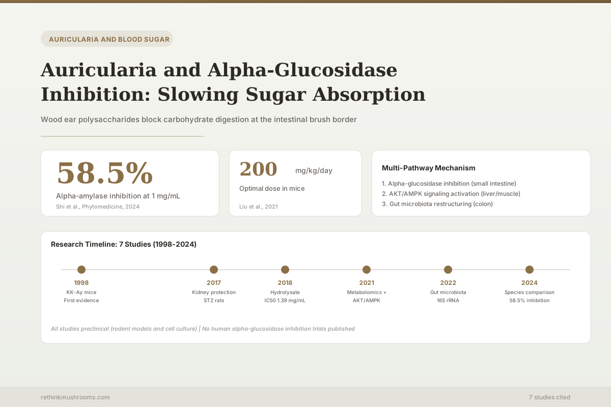
Wood ear polysaccharides lowered blood glucose in seven rodent studies spanning 1998 to 2024. Not one controlled human trial has tested the effect.
10 preclinical studies, 1998–2024
1998
first hypoglycemic study (KK-Ay mice)
2 of 10
measured a carbohydrate enzyme directly
AKT/AMPK
host signaling pathways implicated
10 preclinical studies, 1998–2024
 View as image ↗
Alpha-glucosidase is a brush border enzyme in the small intestine that cleaves complex sugars into absorbable glucose. Inhibiting it slows the rate at which dietary carbohydrates enter the bloodstream, flattening postprandial glucose spikes. This is the mechanism behind acarbose (Precose), a first-line pharmaceutical for type 2 diabetes. Auricularia auricula-judae, the wood ear mushroom consumed across East Asia for centuries, is widely described as working the same way.
The evidence is more interesting, and more limited, than that description suggests. Wood ear polysaccharides do lower blood glucose — reliably, across seven independent rodent studies running from 1998 to 2024. But the direct enzyme-inhibition assays that give this article its title were run on a different species of Auricularia, and none of the studies cited here put A. auricula-judae polysaccharide into an alpha-glucosidase assay and published the number. For the broader picture of how the polysaccharides affect glucose handling overall, see the companion review on Auricularia polysaccharides and glucose metabolism.
Key Takeaways
- No controlled human trial has tested Auricularia for blood glucose. Every finding below comes from rodents, cell lines, or enzymes in a tube.
- In the studies cited here, direct alpha-glucosidase and alpha-amylase inhibition was measured in Auricularia polytricha (cloud ear), not in A. auricula-judae (wood ear) — the species this article is named after (Wu et al., 2014; Ng et al., 2019)
- Water-soluble polysaccharide from A. auricula-judae lowered plasma glucose, insulin, and urinary glucose and improved glucose tolerance in genetically diabetic KK-Ay mice, with a food-restricted control group ruling out appetite suppression as the explanation (Yuan et al., 1998)
- Simulated digestion moved the two carbohydrate enzymes in opposite directions: anti-alpha-glucosidase activity significantly increased while anti-alpha-amylase activity significantly decreased (Ng et al., 2019)
- Rodent doses disagree. Shi et al. ran 100, 200 and 300 mg/kg/day for eight weeks and found 100 mg/kg best; Liu et al. reported 200 mg/kg/day as optimal in two separate studies (Shi et al., 2024; Liu et al., 2021; Liu et al., 2022)
- The frequently quoted "Firmicutes down, Bacteroidetes up" result belongs to Liu et al. (2022). Xu et al. (2021) reported genus-level changes — increased Lactobacillus and Bacteroides, decreased Clostridium and Allobaculum — alongside AKT/AMPK activation
- Auricularia polysaccharides reduced blood urea nitrogen and creatinine in diabetic rats, performing comparably to metformin as a positive control (Hu et al., 2017)
What Alpha-Glucosidase Does and Why Inhibiting It Matters
Alpha-glucosidase sits on the brush border of intestinal epithelial cells and performs the final step of carbohydrate digestion: splitting disaccharides and oligosaccharides into monosaccharides that can cross into the blood. Without this enzyme, complex carbohydrates pass through largely undigested.
Pharmaceutical alpha-glucosidase inhibitors like acarbose are prescribed for type 2 diabetes precisely because slowing this step reduces the glycemic spike after meals. Their characteristic side effects — bloating, flatulence, diarrhea — result from undigested carbohydrate reaching the colon, where bacteria ferment it. Any natural alpha-glucosidase inhibitor faces the same trade-off in principle: effective inhibition reduces glucose absorption but may increase gastrointestinal symptoms.
There is a structural reason the mushroom research is worth taking seriously. Unlike synthetic inhibitors, Auricularia polysaccharides are themselves complex carbohydrates. They resist digestion by human enzymes, which is what allows them to reach the brush border at all, and they then continue into the colon where they become fermentation substrate. That dual identity — potential enzyme inhibitor in the small intestine, prebiotic in the large — is mechanistically distinct from any pharmaceutical approach, and it runs through most of the findings below.
What Was Actually Measured
Before walking through the studies, it is worth being precise about which claim rests on which evidence, because two different things are routinely merged into one.
What Was Actually Measured
The Enzyme Data Comes From a Different Mushroom
Two claims often get merged into one. Wood ear (A. auricula-judae) reliably lowers blood glucose in diabetic rodents. Carbohydrate-digesting enzymes were directly inhibited in lab assays — but those assays used A. polytricha, a relative. In the studies cited here, no one put A. auricula-judae polysaccharide in an alpha-glucosidase assay and reported the result.
Why this matters: different Auricularia species build structurally different polysaccharides — different molecular weights, branching, and sugar composition. Enzyme-binding potency is sensitive to exactly those features, so a result in one species does not automatically transfer to another. Treat alpha-glucosidase inhibition as a plausible and partially supported mechanism for wood ear, not a demonstrated one.
Assignment of assay type to species read from the referenced studies [1]–[10]
This is not a technicality. Polysaccharides from different Auricularia preparations differ measurably in the features that govern enzyme binding — Lu et al. resolved a six-sugar composition with a specific molar ratio, while Shi et al. reported a weight-averaged molecular weight of 6.38 × 10⁴ Da and a particular set of glycosidic linkages.[4][10] Potency is sensitive to exactly those properties, so a result obtained in one species does not automatically transfer to another. The honest framing is that alpha-glucosidase inhibition is a plausible and partially supported mechanism for wood ear — demonstrated directly in a relative, and supported indirectly, through downstream glucose lowering, in wood ear itself.
The In Vitro Evidence: Enzymes, Fibre, and Viscosity
The earliest direct enzyme work in this set is also the most mechanistically revealing, because it tested more than one mechanism at once.
Wu, Chiou, Weng, Yu & Wang (2014) · International Journal of Food Sciences and Nutrition
- Population
- In vitro assays — no animals or humans
- Intervention
- Hot water extract of Auricularia polytricha (wood ear mushroom), highly viscous
- Comparator
- Psyllium and oat fibre (soluble); cellulose (insoluble)
- Finding
- The extract adsorbed glucose about as well as psyllium and slowed glucose diffusion in a dose-dependent way, and it suppressed alpha-amylase activity. Two distinct mechanisms — physical entrapment and enzyme inhibition — operating together.
preliminary Benchtop assays only. Establishes plausibility, not effect size in a person.
Wu et al., 2014 [2]
The viscosity finding deserves more attention than it usually gets. A viscous soluble fibre slows sugar absorption simply by making the intestinal contents harder to mix and diffuse through — no enzyme involvement required. Wu et al. reported that the wood ear extract adsorbed glucose about as well as psyllium and slowed glucose diffusion dose-dependently, in addition to suppressing alpha-amylase.[2] Because the extract behaves like psyllium in this respect, some of the glucose-flattening effect attributed to "alpha-glucosidase inhibition" may in fact be ordinary soluble-fibre physics. Both mechanisms are real; they are not the same claim, and they have different implications for whether a purified extract or the whole food is the better vehicle.
The second piece of direct enzyme evidence produced a genuinely counterintuitive result.
Digestion Changes the Target
One Process, Two Enzymes, Opposite Directions
It is tempting to assume digestion simply destroys or simply activates a mushroom extract. When Ng et al. ran cooked extracts through a simulated digestion, neither happened. Anti-alpha-glucosidase activity went up; anti-alpha-amylase activity went down.
Digestion made the extracts better at blocking α-glucosidase
In vitro digestion significantly improved anti-alpha-glucosidase activity in the cooked mushroom extracts. This is the enzyme that performs the final cleavage step at the intestinal brush border — the acarbose target.
Ng et al. 2019, five edible mushrooms including A. polytricha [6]
The same digestion made them worse at blocking α-amylase
Anti-alpha-amylase activity significantly decreased after in vitro digestion. Alpha-amylase acts earlier, in the mouth and small-intestinal lumen, breaking starch into shorter chains. Gains at one enzyme came with losses at the other.
Ng et al. 2019, same extracts, same digestion protocol [6]
Corroborating direction for wood ear: in A. auricula-judae specifically, acid-hydrolyzed polysaccharide (AAPs-F) suppressed advanced glycation end-product formation at 0.2 mg/mL where the native polysaccharide needed 0.5 mg/mL, and AAPs-F at 0.5 mg/mL raised glucose absorption in HepG2 liver cells by 24.4% [5]. Breaking the polysaccharide into smaller fragments tends to increase activity — but that was measured on glycation and cell glucose uptake, not on alpha-glucosidase.
Hard limit: every result here is a benchtop assay — enzymes in a tube and immortalized cell lines, not a person eating a meal. Ng et al. tested A. polytricha, not A. auricula-judae, and neither study measured whether the effect survives a real digestive tract at food-realistic doses.
Directional split and variance percentages from Ng et al. 2019 [6]; degraded-fraction figures from Shen et al. 2019 [5]
The Animal Evidence: 1998 to 2024
The Original Finding (1998)
Yuan et al. published the first study on Auricularia and blood sugar. Using genetically diabetic KK-Ay mice, which develop type 2 diabetes spontaneously, they fed a water-soluble polysaccharide fraction (FA) from A. auricula-judae fruiting bodies at 30 g/kg of diet from 10 to 14 weeks of age. Compared to controls, FA supplementation significantly lowered plasma glucose, insulin, and urinary glucose, increased tolerance to intraperitoneal glucose loading, and raised hepatic glycogen content.[1]
The design detail that makes this study hold up is the third arm. FA supplementation also reduced food intake, which on its own could explain lower glucose. So the authors added a food-restricted control group, fed the control diet at the level the FA group ate. That group showed slightly lower plasma and urinary glucose — but no improvement in hyperinsulinemia or glucose tolerance. Twenty-eight years on, that remains one of the cleaner pieces of evidence in the whole literature that the effect is metabolic rather than an artifact of eating less.
Dose, Duration, and the Disagreement Between Groups
Six further rodent studies followed. Rather than narrate each in isolation, it is more useful to see what they collectively do and do not agree on.
Every In Vivo Dose, Side by Side
Seven Rodent Studies, No Agreed Dose
Any dose recommendation for wood ear has to survive this table. The studies disagree on how much, for how long, and even on what material is being dosed — and the two groups that ran a proper dose ladder picked different winners.
| Study | Model | Preparation | Dose(s) given | Duration | Authors' optimal |
|---|---|---|---|---|---|
| Yuan 1998[1] | KK-Ay mice (genetic) | FA water-soluble fraction | 30 g/kg of diet | 4 weeks (age 10→14 wks) | single dose tested |
| Hu 2017[3] | diet/STZ rats | AAP (separated) | 100, 400 mg/kg | 4 weeks | not designated |
| Lu 2018[4] | STZ rats | APSHs simulated hydrolysate | 0.15 g/kg | 7 weeks | single dose tested |
| Liu 2021[7] | HFD/STZ mice | AAP | 200 mg/kg/day | not stated in abstract | 200 mg/kg/day |
| Xu 2021[8] | HFD/STZ C57BL/6J mice | AAPs | 50, 100 mg/kg | not stated in abstract | not designated |
| Liu 2022[9] | HFD/STZ mice | AAP (low/mid/high) | AAP-M = 200 mg/kg/day | 5 weeks | 200 mg/kg/day |
| Shi 2024[10] | STZ mice | acid hydrolysate | 100, 200, 300 mg/kg/day | 8 weeks | 100 mg/kg/day |
The disagreement is the finding: Shi et al. ran 100/200/300 mg/kg/day for eight weeks and concluded 100 was best. Liu et al. (2021 and 2022) landed on 200. A dose ladder where the lowest rung wins is a signal that the dose–response curve is not simply rising — but with different preparations, different models and different durations across these studies, the disagreement cannot be resolved from the published abstracts alone.
Hard limit: none of these doses has been tested in a human being. Allometric conversion from rodent to human is an approximation, not a prescription, and it cannot tell you how much active polysaccharide is in any particular wood ear product or meal.
Doses, models, durations and author-designated optima read from refs [1], [3], [4], [7], [8], [9], [10]
Two findings are worth drawing out of that table.
The metabolic effect is broad, not narrow. Liu et al. (2021) applied metabolomics to HFD/STZ diabetic mice and screened 45 differential metabolites as biomarkers of AAP treatment, with effects spanning amino acid, unsaturated fatty acid, bile acid, and glycerophospholipid metabolism.[7] That breadth is more consistent with a prebiotic or signalling-based mechanism than with simple enzyme blockade — a single-target intestinal inhibitor would not be expected to reorganise bile acid and lipid metabolism.
A lower dose winning is a real signal. Shi et al. (2024) tested acid hydrolysate at 100, 200, and 300 mg/kg/day for eight weeks in STZ diabetic mice and concluded that 100 mg/kg/day was the best intervention concentration. Alongside reduced blood glucose and serum lipids, they reported enhanced antioxidant enzyme activity, increased GLP-1 and insulin secretion, improved insulin sensitivity, and — via transcriptomics and metabolomics — modulation of glucokinase expression and the glutathione metabolism pathway.[10] When the lowest rung of a dose ladder wins, the dose–response curve is not simply rising, and "more is better" reasoning fails.
Two studies also used preparations that had already been broken down. Lu et al. (2018) gave simulated hydrolysates of A. auricular polysaccharide to STZ diabetic rats at 0.15 g/kg for seven weeks, reporting significantly reduced fasting blood glucose, improved glucose tolerance, prevention of body-weight loss, increased hepatic glycogen and pancreatic insulin, and reduced serum triglycerides and LDL-C — with no significant change to total cholesterol or HDL-C.[4] Shen et al. (2019) found that acid-hydrolyzed polysaccharide (AAPs-F) suppressed advanced glycation end-product formation at 0.2 mg/mL where native polysaccharide required 0.5 mg/mL, and raised glucose absorption in HepG2 liver cells by 24.4%.[5] Both point the same direction: smaller fragments, more activity.
Signalling and the Gut Microbiome
Xu et al. (2021) looked for the host-side mechanism. In HFD/STZ-induced C57BL/6J mice, AAPs at 50 and 100 mg/kg significantly reduced inflammation, liver injury, and insulin resistance, and improved glycolipid metabolism by activating the AKT and AMPK signalling pathways — two kinase cascades that govern glucose uptake and fatty acid oxidation. 16S rDNA sequencing showed elevated microbiota diversity and a restructured community.[8]
Liu et al. (2022) focused on the microbiome directly, and reported the phylum-level shift that now circulates as the headline result for this mushroom.[9] The two studies are usually summarised as having found the same thing. They did not.
Who Found What
Two Microbiome Studies, Two Different Resolutions
These two papers are routinely summarised as if they found the same thing. They did not. The widely quoted “Firmicutes down, Bacteroidetes up” result comes from Liu et al. only. Xu et al. reported at genus level — and Bacteroides (a genus) is not Bacteroidetes (a phylum).
Xu et al. 2021
[8]HFD/STZ C57BL/6J mice · AAPs 50 and 100 mg/kg
Reported at genus level
- Lactobacillus increased
- Bacteroides increased
- Clostridium decreased
- Allobaculum decreased
Also raised overall microbiota diversity, and reported AKT and AMPK pathway activation as the host-side mechanism. No phylum ratio reported.
Liu et al. 2022
[9]HFD/STZ mice · AAP-M 200 mg/kg/day · 5 weeks
Reported at phylum and genus level
- Bacteroidetes (phylum) increased
- Firmicutes (phylum) decreased
- Faecalibaculum, Dubosiella increased
- Desulfovibrio, Helicobacter decreased
Also raised T-SOD, CAT and GSH-Px activity and lowered MDA, and increased Alloprevotella and Lachnospiraceae members while decreasing Enterorhabdus.
What the convergence actually supports: two independent groups, using different doses and reporting at different taxonomic ranks, both found that Auricularia polysaccharide measurably restructures the gut community in diabetic mice alongside improved glucose markers. That is a real, replicated observation. It is not evidence that the microbiome change causes the glucose change — no study here manipulated the microbiome independently to test direction.
Taxa, ranks, doses and durations read from Xu et al. 2021 [8] and Liu et al. 2022 [9]
The convergence that survives scrutiny is still meaningful: two independent groups, different doses, different taxonomic resolutions, both found that Auricularia polysaccharide restructures the gut community in diabetic mice alongside improved glucose markers. What neither established is direction. Polysaccharides that resist human digestion are classic prebiotics — they reach the colon intact and feed bacteria that produce short-chain fatty acids, which can improve insulin sensitivity. That is a coherent story, but no study here manipulated the microbiome independently to show it causes the glucose change rather than accompanying it. For more on that relationship, see Auricularia and gut microbiome modulation.
Kidney Protection
Hu et al. (2017) examined a downstream consequence of chronic hyperglycemia. In diet/STZ-induced diabetic Sprague-Dawley rats treated for four weeks, Auricularia polysaccharide at 100 and 400 mg/kg reduced blood glucose by promoting glucose metabolism — similarly to the metformin positive control — while modulating superoxide dismutase, glutathione peroxidase, reactive oxygen species, and malondialdehyde. It also regulated blood urea nitrogen, creatinine, urinary protein, and inflammatory factors, which the authors read as protection against diabetic nephropathy, partly via NF-κB-related signalling.[3]
This matters for the article's central question in a specific way: alpha-glucosidase inhibition alone would affect postprandial glucose spikes, not organ damage from chronic hyperglycemia. Renal protection implies systemic activity beyond the intestinal lumen — which again suggests the enzyme mechanism is at most part of the story. The metformin comparator is also the closest thing in this literature to a benchmark against a real drug, though metformin is not itself an alpha-glucosidase inhibitor, so it does not answer how wood ear compares to acarbose.
Where This Sits on the Evidence Ladder
- Cell & enzyme studiescompleted
Enzyme inhibition assays, HepG2 glucose absorption, glucose adsorption and diffusion, simulated digestion
- Animal studiescurrent
Seven rodent studies, 1998–2024, across genetic (KK-Ay) and HFD/STZ models, with metabolomics, transcriptomics and 16S sequencing
- Small human trialsmissing
None published for blood glucose
- Large randomized trialsmissing
None
- Meta-analysismissing
Nothing to pool
The preclinical layer is unusually deep for a culinary mushroom — replicated across decades, models, research groups and methods. The clinical layer is entirely empty. Depth at one level does not substitute for absence at the next.
Refs [1]–[10]
Limitations
No human trials of any kind for blood glucose. All data comes from rodent models, cell lines, and benchtop assays. This is the limitation that governs every other statement in this article.
The enzyme claim is species-displaced. In the studies cited here, direct alpha-glucosidase inhibition was measured in A. polytricha, not A. auricula-judae. Newer work on degraded wood ear polysaccharide fractions is beginning to report alpha-glucosidase figures for the species directly, but it has not yet been assessed for this review. Anyone citing an alpha-glucosidase inhibition number for wood ear should check which species and which preparation produced it.
Dose translation is guesswork. Allometric scaling from a rodent dose to a human dose is an approximation built on body-surface-area assumptions, and it says nothing about how much active polysaccharide any given commercial product or portion of mushroom contains. The rodent doses themselves do not agree.
Polysaccharide standardization. Extraction method — hot water, alkaline, acid hydrolysis, simulated digestion — produces materially different fractions with different molecular weights and activities. The studies here used at least four distinct preparations, so cross-study comparison is imprecise by construction. See wood ear bioactive compounds for what those fractions contain.
No head-to-head against acarbose. No study compared Auricularia to the pharmaceutical alpha-glucosidase inhibitor. Without that comparison, inhibition potency cannot be placed on a clinically meaningful scale.
Genetic versus diet-induced models. The 1998 study used KK-Ay mice, which carry inherent insulin resistance; later work used HFD/STZ models combining dietary obesity with chemically induced beta-cell damage. Neither reproduces the slow, multifactorial progression of human type 2 diabetes, and results are not interchangeable between them.
Food versus extract. People eat wood ear as food, not as dosed polysaccharide. Cooking method measurably changes carbohydrate-enzyme inhibition — in Ng et al., cooking accounted for 19.7% of the variance in observed activity, with boiling best for A. polytricha.[6] How that maps onto a bowl of soup is unstudied. See the wood ear preparation guide for practical context.
Before acting on any of this
ModerateConcentrated extracts and glucose-lowering medication
Concern
Wood ear as a food has a long culinary history. Concentrated polysaccharide extracts taken specifically to lower blood glucose are a different proposition, and they have never been tested for safety or efficacy in humans. Anything that genuinely lowers blood glucose can, in principle, compound the effect of glucose-lowering medication.
What the research says
Nothing in this literature supports using wood ear or its extracts to treat, manage, or replace treatment for diabetes or prediabetes. There is no established human dose because there are no human studies.
Particularly relevant for: Anyone taking insulin, sulfonylureas, metformin, or other glucose-lowering medication; anyone with diagnosed diabetes or prediabetes
What to do
Talk to the clinician managing your diabetes before adding any concentrated mushroom extract, and do not change prescribed medication on the basis of preclinical research.
Editorial safety note — no human safety or interaction data exists in the cited literature
This article is research education, not medical advice.
The Bottom Line
Auricularia polysaccharides lower blood glucose across seven rodent studies spanning 1998 to 2024, working through several mechanisms at once: AKT/AMPK signalling activation, gut microbiota restructuring, antioxidant enzyme support, increased GLP-1 and insulin secretion, and — in a related species — direct inhibition of carbohydrate-digesting enzymes plus fibre-like physical slowing of glucose diffusion. Degraded and hydrolyzed fractions are consistently more active than native ones. Two caveats govern all of it. First, the direct alpha-glucosidase and alpha-amylase inhibition data comes from Auricularia polytricha, not from A. auricula-judae, so the enzyme mechanism is supported by analogy rather than measured in wood ear itself. Second, there are no human trials at all — no dose, no efficacy, no safety, no comparison to acarbose. The preclinical case is genuinely strong; the clinical case does not yet exist.
Frequently Asked Questions
Sources & References
- 1RTHM-00028·Yuan, Z et al. (1998). “Hypoglycemic effect of water-soluble polysaccharide from Auricularia auricula-judae Quel. on genetically diabetic KK-Ay mice..” Bioscience.Study breakdown →PubMed →↩
- 2RTHM-00657·Wu, Ni-Jung et al. (2014). “In vitro hypoglycemic effects of hot water extract from Auricularia polytricha (wood ear mushroom)..” International journal of food sciences and nutrition.Study breakdown →PubMed →↩
- 3RTHM-00474·Hu, Xinyu et al. (2017). “Hpyerglycemic and anti-diabetic nephritis activities of polysaccharides separated from Auricularia auricular in diet-streptozotocin-induced diabetic rats..” Experimental and therapeutic medicine.Study breakdown →PubMed →↩
- 4RTHM-00522·Lu, Aoxue et al. (2018). “Preparation of the Auricularia auricular polysaccharides simulated hydrolysates and their hypoglycaemic effect..” International journal of biological macromolecules.Study breakdown →PubMed →↩
- 5RTHM-00586·Shen, Meng et al. (2019). “Hypoglycemic Effect of the Degraded Polysaccharides from the Wood Ear Medicinal Mushroom Auricularia auricula-judae (Agaricomycetes)..” International journal of medicinal mushrooms.Study breakdown →PubMed →↩
- 6RTHM-02355·Ng, Zhi Xiang et al. (2019). “In vitro digestion and domestic cooking improved the total antioxidant activity and carbohydrate-digestive enzymes inhibitory potential of selected edible mushrooms..” Journal of food science and technology.Study breakdown →PubMed →↩
- 7RTHM-00704·Liu, Nannan et al. (2021). “Hypoglycemic effects of Auricularia auricula polysaccharides on high fat diet and streptozotocin-induced diabetic mice using metabolomics analysis..” Food & function.Study breakdown →PubMed →↩
- 8RTHM-00728·Xu, Nuo et al. (2021). “Auricularia auricula-judae (Bull.) polysaccharides improve type 2 diabetes in HFD/STZ-induced mice by regulating the AKT/AMPK signaling pathways and the gut microbiota..” Journal of food science.Study breakdown →PubMed →↩
- 9RTHM-00780·Liu, Nannan et al. (2022). “Effects of Auricularia auricula Polysaccharides on Gut Microbiota Composition in Type 2 Diabetic Mice..” Molecules (Basel.Study breakdown →PubMed →↩
- 10RTHM-00909·Shi, Qianwen et al. (2024). “Effects of Auricularia auricula-judae (Bull.) Quél. polysaccharide acid hydrolysate on glucose metabolism in diabetic mice under oxidative stress..” Phytomedicine : international journal of phytotherapy and phytopharmacology.Study breakdown →PubMed →↩