Lion's Mane and MS
3 key studies
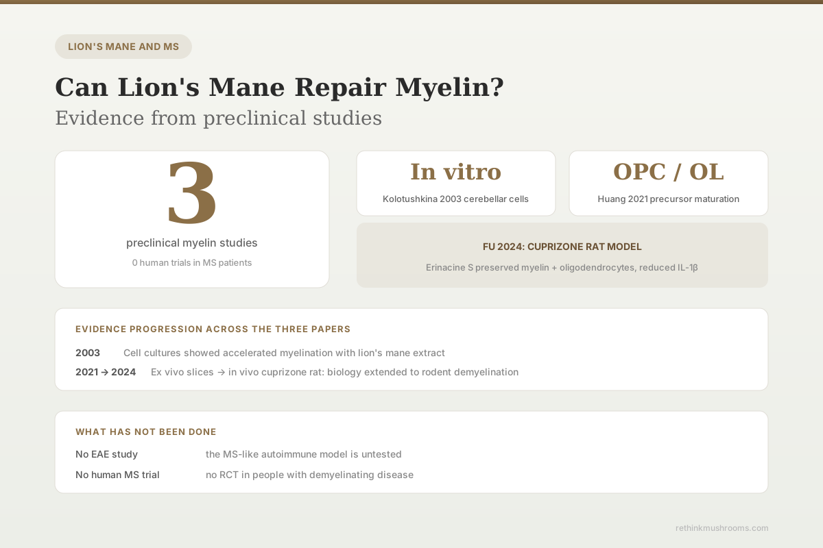
Kolotushkina 2003 showed accelerated myelination in cultured cerebellar cells; Huang 2021 showed oligodendrocyte maturation and higher myelin basic protein; Fu 2024 showed myelin and oligodendrocyte preservation in cuprizone-exposed rats.
Kolotushkina 2003; Huang 2021; Fu 2024
In vitro
Kolotushkina used cerebellar cell cultures
OPC / OL
Huang tracked oligodendrocyte precursor maturation
Cuprizone
Fu used the standard rodent demyelination model
Kolotushkina 2003; Huang 2021; Fu 2024
 View as image ↗
Myelin repair is one of the most aspirational targets in multiple sclerosis research, and lion's mane keeps showing up in preclinical papers that touch it. The question of whether Hericium erinaceus can "repair myelin" has a more specific answer than most supplement marketing admits: three preclinical studies across two decades have each tested a different layer of the question. None of them were done in humans. All three found measurable effects on myelin or the cells that build it, but the jump from in vitro cerebellar cells to rodent demyelination models to a clinical MS patient is a long one, and only the first two steps have been taken. This article walks through each study in full, explains why the cuprizone model matters, and says what the evidence does and does not support. For the broader MS picture, see Lion's Mane and Multiple Sclerosis: Remyelination Research.
Key Takeaways
- Kolotushkina et al. 2003 cultured cerebellar cells from neonatal rats and observed that Hericium erinaceus extract accelerated the myelination process, with myelination starting earlier and proceeding faster than in control cultures, while showing no cytotoxicity[1]
- Huang et al. 2021 tested Hericium erinaceus mycelium extract and its small active compounds on oligodendrocyte precursor cells and ex vivo cerebellar slices, showing that the extract promoted OPC differentiation into mature oligodendrocytes and increased myelin basic protein levels on neuronal fibers[2]
- Fu et al. 2024 tested erinacine S (a small active component of Hericium erinaceus) in cuprizone-fed rats and found that it preserved myelin and oligodendrocytes in the corpus callosum, reduced microglia and astrocyte activation, and downregulated IL-1β expression[3]
- The cuprizone model used by Fu 2024 is the standard rodent assay for testing demyelination and remyelination because it produces reproducible loss of oligodendrocytes in specific brain regions, which makes it a gatekeeper for any compound proposed as a remyelinating agent
- No randomized controlled trial has tested lion's mane in people with multiple sclerosis or any other demyelinating disease, so claims of myelin repair in humans are not supported by clinical evidence
Why myelin matters, and what "repair" actually means
Myelin is the fatty insulating sheath wrapped around neuronal axons by specialized cells called oligodendrocytes in the central nervous system. Without intact myelin, electrical signals slow, misfire, or fail entirely, which is the mechanistic basis of most symptoms in multiple sclerosis and other demyelinating diseases. Myelin "repair" in a biological sense means either preserving existing myelin when something is damaging it, or regrowing new myelin where it has been lost. These are two different endpoints and the research literature distinguishes them carefully.
Researchers test myelin outcomes at three levels of increasing realism. The first is in vitro, growing nerve and glial cells in a dish and measuring whether myelination proceeds normally. The second is in oligodendrocyte precursor cell (OPC) cultures or ex vivo tissue slices, which adds realism but still sits outside a living animal. The third is in vivo, using a rodent model in which demyelination is induced chemically or immunologically, and then testing whether a candidate compound protects against the damage or accelerates recovery. The three published lion's mane myelin studies happen to span exactly those three levels, which makes them unusually easy to compare.
Kolotushkina 2003: the original in vitro observation
The first published report of lion's mane affecting myelin came from Kolotushkina and colleagues at the Bogomoletz Institute of Physiology in Kyiv, published in 2003 in Fiziolohichnyi Zhurnal.[1] The team cultured cerebellar cells from neonatal rats and added Hericium erinaceus extract to the culture medium. They then used standard histological methods to track whether and how quickly myelin formed on neuronal processes.
Two findings stood out. First, myelination began earlier in the lion's mane cultures than in controls. Second, the rate of myelination, measured as the proportion of myelinated fibers over time, was higher. The authors noted no cytotoxic or pathological effects on either the nerve cells or the supporting glial cells, which is an important boundary condition: a compound that accelerates myelination but kills cells would not be useful. The study did not identify which component of the extract was responsible, and the extract itself was not standardized in a way that allows modern researchers to reproduce the exact preparation.
This is a classic first-observation paper. It said something real about lion's mane and myelination but left open every specific question about mechanism, dose, and active compound. For almost two decades, no one followed it up in a major way.
Huang 2021: oligodendrocyte precursors and myelin basic protein
Huang and colleagues published the second substantive myelin paper in Scientific Reports in 2021.[2] Their design was built around the oligodendrocyte lineage directly. They used OPC cultures and an ex vivo cerebellar slice system, which preserves the three-dimensional architecture of brain tissue in a way that flat dish cultures cannot. They tested both whole Hericium erinaceus mycelium extract and two identified small bioactive compounds isolated from it.
Three findings emerged. First, the mycelium extract promoted OPC differentiation into mature oligodendrocytes in a differentiation medium, meaning the precursor cells committed to becoming myelin-making cells at a higher rate than in controls. Second, the extract increased myelin basic protein (MBP) levels on neuronal fibers in the cerebellar slices. MBP is one of the most important structural proteins of the myelin sheath and a standard biomarker for myelin presence. Third, the small bioactive compounds from the mycelium were sufficient to reproduce key effects, which means the biological activity can be narrowed to specific molecules rather than the whole extract.
Huang 2021 is the paper that reopened the lion's mane and myelin conversation. Its authors explicitly framed the work as preliminary and called for further preclinical studies in remyelination models, which set up the next paper in the sequence. For more on the cellular biology, see Hericium erinaceus and Oligodendrocyte Differentiation.
Fu 2024: the cuprizone model and erinacine S
Fu and colleagues published the most clinically suggestive myelin paper in 2024.[3] Their study used the cuprizone model, which is the standard rodent assay for demyelination research. Cuprizone is a copper chelator that selectively damages oligodendrocytes when fed in the diet, producing reproducible myelin loss in specific brain regions, including the corpus callosum. Remove the cuprizone and myelin regrows over weeks. Compounds that preserve myelin during cuprizone exposure or that accelerate its return afterward are considered candidates for clinical demyelination therapies.
The Fu team tested erinacine S, one of the small active compounds from Hericium erinaceus mycelium, in cuprizone-fed rats. The treated animals showed preserved myelin and oligodendrocytes in the corpus callosum compared with untreated cuprizone controls. The researchers also found reduced activation of microglia and astrocytes, which are the central nervous system's resident immune cells, and downregulated expression of interleukin-1 beta (IL-1β), a pro-inflammatory cytokine involved in demyelinating disease. Finally, the treated animals showed improvement in mood-related behavioral measures, which is consistent with the known comorbidity of mood symptoms in MS.
This is the first lion's mane myelin study to combine a preserved-myelin biomarker with reduced neuroinflammation and a behavioral outcome in a standard rodent demyelination model. It is the strongest preclinical evidence currently available for the "lion's mane protects myelin" claim, and it ties directly into the larger neuroinflammation literature. See Lion's Mane Neuroinflammation: The Root of MS Progression for the inflammation side of the story.
Why preclinical myelin data is not the same as clinical MS evidence
A reader looking at the three studies above might reasonably ask why a supplement with measurable effects on myelin in a standard rodent model is not already a recommended MS therapy. The answer has to do with how preclinical and clinical evidence differ and why the translation gap is so large in MS specifically.
First, the cuprizone model is a chemical demyelination assay, not a model of MS per se. MS in humans is an autoimmune disease driven by T cell attack on myelin antigens; cuprizone simply kills oligodendrocytes with a copper chelator. A compound that helps in cuprizone does not automatically help in MS, because the upstream cause of damage is different. Most candidate remyelinating drugs are tested in both cuprizone and the experimental autoimmune encephalomyelitis (EAE) model, which is closer to the autoimmune mechanism of MS. No published study has yet tested lion's mane or its active compounds in EAE.
Second, preclinical dose translation to humans is rarely straightforward. Rodent studies use body-weight-adjusted doses that can be many times higher than what human supplement protocols deliver. The Huang and Fu studies used isolated compounds at experimental concentrations, not whole-mushroom powder at consumer doses. A commercial lion's mane capsule and a purified erinacine S injection in a rat are not the same intervention.
Third, MS is a heterogeneous disease with relapsing-remitting, secondary progressive, and primary progressive forms, each with different inflammation and remyelination profiles. Any drug developed for MS goes through very specific clinical pathways precisely because MS patients vary so widely. Lion's mane has not entered any of those pathways. For more on the clinical symptom side, see Lion's Mane for MS Fatigue and Cognitive Symptoms.
What a reader should take from the three studies together
The three papers, taken as a sequence, tell a reasonably consistent story. Lion's mane extract accelerated myelination in cultured cerebellar cells two decades ago. Lion's mane mycelium and its small active compounds promoted oligodendrocyte precursor maturation and increased MBP in a more advanced culture system in 2021. Erinacine S protected myelin in a standard rodent demyelination model and reduced neuroinflammatory markers in 2024. Each step supports the biological plausibility of the next, and each paper uses a methodology closer to the clinic than the last.
What is missing is a published in-vivo EAE study, any clinical trial in MS patients, and any standardized commercial preparation that matches the compounds tested in the preclinical work. A reader who is told that lion's mane can "repair myelin" should understand that the claim is grounded in real preclinical data but has not been tested in humans with demyelinating disease. The rodent data suggest that this is a legitimate direction for further research, not a reason to change clinical management.
The erinacine question and the fruiting body gap
One of the most important details in the preclinical myelin literature is which part of the Hericium erinaceus organism was tested. The Huang 2021 and Fu 2024 papers both used mycelium-derived compounds, not fruiting body powder. Erinacine S and other erinacines are concentrated in the mycelium and are largely absent from the fruiting body. Hericenones, by contrast, are found in the fruiting body. Most commercial lion's mane products sold for general health use fruiting body powder or a mixed extract, not purified erinacine S, because erinacine extraction requires specialized mycelial culture.
This creates a real alignment problem for consumers. The clinical-grade preclinical myelin evidence points toward mycelium-derived erinacines as the active components, while most supplements deliver fruiting body material. Until a trial directly compares the two preparations on a myelin outcome, readers should not assume that taking a fruiting body capsule reproduces what was tested in the Fu 2024 rat study. For reference, the Kolotushkina 2003 paper used a whole extract that was not separated into mycelium and fruiting body fractions, so it is informative about activity but not about which part of the mushroom is responsible.
A useful parallel is curcumin. The preclinical literature on curcumin is enormous and repeatedly impressive, but bioavailability and preparation issues have made clinical translation hard. Lion's mane is at an earlier stage than curcumin but faces a similar structural problem: the compounds that look most interesting in the lab are not always the ones in the bottle on the shelf.
The Bottom Line
Three preclinical studies have tested lion's mane on myelin: Kolotushkina 2003 (in vitro acceleration of myelination), Huang 2021 (oligodendrocyte precursor differentiation and higher MBP), and Fu 2024 (myelin preservation in cuprizone-fed rats with reduced neuroinflammation). The sequence builds biological plausibility for lion's mane as a candidate remyelinating agent, but no clinical trial has been run in people with MS or any demyelinating disease. The claim that lion's mane repairs myelin is preclinical evidence, not clinical proof.
Frequently Asked Questions
Sources & References
- 1RTHM-00058·Kolotushkina, E V et al. (2003). “The influence of Hericium erinaceus extract on myelination process in vitro..” Fiziolohichnyi zhurnal (Kiev.Study breakdown →PubMed →↩
- 2RTHM-00692·Huang, Hui-Ting et al. (2021). “Hericium erinaceus mycelium and its small bioactive compounds promote oligodendrocyte maturation with an increase in myelin basic protein..” Scientific reports.Study breakdown →PubMed →↩
- 3RTHM-00878·Fu, Jing-Ting et al. (2024). “Erinacine S, a small active component derived from Hericium erinaceus, protects oligodendrocytes and alleviates mood abnormalities in cuprizone-exposed rodents..” Biomedicine & pharmacotherapy = Biomedecine & pharmacotherapie.Study breakdown →PubMed →↩