Lion's Mane & Multiple Sclerosis / Remyelination
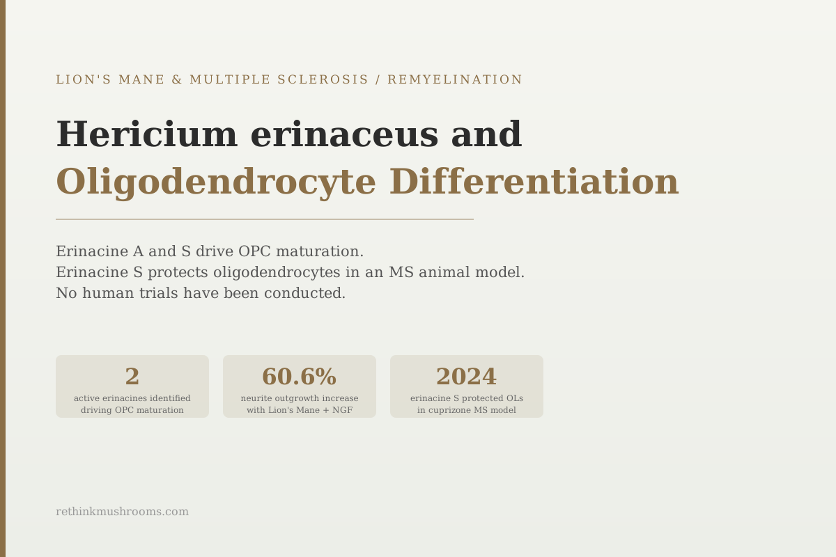
2 active erinacines
Erinacine A and erinacine S, two compounds from Lion's Mane mycelium, were the most potent agents driving oligodendrocyte precursor cells to mature into myelin-producing cells in a 2021 Scientific Reports study.
Huang et al., Scientific Reports, 2021
60.6%
peak neurite outgrowth increase when 1 µg/mL Lion's Mane extract was added to 10 ng/mL NGF (Lai 2013)
2024
year erinacine S was shown to preserve oligodendrocytes and myelin in cuprizone-fed rodents
0
human trials measuring myelin or oligodendrocyte outcomes for Lion's Mane
Huang et al., Scientific Reports, 2021
 View as image ↗
The nerve fibers that carry signals through the brain and spinal cord are wrapped in a protective insulating layer called myelin. The cells that build and maintain this layer are called oligodendrocytes. When oligodendrocytes die or fail to develop properly, the myelin sheath degrades. The result, in multiple sclerosis, is nerve signal disruption, progressive neurological damage, and the symptoms that define the disease.
Research on Lion's Mane mushroom (Hericium erinaceus) and oligodendrocyte differentiation has been building since 2003, with the most mechanistically precise work published between 2021 and 2025. This article covers what that research found, which specific compounds drive the effect, who produced the evidence, and where it currently stops. The broader context for Lion's Mane's relationship to MS and remyelination is covered in can Lion's Mane repair myelin: evidence from preclinical studies, and the overview of Lion's Mane biology is in what is Lion's Mane mushroom: a science-based overview.
Key Takeaways
- A 2021 Scientific Reports study by Huang and colleagues found that Lion's Mane mycelium extract pushed oligodendrocyte precursor cells to mature into myelin-producing oligodendrocytes, with erinacine A and erinacine S the most potent of the isolated compounds tested
- After 7 days of oral mycelium extract, neonatal rat pups showed increased myelin basic protein and more oligodendrocytes in the corpus callosum, the fiber bundle connecting the brain's two hemispheres
- In 2024, erinacine S preserved myelin and oligodendrocytes in cuprizone-fed rodents and reduced anxious and depressive-like behavior — but erinacine A, which had worked in the 2021 differentiation assay, was not reported among the compounds that protected myelin under active injury
- A second cuprizone study in 2022 found Lion's Mane fruiting bodies suppressed demyelination but, alone among four mushroom species tested, did not significantly improve motor function
- Both foundational oligodendrocyte studies come from the same laboratory and share four co-authors employed by the company that commercializes erinacine-enriched Lion's Mane mycelium — the finding has never been independently replicated
- All current evidence is preclinical. No human trial has measured oligodendrocyte or myelin outcomes for Lion's Mane in any population
What Oligodendrocytes Do and Why They Matter for Myelin Research
Myelin is not a passive coating. It is a metabolically active structure that the nervous system must constantly maintain and repair. Oligodendrocytes are the cells responsible for producing and sustaining it in the brain and spinal cord. Each mature oligodendrocyte extends dozens of arms that wrap repeatedly around nearby nerve fibers, forming the layered sheath that allows nerve signals to travel quickly and efficiently.
Before becoming mature, oligodendrocytes exist as oligodendrocyte precursor cells, or OPCs — immature cells distributed through the brain that can be recruited to repair damaged myelin. The process by which an OPC becomes a mature, myelin-producing oligodendrocyte is called differentiation. This step is a key bottleneck in remyelination: OPCs are often present in abundance at sites of myelin damage in MS, but they fail to differentiate, leaving the repair stalled.
A critical marker of successful maturation is myelin basic protein, or MBP, a structural protein that holds the layers of the myelin sheath together. When researchers measure MBP, they are measuring how much myelin-forming activity is actually happening.
Understanding these three elements — OPCs, differentiation, and MBP — is the prerequisite for reading the Lion's Mane research in this area. It also matters for reading it critically, because different studies stop at different points along that chain.
Evidence Mapping
Remyelination Is a Four-Step Chain. Every Study Stops Somewhere Along It.
“Lion’s mane repairs myelin” compresses four separate biological steps into one claim. Laid out separately, the research covers the middle of the chain well and the end of it barely — and the single study that tested motor function found lion’s mane did not move it.
“Partial” marks a step the study touched but did not measure in full: Fu 2024 shows oligodendrocytes preserved under injury rather than newly differentiated, and its behavioural endpoint is anxiety and depression-like behaviour rather than motor function. The distinction matters because preserving cells that already exist and restarting a stalled repair process are different therapeutic problems — and MS involves the second one.
Endpoints from refs [1][2][3][4] (see references)
The 2021 Scientific Reports Study: Erinacines A and S Drive OPC Maturation
Huang and colleagues published a study in Scientific Reports in 2021 that remains the most detailed mechanistic investigation of how Lion's Mane mycelium affects oligodendrocyte biology.[1] The study worked across three tiers: isolated cell cultures, ex vivo brain tissue slices, and living animals.
In OPC cultures, Hericium erinaceus mycelium extract (HEM) both promoted the differentiation of OPCs into mature oligodendrocytes and increased the level of MBP on neuronal fibers. In cultured cerebellar tissue slices — intact brain tissue kept alive outside the body, a step closer to real anatomy than dissociated cells — the overlap between MBP and neuronal fibers increased as well.
The study then asked which compounds were responsible. It evaluated erinacine A (HeA) and erinacine C (HeC), which are diterpenoid xylosides, alongside erinacine S (HeS), which is structurally different: a sesterterpene built from five isoprene units. Of these, erinacine A and erinacine S more potently stimulated MBP expression and increased the number of oligodendrocytes.[1]
The in vivo component tested whether this translated to a living system. Neonatal rat pups received daily oral HEM for seven days. MBP expression and oligodendrocyte numbers increased in the corpus callosum — the bundle connecting the brain's hemispheres, and one of the structures most affected by demyelination in MS.[1]
Huang, Ho, Sung et al. (2021) · Scientific Reports
- Population
- Rat oligodendrocyte precursor cell cultures, ex vivo cerebellar tissue slices, and neonatal rat pups
- Intervention
- Hericium erinaceus mycelium extract (HEM); isolated erinacine A, erinacine C and erinacine S; 7 days of daily oral HEM in pups
- Finding
- HEM promoted OPC-to-oligodendrocyte differentiation and raised MBP on neuronal fibers. Erinacine A and erinacine S were the most potent isolated compounds. Oral HEM increased MBP and oligodendrocyte number in the corpus callosum of neonatal rats.
positive Genuinely three-tiered and internally consistent. But the in vivo arm is a healthy, actively myelinating newborn brain — a developmental system, not a damaged adult one. Promoting myelination where it is already happening is a different problem from restarting it where it has failed.
Ref [1] Huang et al., 2021 (PMID 33753806)
This paper is the foundational reference for Lion's Mane and OPC differentiation. It named candidate active compounds, demonstrated the effect across three preparations, and confirmed an in vivo tissue change.
Erinacine S in a Live MS Model: The 2024 Cuprizone Data
The cuprizone model is a standard method for studying demyelination in rodents. Feeding animals cuprizone, a copper-chelating agent, causes reliable and reproducible damage to myelin and to the oligodendrocytes that produce it, concentrated in the corpus callosum.
A 2024 study by Fu and colleagues tested this directly.[2] Rats were fed a cuprizone diet while simultaneously receiving daily oral HEM, erinacine A, or erinacine S for three weeks. HEM and erinacine S preserved myelin and oligodendrocytes in the corpus callosum, reduced microglial and astrocyte activation, and downregulated IL-1β, an inflammatory signaling protein.
The study then ran a harder test. In mice with acute (6-week) or chronic (12-week) cuprizone-induced demyelination, erinacine S was given only during the final four weeks — treating damage already established rather than preventing it. It still preserved myelin, and it inhibited anxious and depressive-like behaviors in both models.[2]
Fu, Yang, Lee et al. (2024) · Biomedicine & Pharmacotherapy
- Population
- Cuprizone-fed rats (3 weeks) and mice with acute 6-week or chronic 12-week demyelination
- Intervention
- Daily oral HEM, erinacine A, or erinacine S; plus post-treatment erinacine S given only in the final 4 weeks
- Finding
- HEM and erinacine S preserved myelin and oligodendrocytes, reduced microglial and astrocyte activation, and downregulated IL-1β. Post-treatment erinacine S preserved myelin in both acute and chronic models and reduced anxious and depressive-like behavior.
positive The strongest result in this literature: injury, repair and behavior measured together. Two caveats matter — erinacine A is not reported among the compounds that preserved myelin here despite scoring in 2021, and the model has no autoimmune component.
Ref [2] Fu et al., 2024 (PMID 38394854)
The compound-level detail is the part most often lost in summaries. Erinacine A was one of the two standouts in the 2021 differentiation work, but in the 2024 injury model it is not reported among the compounds that preserved myelin and oligodendrocytes — only HEM and erinacine S are. Driving maturation in a healthy developing brain and protecting existing cells under active chemical attack are different tasks, and the same compound did not clear both.
Compound Specificity
Building Myelin and Defending It Were Not the Same Job
“Erinacines” is a plural that hides real chemistry — A and C are diterpenoid xylosides, S is a structurally different sesterterpene. Erinacine A drove differentiation in a healthy system and uniquely induced Nrf2, yet it is absent from the compounds reported to have protected myelin under active injury. Erinacine S is the only one that scored in both contexts.
“Given, not reported” means erinacine A was administered in that experiment but does not appear among the compounds the authors report as producing the effect. That is an absence in the published record, not a demonstrated null result, and it should not be read as evidence that erinacine A fails under injury. Note also that all three compounds are mycelium-derived; fruiting-body products contain hericenones and hericerins instead, and were not tested in either study.
Endpoints from refs [1][2][8] (see references)
A Second Cuprizone Study, and a Result That Complicates the Picture
Fu and colleagues are not the only group to have put Lion's Mane through the cuprizone model. Yamashina and colleagues published a 2022 study testing fruiting bodies of four medicinal mushroom species, each fed at 5% of the diet alongside cuprizone for five weeks, measuring body weight, motor function by rotarod, and myelin by Black-Gold II staining.[3]
All four species, including Hericium erinaceus, suppressed cuprizone-induced demyelination. But Lion's Mane was the only one of the four that did not significantly improve motor dysfunction — the endpoint closest to real-world disability. Pleurotus eryngii, Ganoderma lucidum and Flammulina velutipes all did.[3]
This does not overturn the Fu findings; the studies used different preparations (fruiting bodies rather than erinacine-rich mycelium), different species of animal and different endpoints. But it is a useful corrective to the assumption that a tissue-level myelin result automatically produces a functional one.
The Historical Anchor: What a 2003 Cell Study Established First
Nearly two decades before the compound identification, a much simpler experiment pointed in the same direction. Kolotushkina and colleagues tested whether Hericium erinaceus extract affected myelination in cultured cerebellar cells.[4]
Myelination began earlier in the presence of the extract and proceeded at a higher rate. Nerve and glial cells grew normally, cell ultrastructure remained intact, and no toxic effect was observed. This was an early finding without compound-level specificity, published in a Ukrainian physiology journal well before the erinacine biology had been worked out, but it established the basic phenomenon that the later studies would attempt to explain: something in Lion's Mane accelerates myelination in neural cell culture.
Two Pathways Beyond OPC Differentiation
The OPC differentiation pathway is not the only way Lion's Mane compounds interact with neural biology. Two additional mechanisms operate in parallel, and neither is specific to oligodendrocytes.
The first is NGF synergy. Nerve growth factor signals neurons to survive, grow and form connections. Lai and colleagues showed in 2013 that combining 1 µg/mL Lion's Mane aqueous extract with 10 ng/mL NGF produced a 60.6% increase in neurite outgrowth in NG108-15 cells — the largest increase among the combinations tested.[5] The extract also induced NGF secretion from the cells independently. Two limits are worth stating plainly: NG108-15 is a neuroblastoma-glioma line used for neurite outgrowth, not an oligodendrocyte assay, and in the same study the extract failed to protect those cells against oxidative stress in either pre-treatment or co-treatment.[5] Any link from this result to OPC maturation is an inference about shared signaling, not a measured finding.
The second is a neurotrophic pathway that bypasses the standard neurotrophin receptor. Martínez-Mármol and colleagues showed in 2023 that N-de phenylethyl isohericerin (NDPIH), a hericerin-derived compound from Lion's Mane, activated ERK1/2 signaling in cells lacking the TrkB receptor that NGF and BDNF normally work through.[6] Hericene A, the hydrophobic derivative of NDPIH, drove extensive axon outgrowth in hippocampal neurons, and mice fed Lion's Mane extract and hericene A showed increased neurotrophin signaling and significantly improved hippocampal memory.[6] These are hericerin-class compounds from fruiting bodies, structurally distinct from the mycelial erinacines — a reminder that different parts of the mushroom contribute different chemistry.
For peripheral nervous system context, Wong and colleagues published a 2012 paper presenting rat peroneal-nerve crush-injury data alongside a review of the area. Aqueous fruiting-body extract produced earlier return of hind limb function and better axon regeneration than the untreated control group, with mecobalamin (a vitamin B12 derivative used clinically for nerve disorders) as an active comparator.[7] Peripheral nerve regeneration is a genuinely different process from central remyelination — peripheral myelin is made by Schwann cells, not oligodendrocytes — so this is background context rather than direct evidence.
The 2025 Systematic Review: What It Actually Concluded
A 2025 PRISMA systematic review by Spangenberg and colleagues synthesized the preclinical evidence on erinacine compounds across cellular and rodent models.[8] Its stated conclusions were that Hericium erinaceus mycelial formulations and their derived erinacines produce dose-dependent benefits in motor, cognitive and depression-like behaviors in animal models, and that both the extracts and the isolated compounds induce antioxidant responses and activate pro-survival signaling pathways. Erinacine A and erinacine C were singled out for uniquely inducing accumulation of Nrf2, a master regulator of the antioxidant response. The review also reported anti-inflammatory activity, enhanced neurogenesis and improved cell survival.[8]
It is worth being precise here, because this review is often summarized more strongly than it reads. It did not designate oligodendrocyte protection as a headline mechanism, and it did not present a settled three-mechanism taxonomy. It included the 2021 oligodendrocyte maturation work within a broader preclinical evidence base whose dominant themes were antioxidant, anti-inflammatory and pro-survival signaling. The review's own framing is that erinacines show promise, not that any mechanism is established.
Who Produced This Evidence, and Why It Matters
The oligodendrocyte findings rest on a narrower base than the publication dates suggest. The 2021 study and the 2024 cuprizone study share the same senior author and the same institution, and four co-authors on both papers are employed by Grape King Biotech, the company that produces and commercializes erinacine-enriched Hericium erinaceus mycelium.[1][2] Both papers carry declarations of no competing interests.
This is not an allegation of misconduct, and industry-affiliated research is normal in this field. But it changes how the evidence should be weighted. The 2024 paper is best read as the same group extending its own 2021 finding into an injury model — a natural and valuable progression, but not independent replication. No unaffiliated laboratory has reproduced the OPC differentiation result. The one clearly independent cuprizone study, Yamashina 2022, used fruiting bodies rather than erinacine-rich mycelium and produced a partially discordant functional result.[3]
Evidence Provenance
Two Studies, Three Years Apart — One Laboratory
“A 2021 study and a 2024 study both found…” reads like corroboration. Here the two papers share a senior author, an institution, and four co-authors employed by the company that commercializes erinacine-enriched mycelium. The oligodendrocyte finding has never been reproduced outside this group.
Affiliations from PubMed records, refs [1][2][3][4]
Where the Evidence Stops
- Cell and tissue studiescompleted
OPC differentiation and MBP increases in culture and ex vivo cerebellar slices (Huang 2021); myelination accelerated in cerebellar culture (Kolotushkina 2003)
- Animal modelscurrent
Myelin and oligodendrocytes preserved in cuprizone rodents with reduced gliosis and IL-1β (Fu 2024); demyelination suppressed but motor function not improved (Yamashina 2022). No autoimmune MS model tested
- Human case reports with myelin outcomesmissing
None published
- Small human trials with myelin or OPC endpointsmissing
None registered or published. The only Lion's Mane trial in this species measuring cognition (Mori 2009) used a dementia rating scale, not myelin imaging or markers
- Large randomized trialsmissing
None
- Meta-analysismissing
Not possible — no human trials with these endpoints exist to pool
The evidence stops at rodent chemical-demyelination models, and the central finding has not been reproduced outside the originating laboratory. Every rung above animal models is empty.
Refs [1][2][3][4][9] (see references)
The honest picture is that this body of research is preclinical throughout. Cell and tissue studies show that erinacines promote OPC differentiation. Rodent studies show that erinacine S preserves oligodendrocytes under chemical demyelination. What is missing is human evidence. No human trial has tested Lion's Mane in MS patients, in people with myelin damage, or with any oligodendrocyte or myelin outcome measured.
The closest human data point is unrelated to myelin. A 2009 double-blind, placebo-controlled trial randomized 30 Japanese adults aged 50 to 80 with mild cognitive impairment to Lion's Mane dry powder or placebo for 16 weeks; the treated group scored significantly higher on a dementia rating scale at weeks 8, 12 and 16, and those gains faded significantly four weeks after they stopped taking it.[9] It is a small, short trial using a cognitive scale, and it says nothing about oligodendrocytes.
Safety
CriticalDo not treat this research as a myelin repair therapy
Concern
No human being has been shown to gain myelin, oligodendrocytes or neurological function from Lion's Mane. Substituting or delaying a disease-modifying therapy for MS on the strength of rodent data risks relapses and permanent, irreversible disability progression. Some MS therapies also carry rebound-disease risk if stopped.
What the research says
If used at all, treat Lion's Mane as something layered on top of a physician-managed plan — never as a replacement for one, and never as a reason to postpone starting treatment.
Particularly relevant for: Anyone with MS, clinically isolated syndrome, or other demyelinating disease, and anyone taking immunomodulating or immunosuppressive medication
What to do
Raise it with your neurologist before starting. Lion's Mane has documented immune-modulating activity, which is a specific reason to disclose it to the clinician managing your immunotherapy rather than adding it silently.
Evidence gap: no human trials with myelin or oligodendrocyte endpoints exist (refs [1][2][3])
This article is research education, not medical advice. Nothing here is a recommendation to start, stop or change any treatment or supplement.
For further reading on the specific erinacine compounds and how they cross the blood-brain barrier, see erinacines: the compounds that cross the blood-brain barrier. The NGF mechanism background is covered in hericenones and NGF synthesis: how Lion's Mane stimulates nerve growth and Lion's Mane and nerve growth factor: the complete research guide. For the broader MS research landscape, see Lion's Mane and multiple sclerosis: remyelination research and Lion's Mane and neuroinflammation: targeting the root of MS progression.
The Bottom Line
The research on Hericium erinaceus and oligodendrocyte differentiation centers on erinacine A and erinacine S, identified in a 2021 Scientific Reports study by Huang and colleagues as the most potent isolated compounds driving OPC-to-oligodendrocyte maturation, with increased myelin basic protein in cell culture, ex vivo cerebellar slices, and the corpus callosum of neonatal rats given seven days of oral extract. A 2024 study from the same laboratory found that erinacine S preserved myelin and oligodendrocytes in cuprizone-fed rodents and reduced anxious and depressive-like behavior, though erinacine A was not reported among the protective compounds in that injury model. An independent 2022 cuprizone study found Lion's Mane suppressed demyelination but, alone among four species tested, did not improve motor function. A 2025 systematic review characterized erinacines primarily in terms of antioxidant, anti-inflammatory and pro-survival signaling rather than oligodendrocyte protection. The evidence base is preclinical, concentrated in one laboratory with industry co-authorship, and has never been tested in humans for any myelin or oligodendrocyte outcome.
Frequently Asked Questions
Sources & References
- 1RTHM-00692·Huang, Hui-Ting et al. (2021). “Hericium erinaceus mycelium and its small bioactive compounds promote oligodendrocyte maturation with an increase in myelin basic protein..” Scientific reports.Study breakdown →PubMed →↩
- 2RTHM-00878·Fu, Jing-Ting et al. (2024). “Erinacine S, a small active component derived from Hericium erinaceus, protects oligodendrocytes and alleviates mood abnormalities in cuprizone-exposed rodents..” Biomedicine & pharmacotherapy = Biomedecine & pharmacotherapie.Study breakdown →PubMed →↩
- 3RTHM-01637·Yamashina, Kota et al. (2022). “Suppressive Effect of Fruiting Bodies of Medicinal Mushrooms on Demyelination and Motor Dysfunction in a Cuprizone-Induced Multiple Sclerosis Mouse Model..” International journal of medicinal mushrooms.Study breakdown →PubMed →↩
- 4RTHM-00058·Kolotushkina, E V et al. (2003). “The influence of Hericium erinaceus extract on myelination process in vitro..” Fiziolohichnyi zhurnal (Kiev.Study breakdown →PubMed →↩
- 5RTHM-00295·Lai, Puei-Lene et al. (2013). “Neurotrophic properties of the Lion's mane medicinal mushroom, Hericium erinaceus (Higher Basidiomycetes) from Malaysia..” International journal of medicinal mushrooms.Study breakdown →PubMed →↩
- 6RTHM-01746·Martínez-Mármol, Ramón et al. (2023). “Hericerin derivatives activates a pan-neurotrophic pathway in central hippocampal neurons converging to ERK1/2 signaling enhancing spatial memory..” Journal of neurochemistry.Study breakdown →PubMed →↩
- 7RTHM-00500·Wong, Kah-Hui et al. (2012). “Neuroregenerative potential of lion's mane mushroom, Hericium erinaceus (Bull.: Fr.) Pers. (higher Basidiomycetes), in the treatment of peripheral nerve injury (review)..” International journal of medicinal mushrooms.Study breakdown →PubMed →↩
- 8RTHM-02184·Spangenberg, E T et al. (2025). “Unveiling the role of erinacines in the neuroprotective effects of Hericium erinaceus: a systematic review in preclinical models..” Frontiers in pharmacology.Study breakdown →PubMed →↩
- 9RTHM-00165·Mori, Koichiro et al. (2009). “Improving effects of the mushroom Yamabushitake (Hericium erinaceus) on mild cognitive impairment: a double-blind placebo-controlled clinical trial..” Phytotherapy research : PTR.Study breakdown →PubMed →↩