Lion's Mane and MS
IL-1b + gliosis reduced in brain
In cuprizone-fed rats, lion's mane mycelium extract and erinacine S preserved myelin and oligodendrocytes while reducing microglial and astrocyte activation and downregulating IL-1-beta. This is the strongest MS-relevant neuroinflammation evidence lion's mane has.
Fu et al., Biomed Pharmacother, 2024
Brain tissue
erinacine S cut gliosis and IL-1b inside a demyelinating brain, not just a dish (2024)
Adipose model
the famous TNF-a / IL-6 result came from a fat-tissue model, not the CNS (2015)
No human MS trials
every finding here is cell or animal; DMTs remain standard care
Fu et al., Biomed Pharmacother, 2024
 View as image ↗
Multiple sclerosis is often described as an autoimmune disease. That description is accurate, but it can obscure what is actually happening inside the brain: a sustained wave of inflammatory damage that kills the cells that make myelin, strips the protective coating from nerve fibers, and leaves axons unable to transmit signals reliably. Neuroinflammation is not just a feature of MS. It is the mechanism through which the disease destroys neural tissue.
Lion's mane mushroom has published evidence for suppressing parts of this inflammatory cascade, and — in one 2024 study — for doing so inside the brain of an animal actively losing myelin. The lion's mane neuroinflammation connection is mechanistically specific, not just a general anti-inflammatory claim. But it is also entirely preclinical, and the single most-quoted piece of evidence for it comes from a study of fat tissue, not brain. Understanding both the strength and the limits of this research is essential for anyone with MS trying to evaluate it.
For broader context on what lion's mane research shows overall, see What Is Lion's Mane Mushroom? A Science-Based Overview. For the complementary myelin repair evidence, see Can Lion's Mane Repair Myelin? Evidence from Preclinical Studies.
Key Takeaways
- Neuroinflammation in MS kills oligodendrocytes, the myelin-producing cells, through cytokines including TNF-alpha, IL-1-beta and IL-6 released by activated microglia and macrophages
- The strongest MS-relevant finding: in cuprizone-fed rats, lion's mane mycelium extract and erinacine S preserved myelin and oligodendrocytes while reducing microglial and astrocyte activation and downregulating IL-1-beta (Fu et al., 2024)
- The widely cited TNF-alpha and IL-6 result is real but comes from an adipocyte-macrophage co-culture built to model obesity-related fat-tissue inflammation, not brain tissue (Mori et al., 2015)
- Lion's mane mycelium extract and the erinacines A and S drove oligodendrocyte precursor cells to mature and raised myelin basic protein in culture and in neonatal rat brain (Huang et al., 2021)
- In a four-species cuprizone comparison, lion's mane suppressed demyelination but was the only species that did not significantly improve motor function (Yamashina et al., 2022)
- No human MS trials exist for lion's mane, and nothing here addresses the autoimmune trigger; this evidence does not support replacing disease-modifying therapies
What Neuroinflammation Actually Does to the Brain in MS
Multiple sclerosis starts as an immune problem. T cells, a type of immune cell that normally helps fight infections, lose their ability to recognize myelin as "self" and attack it instead. This autoimmune attack breaches the blood-brain barrier, the protective layer that normally keeps most immune cells out of the brain, allowing inflammatory cells to enter central nervous system tissue.
Once inside, these cells activate the brain's resident immune cells, called microglia. Microglia in their resting state monitor for problems. When activated by inflammatory signals, they switch to an aggressive state that produces large amounts of inflammatory signaling proteins called cytokines. Astrocytes, another resident brain cell type, become reactive alongside them — a combined response known as gliosis.
Several cytokines matter here, and they do not all behave the same way. TNF-alpha (tumor necrosis factor alpha) and IL-1-beta (interleukin-1-beta) are consistently damaging to oligodendrocytes, the cells whose entire purpose is to wrap myelin around axons. IL-6 (interleukin-6) is more complicated: it is elevated in MS, but it is a pleiotropic cytokine that can be either damaging or protective depending on which signaling route it takes and what else is happening in the tissue. Describing IL-6 as a straightforward oligodendrocyte killer overstates a genuinely contested piece of biology.
When oligodendrocytes die, myelin is not replaced. The axon they were supporting becomes demyelinated, meaning it loses its protective sheath. Demyelinated axons transmit electrical signals slowly and unreliably. Over time, the axons themselves begin to degenerate. This progression, from inflammation to oligodendrocyte death to demyelination to axon loss, is why MS disability accumulates.
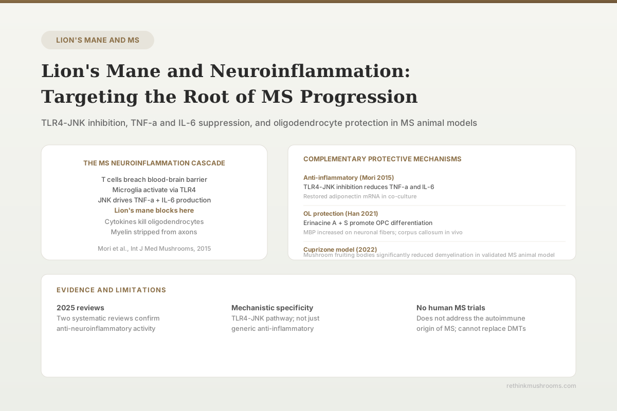
One key molecular driver of TNF-alpha and IL-6 production is a receptor called TLR4, or toll-like receptor 4. TLR4 sits on the surface of macrophages and microglia. When it detects the molecular patterns it is tuned for — bacterial products, and also damage signals released by injured tissue — it activates an internal signaling cascade called JNK, which then drives expression of these cytokines. Blocking TLR4-JNK breaks that particular chain.
That is the pathway a 2015 lion's mane study landed on. It is worth understanding precisely, because it is also the piece of this literature most often stretched beyond what it shows.
The TNF-alpha and IL-6 Result — and Why the Model Matters
Mori and colleagues in 2015 tested an ethyl acetate soluble fraction of lion's mane (abbreviated EAHE) in a co-culture of 3T3-L1 adipocytes — fat cells — and RAW264 macrophages.[1] They used LPS, a bacterial compound that strongly activates TLR4, to trigger the inflammatory response.
The findings were clear. EAHE significantly suppressed LPS-induced TNF-alpha and IL-6 production in the macrophages. It caused notable inhibition of JNK activation, which the authors identified as the likely route to that cytokine suppression. It also prevented the LPS-induced drop in adiponectin messenger RNA in the fat cells — adiponectin being an anti-inflammatory protein that normally falls in an inflammatory state.
Two points of precision are worth holding onto. First, JNK inhibition was directly measured; the TLR4 step was an interpretation. In the authors' own framing, they attributed the anti-inflammatory effect to suppression of TLR4 signaling rather than demonstrating TLR4 blockade as a separate measured endpoint. Second — and more consequential — the study was designed to investigate adipose tissue inflammation in obesity. Its stated conclusion is that lion's mane may prevent or ameliorate the fat-tissue inflammation associated with obesity. It says nothing about brain, microglia, oligodendrocytes, or MS.
Mori, Ouchi & Hirasawa (2015) · International Journal of Medicinal Mushrooms
- Population
- 3T3-L1 adipocytes co-cultured with RAW264 macrophages (cell culture)
- Intervention
- Ethyl acetate soluble fraction of Hericium erinaceus (EAHE), with LPS stimulation
- Finding
- EAHE suppressed LPS-induced TNF-alpha and IL-6 production in macrophages, inhibited JNK activation, and prevented the LPS-induced fall in adiponectin mRNA. The authors attributed the effect to suppression of TLR4 signaling.
narrow-scope Real and well-conducted, but the system models obesity-related adipose inflammation. Extending it to the MS brain is an inference, not a finding — macrophages and microglia are both myeloid cells using overlapping pathways, which makes the reasoning plausible but untested here.
Ref [1] Mori et al., 2015 (PMID 26559695)
Macrophages and microglia are both myeloid immune cells and share much of the TLR4-JNK machinery, so reasoning from one to the other is not unreasonable. But it is reasoning, not evidence. The good news for this article is that we no longer have to rely on it, because a 2024 study did the experiment in brain tissue.
For more on the specific erinacine and hericenone compounds driving these effects, see Erinacines: The Compounds That Cross the Blood-Brain Barrier.
The Strongest Evidence: Erinacine S Inside a Demyelinating Brain
Fu and colleagues in 2024 published the study that most directly tests the article's central question.[2] Rats were fed a cuprizone-containing diet — cuprizone is a copper-chelating agent that kills oligodendrocytes and induces demyelination in the corpus callosum — and simultaneously given daily oral lion's mane mycelium extract (HEM), erinacine A, or erinacine S for three weeks.
HEM and erinacine S preserved myelin and oligodendrocytes in the corpus callosum. Alongside that structural protection, the authors report reduced microglial and astrocyte activation and downregulated IL-1-beta expression. That combination is what makes this study matter: it is anti-neuroinflammatory activity measured in the actual tissue, in an animal actively losing myelin, with the myelin outcome measured in the same experiment.
The study went further. In separate post-treatment arms, mice with acute (6-week) or chronic (12-week) cuprizone-induced demyelination received erinacine S only during the final four weeks — that is, after damage was already underway. Erinacine S preserved myelin in the corpus callosum in both, and inhibited anxious and depressive-like behaviors in the treated mice.
Fu, Yang, Lee et al. (2024) · Biomedicine & Pharmacotherapy
- Population
- Cuprizone-fed rats (3 weeks) and mice with acute 6-week or chronic 12-week demyelination
- Intervention
- Daily oral lion's mane mycelium extract (HEM), erinacine A, or erinacine S
- Finding
- HEM and erinacine S preserved myelin and oligodendrocytes in the corpus callosum, reduced microglial and astrocyte activation, and downregulated IL-1-beta. Erinacine S given only in the final 4 weeks still preserved myelin in both acute and chronic models, and reduced anxious and depressive-like behavior.
positive The most MS-relevant result in the lion's mane literature: neuroinflammation, myelin and behavior measured together in CNS tissue. Still a rodent chemical-toxicity model with no autoimmune component, and no human data follows it.
Ref [2] Fu et al., 2024 (PMID 38394854)
The post-treatment design is the underrated part. Most preclinical work gives the compound at the same time as the insult, which answers a prevention question. Giving erinacine S only after demyelination is established asks something closer to the question a person with MS would actually ask.
Where the Evidence Actually Sits
The MS Cascade, Step by Step — and What Lion’s Mane Has Been Shown to Touch
Reading the cascade top to bottom shows the shape of the evidence clearly. Lion’s mane has brain-tissue data on the middle and downstream steps and none at all on the upstream autoimmune trigger. The most-quoted cytokine finding is real but comes from a fat-tissue model, not the brain.
Every row is drawn from the cited study’s own reported findings. Note the shape: the one step with no evidence at all is the autoimmune trigger — the step that disease-modifying therapies are built to address. Nothing here is a human MS finding; all seven rows are cell or animal data.
Evidence synthesis; steps from refs [1][2][3][4][9] (see references)
The Downstream Payoff: Protecting and Restoring Oligodendrocytes
Reducing inflammatory pressure on oligodendrocytes matters more when the same compounds also help replace the ones already lost.
Huang and colleagues in 2021 published a study in Scientific Reports showing that lion's mane mycelium extract promoted the differentiation of oligodendrocyte precursor cells into mature, myelin-producing oligodendrocytes.[3] These precursor cells, called OPCs, are stem-like cells that can become mature oligodendrocytes if given the right signals. In MS, OPCs are frequently present in lesions but often fail to complete differentiation.
The study worked across three systems. In OPC cultures and ex vivo cerebellar slices, the extract promoted OPC-to-oligodendrocyte differentiation and increased myelin basic protein (MBP) on neuronal fibers. In neonatal rat pups given daily oral extract for seven days, MBP expression and oligodendrocyte numbers rose in the corpus callosum. Among the isolated compounds tested, erinacine A and erinacine S more potently stimulated MBP expression and increased oligodendrocyte numbers than erinacine C did.
Worth noting: the authors framed their own findings around neurodevelopmental disorders, not MS. The MS relevance is a reasonable extension — the cell biology of OPC maturation is the same — but it is our extension, not theirs.
This finding also has a much older precedent. In 2003, Kolotushkina and colleagues cultured cerebellar cells with lion's mane extract and found that myelination began earlier and proceeded at a higher rate than in controls, with no toxic effect on the cells.[9] It is a small, old, in vitro study, but it is direct evidence on myelin genesis and it points the same direction as the modern work.
Compound Specificity
Building Myelin and Defending Myelin Are Not the Same Job
The two studies carrying the MS argument both broke lion’s mane mycelium into individual compounds. Erinacine A drove oligodendrocyte differentiation in a healthy developing system, but is not among the compounds reported to have protected myelin under active injury. Erinacine S is the only one that scored on every endpoint in both studies.
“Not reported” means the compound was administered in that experiment but does not appear among those the authors report as producing the effect. That is an absence in the record, not a demonstrated null result — an important distinction, and a reason not to over-read the erinacine A row. Note too that all three preparations are mycelium-derived; fruiting-body products contain hericenones instead, and were not tested in either study.
Endpoints from refs [2][3] (see references)
Animal Evidence: Lion's Mane in a Four-Species Comparison
The cuprizone model is one of the most widely used animal models for studying MS-like demyelination. Cuprizone specifically kills oligodendrocytes when fed to rodents, producing demyelination in predictable brain regions without the autoimmune component of MS. This makes it useful for studying demyelination and remyelination, and limited for studying MS itself.
Yamashina and colleagues in 2022 fed mice a diet containing 5% of each of four medicinal mushrooms alongside cuprizone for five weeks, and measured body weight, motor function by rotarod, and myelin by Black-Gold II staining.[4]
All four species — king oyster, reishi, enoki and lion's mane — effectively suppressed cuprizone-induced demyelination. Reishi and lion's mane both showed recovery from weight loss. But on motor function, king oyster, reishi and enoki produced significant improvement and lion's mane did not. Reishi, not lion's mane, was the only species to clear all three outcomes.
This is a result the secondary literature tends to smooth over, and it should not be. Motor function is the endpoint in this study closest to what disability means for a person. Lion's mane protected the tissue and missed the function.
One further correction to a claim that circulates about this study: it did not measure neuroinflammation. No cytokine level, microglial activation marker, or other inflammatory readout appears among its reported outcomes. It supports a demyelination claim, not a neuroinflammation claim. The neuroinflammation evidence in this article comes from Fu 2024, not from here.
What the Cuprizone Study Actually Found
Four Mushrooms, Three Outcomes — and Lion’s Mane Missed the One That Matters Most
Yamashina 2022 is usually summarised as “mushrooms reduced demyelination in an MS model.” It did. But the study tested four species against three outcomes, and lion’s mane was the only one that failed to significantly improve motor function — the endpoint closest to actual disability.
Reishi (G. lucidum), not lion’s mane, was the only species to clear all three outcomes. Note also what this study did not measure: no cytokine or microglial-activation marker was reported, so it cannot support a claim that mushrooms “reduced neuroinflammation” here — only that they suppressed demyelination and, for three of four species, improved motor function.
All outcomes from ref [4] Yamashina 2022 (see references)
Where This Stands: What the Reviews Actually Say
Three recent reviews are commonly cited in support of lion's mane for MS. Read carefully, they support less than they are used to support.
A 2025 PRISMA systematic review examined erinacine compounds across cellular and rodent models.[5] It found that lion's mane mycelial formulations and their erinacines produced dose-dependent benefits in motor, cognitive, and depression-like behaviors in animal models, induced antioxidant responses, and activated pro-survival signaling. Erinacines A and C specifically drove accumulation of Nrf2, a master regulator of the antioxidant response, and were anti-inflammatory, enhanced neurogenesis and cell survival, and improved cognitive and behavioral outcomes in vivo. What the review does not do is single out oligodendrocyte protection or NGF induction as headline findings, and it frames its conclusion as promise for aging-related and neurodegenerative conditions — not as established effect, and not specifically for MS.
A 2025 review of medicinal mushrooms for MS surveys the field more broadly.[6] It attributes potential benefit in MS to mushroom polysaccharides, terpenes and peptides acting through antioxidant enzymes, immune modulation, anti-inflammatory effects and nerve cell protection. It is a genuinely MS-focused review, which is rare. It is also notably optimistic in tone — it discusses mushrooms in the language of "curing" MS — and it does not single out lion's mane as the leading candidate species. Treat it as a map of a research area, not as an evidence appraisal.
A 2023 review of Hericium erinaceus in neurodegenerative disease provides the most useful corrective.[7] It surveys the preclinical work across Alzheimer's disease, depression, Parkinson's disease and spinal cord injury — MS is not among the conditions it covers — and correlates erinacine effects with increased neurotrophic factor production. Its central conclusion is about the state of the field rather than the compound: despite promising preclinical results, only a limited number of clinical trials have been carried out, and there is an urgent need for wider trials to establish safety and efficacy.
For additional mechanistic depth on how NGF stimulation fits into the neuroinflammation picture, see Hericenones and NGF Synthesis: How Lion's Mane Stimulates Nerve Growth and NGF Deficiency and Neurological Disease: Can Lion's Mane Help?.
What the NGF Connection Adds — and Where It Stops
Lion's mane is well known for stimulating nerve growth factor (NGF), and NGF is often described as having anti-inflammatory and pro-survival roles in the central nervous system. It is tempting to fold this into the MS argument as a second protective arm. The primary evidence urges caution.
Lai and colleagues in 2013 tested an aqueous lion's mane extract in NG108-15 neuroblastoma-glioma cells.[8] Combining 10 ng/mL NGF with 1 microgram/mL of extract produced the largest increase in neurite outgrowth, 60.6%, and the extract induced extracellular NGF secretion on its own. So far this supports the neurotrophic story.
But the same study also tested whether the extract protected those cells against oxidative stress, in both pre-treatment and co-treatment modes — and it failed to do so. The authors' own conclusion is explicit: the aqueous preparation had neurotrophic but not neuroprotective activities.
That is an important limit. Stimulating growth signaling is not the same as shielding cells from inflammatory or oxidative injury, and in this experiment lion's mane did the first and not the second. The cell-protection evidence in this article rests on Fu 2024's oligodendrocyte and myelin preservation, not on the NGF literature. It is also worth flagging that while reduced NGF signaling has been discussed in MS, that is background neurobiology rather than something the lion's mane studies cited here established.
- Cell and tissue studiescompleted
OPC differentiation and MBP increases (Huang 2021); myelination accelerated in cerebellar culture (Kolotushkina 2003); cytokine suppression in an adipose-macrophage co-culture (Mori 2015)
- Animal modelscurrent
Cuprizone rodents: myelin and oligodendrocytes preserved, gliosis and IL-1-beta reduced (Fu 2024); demyelination suppressed but motor function not improved (Yamashina 2022). No autoimmune MS model tested
- Human case reports in MSmissing
None published
- Small human MS trialsmissing
None registered or published for any MS endpoint
- Large randomized trialsmissing
None
- Meta-analysis in MSmissing
Not possible — no human MS trials exist to pool
The evidence stops at rodent chemical-demyelination models. Everything above that rung is empty, and the animal work has never been run in a model that reproduces the autoimmune attack that defines MS.
Refs [1][2][3][4][9] (see references)
What Lion's Mane Cannot Do: An Honest Assessment
Several limitations must be stated clearly for anyone with MS evaluating this research.
No human MS trials exist. Every mechanism in this article was demonstrated in cell cultures or animal models. No clinical trial has tested lion's mane in MS patients for any endpoint — neuroinflammation, relapse rate, disability progression, or symptom management. The preclinical evidence is coherent and clinically unvalidated.
Lion's mane does not address the autoimmune origin of MS. The T-cell dysregulation that drives the immune system to attack myelin is a different problem from the downstream inflammation these compounds appear to reduce. Disease-modifying therapies work at that upstream level, and have clinical evidence for reducing relapse rates and slowing disability accumulation. Lion's mane has no evidence of effect on the autoimmune mechanisms that initiate MS.
The cuprizone model is not MS. It produces demyelination through direct chemical toxicity to oligodendrocytes, with no autoimmune component. Evidence in this model demonstrates anti-demyelination activity, not anti-MS activity. Both studies underpinning the strongest claims here — Fu 2024 and Yamashina 2022 — use it.
The tissue result and the function result diverged. In the one study that measured both, lion's mane suppressed demyelination but did not significantly improve motor function, while three other mushroom species did. Protecting tissue in a stain does not automatically translate into an animal that moves better.
The specific form matters enormously. Every oligodendrocyte and myelin finding described here comes from mycelium-derived material and its erinacines. Many commercial products are fruiting-body extracts, which contain hericenones rather than erinacines and were not tested in any of these studies. The anti-inflammatory Mori 2015 data came from an ethyl acetate fraction, not a standardized commercial extract. And within the erinacines, the compounds did not behave identically across studies. Translating any of this to a product on a shelf requires knowing exactly what is in it.
Safety
CriticalDo not substitute lion's mane for disease-modifying therapy
Concern
MS disability accumulation is driven by an autoimmune process that lion's mane has never been shown to affect. Stopping or reducing a disease-modifying therapy in favour of a supplement risks relapses and irreversible disability progression. Some DMTs also carry rebound-disease risk on discontinuation.
What the research says
Treat lion's mane, if used at all, as something layered on top of a physician-managed treatment plan — never as a replacement for it, and never as a reason to delay starting one.
Particularly relevant for: Anyone with diagnosed MS or clinically isolated syndrome, and anyone taking immunomodulating or immunosuppressive medication
What to do
Discuss any supplement with your neurologist before starting. Lion's mane has documented immune-modulating activity, which is a specific reason to raise it with the clinician managing your immunotherapy rather than adding it silently.
Evidence gap: no human MS trials exist for lion's mane (refs [2][4][7])
This article is educational and is not medical advice. Nothing here should be used to diagnose, treat, or manage multiple sclerosis, or to make changes to a prescribed treatment plan. Those decisions belong with your neurologist.
The Bottom Line
Lion's mane has real preclinical evidence for the neuroinflammatory arm of MS pathology, and the best of it is more specific than the version usually quoted. In cuprizone-fed rodents, lion's mane mycelium extract and erinacine S preserved myelin and oligodendrocytes while reducing microglial and astrocyte activation and downregulating IL-1-beta — anti-inflammatory activity measured in brain tissue, with the myelin outcome measured alongside it. Erinacine S also worked when given after demyelination was already established. Separately, the extract and its erinacines pushed oligodendrocyte precursor cells to mature and raised myelin basic protein. The widely cited TNF-alpha and IL-6 finding is genuine but came from a fat-tissue model of obesity, not the brain, and the NGF work explicitly found neurotrophic but not neuroprotective activity. In the one study comparing four mushroom species, lion's mane suppressed demyelination but was the only one that failed to improve motor function. No human MS trials exist, no model tested reproduces the autoimmune attack that defines MS, and disease-modifying therapies remain the evidence-based standard of care.
Frequently Asked Questions
Sources & References
- 1RTHM-00726·Mori, Koichiro et al. (2015). “The Anti-Inflammatory Effects of Lion's Mane Culinary-Medicinal Mushroom, Hericium erinaceus (Higher Basidiomycetes) in a Coculture System of 3T3-L1 Adipocytes and RAW264 Macrophages..” International journal of medicinal mushrooms.Study breakdown →PubMed →↩
- 2RTHM-00878·Fu, Jing-Ting et al. (2024). “Erinacine S, a small active component derived from Hericium erinaceus, protects oligodendrocytes and alleviates mood abnormalities in cuprizone-exposed rodents..” Biomedicine & pharmacotherapy = Biomedecine & pharmacotherapie.Study breakdown →PubMed →↩
- 3RTHM-00692·Huang, Hui-Ting et al. (2021). “Hericium erinaceus mycelium and its small bioactive compounds promote oligodendrocyte maturation with an increase in myelin basic protein..” Scientific reports.Study breakdown →PubMed →↩
- 4RTHM-01637·Yamashina, Kota et al. (2022). “Suppressive Effect of Fruiting Bodies of Medicinal Mushrooms on Demyelination and Motor Dysfunction in a Cuprizone-Induced Multiple Sclerosis Mouse Model..” International journal of medicinal mushrooms.Study breakdown →PubMed →↩
- 5RTHM-02184·Spangenberg, E T et al. (2025). “Unveiling the role of erinacines in the neuroprotective effects of Hericium erinaceus: a systematic review in preclinical models..” Frontiers in pharmacology.Study breakdown →PubMed →↩
- 6RTHM-02163·Pourmohammadi, Golchehr et al. (2025). “New approach of using medicinal mushrooms for managing and curing multiple sclerosis..” Fitoterapia.Study breakdown →PubMed →↩
- 7RTHM-01662·Brandalise, Federico et al. (2023). “Hericium erinaceus in Neurodegenerative Diseases: From Bench to Bedside and Beyond, How Far from the Shoreline?.” Journal of fungi (Basel.Study breakdown →PubMed →↩
- 8RTHM-00295·Lai, Puei-Lene et al. (2013). “Neurotrophic properties of the Lion's mane medicinal mushroom, Hericium erinaceus (Higher Basidiomycetes) from Malaysia..” International journal of medicinal mushrooms.Study breakdown →PubMed →↩
- 9RTHM-00058·Kolotushkina, E V et al. (2003). “The influence of Hericium erinaceus extract on myelination process in vitro..” Fiziolohichnyi zhurnal (Kiev.Study breakdown →PubMed →↩