Chaga and Liver Protection
200 mg/kg Chaga polysaccharide
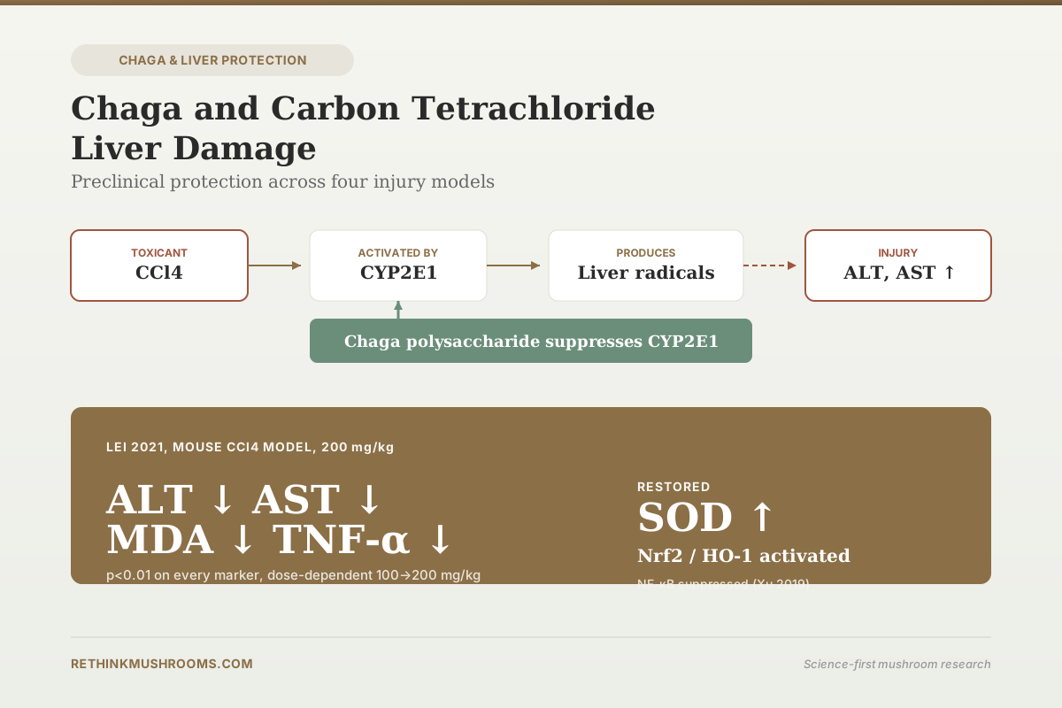
In mice with CCl4-induced acute liver injury, chaga polysaccharide at 200 mg/kg reduced ALT, AST, ALP, MDA, and TNF-α at p<0.01, restored SOD activity, and downregulated liver CYP2E1 mRNA.
Lei et al., Pakistan Journal of Pharmaceutical Sciences, 2021
Dose-dependent
100 and 200 mg/kg both protected, 200 stronger
CYP2E1 ↓
the enzyme that activates CCl4 into reactive metabolites was suppressed
0 Human Trials
no clinical trial of chaga for any liver condition has been published
Lei et al., Pakistan Journal of Pharmaceutical Sciences, 2021
 View as image ↗
Carbon tetrachloride (CCl4) is a toxicological workhorse. Rodent hepatologists use it because it damages the liver in a predictable, dose-responsive way: the CYP2E1 enzyme in hepatocytes converts CCl4 into the trichloromethyl radical, which triggers lipid peroxidation, mitochondrial injury, and inflammatory cytokine release. A natural product that blunts this damage is a candidate hepatoprotective agent, with the caveat that the model produces acute chemical injury rather than the chronic liver diseases most people care about. Chaga (Inonotus obliquus) has been tested against CCl4 in several independent mouse studies. This article walks through what they found, where the mechanism sits, and the hard boundary between preclinical signal and clinical evidence. For the broader picture, see our chaga and liver protection overview.
The short version: chaga polysaccharides and extracts consistently reduce CCl4-induced elevations in ALT and AST at doses of 100-200 mg/kg in mice, with restored antioxidant enzyme activity and suppressed CYP2E1 expression. The effect is dose-dependent and reproduces across multiple toxicant models (CCl4, alcohol, microcystin, Toxoplasma gondii). No human trial exists. A separate line of case report evidence from reishi, not chaga, shows that mushroom supplements can also cause liver injury in humans, which is an important counterweight to the preclinical protection story.
Key Takeaways
- Phaeoporus (Inonotus) obliquus polysaccharide at 100 and 200 mg/kg significantly reduced ALT, AST, ALP, MDA, and TNF-α (p<0.01) and increased SOD activity in CCl4- and alcohol-induced acute liver injury mice, with dose-dependence (Lei et al., Pakistan Journal of Pharmaceutical Sciences, 2021)
- CYP2E1 mRNA expression in liver tissue decreased significantly after chaga polysaccharide treatment compared to the CCl4 model group (p<0.05), suggesting reduced activation of CCl4 to its hepatotoxic radical (Lei et al., Pakistan Journal of Pharmaceutical Sciences, 2021)
- Chaga aqueous extract at 200 mg/kg reversed microcystin-LR-induced SGOT, SGPT, and LDH elevations (1.7-2.2 fold over control) and restored GSH from 19.83 μM back toward 50.83 μM in mouse liver (Ishfaq et al., Current Research in Pharmacology and Drug Discovery, 2022)
- Inonotus obliquus polysaccharide reduced ALT, AST, MDA, and NO and increased SOD and GSH in Toxoplasma gondii-induced liver injury, acting through TLR/NF-κB inhibition and Nrf2/HO-1 activation (Xu et al., International Journal of Biological Macromolecules, 2019)
- Chaga water extract protected rat hepatocytes against tert-butyl hydroperoxide oxidative injury in vitro by quenching free radicals and reducing ALT and AST leakage (Hong et al., International Journal of Medicinal Mushrooms, 2015)
- Zero human trials of chaga for any liver condition have been published; case reports of mushroom-related hepatotoxicity involve reishi powder, not chaga
What the CCl4 Model Actually Tests
Carbon tetrachloride is not a disease. It is a chemical. CCl4 hepatotoxicity follows a specific sequence: the molecule is taken up by hepatocytes, CYP2E1 (a cytochrome P450 enzyme in the endoplasmic reticulum) converts it to the trichloromethyl radical (•CCl3) and then to a peroxyl radical (•OOCCl3), these radicals initiate lipid peroxidation in membrane phospholipids, and the resulting damage releases cytosolic enzymes (ALT, AST, LDH) into the blood. Kupffer cells and infiltrating immune cells amplify the damage with TNF-α, IL-6, and other cytokines. Severe exposure produces hepatocyte necrosis, fatty infiltration, and centrilobular bridging damage.
The model's strength is reproducibility. Give a measured dose of CCl4 by gavage or intraperitoneal injection, and you get predictable biochemical and histological liver damage. This makes CCl4 useful for screening candidate hepatoprotective compounds: if something reduces ALT/AST elevations or restores antioxidant enzyme activity in this model, it has a plausible mechanism.
The model's weakness is that acute chemical injury in mice has limited direct relevance to the liver diseases people actually get: NAFLD, alcoholic liver disease, viral hepatitis, drug-induced injury. Acute CCl4 damage resolves quickly once exposure stops; chronic liver disease involves sustained injury, fibrosis, and altered hepatocyte biology. A compound that protects against CCl4 is a candidate, not a proven therapy. For the chronic metabolic liver angle specifically, see our chaga for fatty liver disease article.
Lei 2021: The Clearest Dose-Response Data
Lei and colleagues' 2021 study is the most directly on-topic chaga-CCl4 paper in the recent literature.[1] They used two acute liver injury models in parallel: CCl4-induced and alcohol-induced. Mice were treated with Phaeoporus obliquus polysaccharide (Phaeoporus obliquus is a taxonomic synonym for Inonotus obliquus) at 100 mg/kg or 200 mg/kg.
Compared to model (untreated CCl4-damaged) mice, the polysaccharide-treated groups showed at both doses, with p<0.01:
- Reduced liver index
- Reduced serum ALT (alanine aminotransferase)
- Reduced serum AST (aspartate aminotransferase)
- Reduced serum ALP (alkaline phosphatase)
- Reduced hepatic MDA (malondialdehyde, a lipid peroxidation marker)
- Reduced serum TNF-α
- Increased hepatic SOD (superoxide dismutase) activity
The effect was dose-dependent: 200 mg/kg produced stronger protection than 100 mg/kg on every marker. Histology showed that hepatocyte necrosis was reduced and, at the high dose, liver architecture approached normal controls.
The most mechanistically informative finding was at the mRNA level: CYP2E1 expression in liver tissue was significantly reduced (p<0.05). This matters because CYP2E1 is the enzyme that converts CCl4 into its toxic radical. If chaga polysaccharide downregulates CYP2E1, it reduces the amount of CCl4 that gets activated in the first place. This is an upstream protective mechanism, distinct from (and additive to) the downstream antioxidant effects.
CYP2E1 is also involved in ethanol metabolism, which is why the same compound protected against alcohol-induced liver injury in the same study. The shared upstream mechanism explains the shared protection. For a look at enzyme-level mechanisms more broadly, see our article on chaga and liver detoxification enzymes.
Ishfaq 2022: Different Toxicant, Same Protection Pattern
Ishfaq and colleagues used microcystin-LR (MC-LR), a cyanobacterial liver toxin, to model a different type of environmental hepatotoxicity.[2] Mice received MC-LR at 20 μg/kg body weight intraperitoneally for 4 weeks. One group also received Inonotus obliquus aqueous extract (IOAE) at 200 mg/kg orally for the same period.
Without IOAE, MC-LR produced:
- 1.9-fold SGOT elevation
- 1.7-fold SGPT elevation
- 2.2-fold LDH elevation
- GSH reduction from normal range to 19.83 ± 3.3 μM
- Reduced catalase activity
- Degenerative histological changes
- Increased caspase-3 expression (a marker of apoptosis)
With IOAE co-treatment:
- SGOT, SGPT, and LDH restored toward control levels
- GSH rose to 50.83 ± 3.0 μM (approaching the normal 52 μM reference)
- Catalase activity restored
- Histological damage reduced
- Caspase-3 expression reduced
The mechanism proposed was anti-inflammatory (via NF-κB inhibition, with molecular docking showing ergosterol peroxide binding to NF-κB-NIK) and antioxidant (via GSH and catalase restoration). The parallel with the CCl4 findings is clear: different toxicant, same pattern of enzyme marker restoration, same antioxidant system recovery, overlapping anti-inflammatory mechanism.
Xu 2019: The Nrf2/HO-1 Pathway
Xu and colleagues used Toxoplasma gondii infection to produce liver injury.[3] This is a biological rather than chemical insult, but the downstream liver damage involves the same oxidative and inflammatory pathways.
Chaga polysaccharide treatment:
- Reduced liver coefficient
- Reduced ALT, AST, MDA, and NO
- Increased SOD and GSH
- Decreased serum TNF-α, IL-6, IL-1β, IFN-γ, IL-4
- Downregulated TLR2 and TLR4
- Reduced phosphorylation of NF-κB p65 and IκBα
- Upregulated Nrf2 and HO-1
The molecular target set (TLRs/NF-κB down, Nrf2/HO-1 up) is the classical hepatoprotective signaling profile. Nrf2 is the master regulator of the cellular antioxidant response, inducing transcription of antioxidant and phase II detoxification enzymes including HO-1, NQO1, and glutathione biosynthesis enzymes. NF-κB drives inflammatory cytokine production. Pushing Nrf2 up while pushing NF-κB down shifts the hepatocyte toward an anti-inflammatory, antioxidant state. This profile has been documented for many hepatoprotective plant and fungal compounds and links chaga's liver effect to a well-characterized signaling framework.
Hong 2015: The In Vitro Confirmation
Hong and colleagues tested chaga water extract in primary cultured rat hepatocytes exposed to tert-butyl hydroperoxide (t-BHP), an oxidative injury chemical.[4] The chaga extract protected hepatocytes by:
- Quenching free radicals
- Inhibiting ALT and AST leakage from cells
- Decreasing MDA formation
In vitro studies do not prove in vivo efficacy, but they do localize the effect to the hepatocyte itself. Combined with the animal data, the picture is consistent: chaga extracts and polysaccharides produce a cell-level antioxidant and anti-inflammatory effect that translates to whole-animal protection against CCl4 and related liver toxicants.
The Consistent Mechanism Across Studies
Four independent studies using four different injury models (CCl4, alcohol, microcystin-LR, Toxoplasma gondii) converged on the same mechanistic profile:
- Downregulated CYP2E1: reducing toxicant activation at the upstream step
- Suppressed TNF-α, IL-6, IL-1β, and related pro-inflammatory cytokines
- Inhibited NF-κB signaling
- Activated Nrf2/HO-1 antioxidant response
- Restored SOD, GSH, and catalase activity
- Reduced MDA (lipid peroxidation marker)
- Reduced caspase-3 expression (apoptosis marker)
The convergence is notable. Different labs, different toxicants, different chaga preparations (aqueous extract, polysaccharide fraction, whole water extract), yet the same signaling and antioxidant shifts. That level of reproducibility is what a preclinical candidate needs before clinical testing becomes a reasonable next step. For a companion piece on chronic chemical liver damage, see our chaga and chemical liver damage article.
The Human Evidence Counterweight
Chaga's preclinical hepatoprotection is clear. The human side of the mushroom-liver literature tells a more complicated story. Case reports of mushroom supplement hepatotoxicity exist, but they involve a different species: reishi (Ganoderma lucidum), not chaga.
Wanmuang and colleagues reported fatal fulminant hepatitis in two patients after 1-2 months of reishi powder, following years of traditionally boiled reishi without problems.[5] Guedikian and colleagues reported acute hepatitis in a 47-year-old who combined reishi powder with alcohol, with significant transaminitis.[6]
These cases are worth citing here for two reasons. First, they establish that "mushroom supplement" is not automatically equivalent to "hepatoprotective," even for species in this broader family. Second, they show that preparation form matters: the Wanmuang and Guedikian patients had used powder forms, not traditional decoctions or standardized extracts, and the adverse events emerged 1-2 months into use rather than acutely.
For chaga specifically, no comparable hepatotoxicity case report has been published. The documented chaga safety signal is renal, not hepatic: case reports link chronic chaga powder to oxalate nephropathy and end-stage renal disease, covered in chaga long-term safety and the broader combination safety article. The absence of a chaga hepatotoxicity report is meaningful but not conclusive; rare events require large populations to detect. For now, the evidence base for chaga on liver is preclinical protection and no reported harm, while the evidence base for reishi on liver is rare documented harm alongside other established benefits.
What Would Confirm the Effect in Humans
A meaningful human trial would require: a standardized chaga preparation with characterized polysaccharide, triterpenoid, and oxalate content; a clinical population with an active liver condition (acute drug-induced injury, early NAFLD, or alcoholic liver disease); duration matched to the natural course of the condition (weeks for acute, months for chronic); and hard endpoints like ALT, AST, GGT, liver stiffness (FibroScan), or histology where feasible. Such a trial has not been published or registered at the time of writing. The preclinical evidence is a reasonable basis for designing one. It is not a substitute for running one. The related ongoing work on general hepatoprotection is catalogued in the chaga and liver protection hub.
The Bottom Line
Chaga (Inonotus obliquus) extracts and polysaccharides consistently reduced CCl4-induced liver injury markers in mice at 100-200 mg/kg, with dose-dependent reductions in ALT, AST, ALP, MDA, and TNF-α, restoration of SOD and GSH, and downregulation of CYP2E1 (the enzyme that activates CCl4 to its toxic radical). The same protective signaling profile - NF-κB suppression, Nrf2/HO-1 activation, antioxidant enzyme restoration - reproduced across alcohol, microcystin-LR, Toxoplasma gondii, and tert-butyl hydroperoxide models. No human trial of chaga for any liver condition has been published. Reishi (not chaga) case reports of human hepatotoxicity exist as a reminder that mushroom-and-liver claims require species-specific evidence.
Frequently Asked Questions
Sources & References
- 1RTHM-00697·Lei, Zhang et al. (2021). “Protective effect of Phaeoporus obliquus polysaccharide against acute liver injury induced by carbon tetrachloride and alcohol in mice..” Pakistan journal of pharmaceutical sciences.Study breakdown →PubMed →↩
- 2RTHM-00768·Ishfaq, Pir Mohammad et al. (2022). “Inonotus obliquus aqueous extract prevents histopathological alterations in liver induced by environmental toxicant Microcystin..” Current research in pharmacology and drug discovery.Study breakdown →PubMed →↩
- 3RTHM-00601·Xu, Lu et al. (2019). “The polysaccharide from Inonotus obliquus protects mice from Toxoplasma gondii-induced liver injury..” International journal of biological macromolecules.Study breakdown →PubMed →↩
- 4RTHM-00374·Hong, Ki Bae et al. (2015). “Hepatoprotective Activity of Water Extracts from Chaga Medicinal Mushroom, Inonotus obliquus (Higher Basidiomycetes) Against Tert-Butyl Hydroperoxide-Induced Oxidative Liver Injury in Primary Cultured Rat Hepatocytes..” International journal of medicinal mushrooms.Study breakdown →PubMed →↩
- 5RTHM-00122·Wanmuang, Harirak et al. (2007). “Fatal fulminant hepatitis associated with Ganoderma lucidum (Lingzhi) mushroom powder..” Journal of the Medical Association of Thailand = Chotmaihet thangphaet.Study breakdown →PubMed →↩
- 6RTHM-00826·Guedikian, Roupen et al. (2023). “Ganoderma lingzhi (Reishi Mushroom)-Induced Acute Liver Injury in the Setting of Alcohol Use: A Case Report and Review of the Literature..” Cureus.Study breakdown →PubMed →↩