Lion's Mane Cancer Research
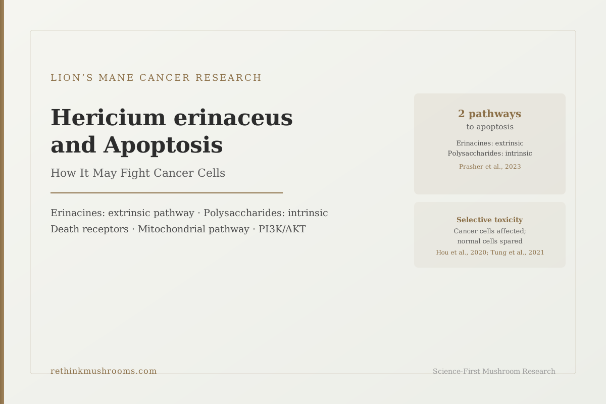
2 pathways to apoptosis
Hericium erinaceus compounds trigger cancer cell death through two mechanistically distinct routes. Erinacine diterpenoids from mycelium activate the extrinsic (death receptor) pathway. Polysaccharides from the fruiting body activate the intrinsic (mitochondrial) pathway. These are separate compound classes, separate mechanisms, and separate evidence bases.
Prasher et al., Biomedicine & Pharmacotherapy, 2023
ROS proven upstream
Adding the antioxidant N-acetyl-cysteine abolished the fruiting body polysaccharides' ability to trigger the mitochondrial cascade, placing reactive oxygen species causally upstream rather than incidental (Hou et al., Food & Function, 2020)
Xenograft confirmed
Both erinacine apoptosis findings were reproduced in xenograft mice, not just cell culture: erinacine A in colorectal (Lee et al., Frontiers in Pharmacology, 2019) and erinacine S in gastric (Tung et al., Food & Function, 2021)
No human trials
All Lion's Mane cancer evidence is preclinical. The 2025 systematic review found the human trial base covers cognition and mood, not cancer (Menon et al., Frontiers in Nutrition, 2025)
Prasher et al., Biomedicine & Pharmacotherapy, 2023
 View as image ↗
Cancer cells are supposed to die. Every normal cell in the body carries a built-in self-destruct mechanism called apoptosis (programmed cell death) that activates when the cell is too damaged to function or when it receives specific signals. Cancer cells survive by disabling this mechanism. They accumulate mutations that break the apoptosis machinery, allowing them to divide uncontrollably instead of dying as they should.
When researchers look for compounds that fight cancer, one of the things they look for is whether the compound can restore or bypass this broken apoptosis system and get cancer cells dying again.
Hericium erinaceus, the fungus known as lion's mane, turns out to contain two distinct compound classes that trigger cancer cell apoptosis through two different biological routes; those routes are mechanistically unrelated to each other.
The first route runs through erinacines, a class of diterpenoid compounds found primarily in the mycelium. Erinacines trigger what researchers call the extrinsic apoptosis pathway: the cell receives death signals from outside its surface, which activate a protein cascade that dismantles the cell from within.
The second route runs through polysaccharides found in the fruiting body. These compounds trigger the intrinsic apoptosis pathway: internal damage signals cause the cell's own mitochondria (its power-generating structures) to release death-inducing molecules, activating a different but equally lethal cascade.
All of this is preclinical evidence from cell lines and animal models. No human clinical trials have tested lion's mane for cancer. But the mechanistic specificity of the research is considerably more sophisticated than most coverage suggests. For a broader overview of the cancer research across multiple cancer types, see lion's mane and cancer: what the research shows. This article covers specifically how those mechanisms work at the molecular level.
Key Takeaways
- Lion's Mane contains two distinct compound classes that trigger cancer cell apoptosis through unrelated biological mechanisms: erinacine diterpenoids (from mycelium) work via the extrinsic death receptor pathway; polysaccharides (from fruiting body) work via the intrinsic mitochondrial pathway. The two cascades share no step until caspase-3
- Erinacine A induces extrinsic apoptosis in DLD-1 colorectal cancer cells by increasing death receptor proteins (TNFR, Fas, FasL) through JNK/p300/NFκB p50 signaling, which writes an activating histone H3K9K14 acetylation mark on those genes' promoters; confirmed in xenograft mice (Lee et al., Frontiers in Pharmacology, 2019)
- Erinacine S, a distinct molecule from erinacine A, is also epigenetic but uses a different mark: H3K4 trimethylation (not acetylation) on the FasL and TRAIL promoters in AGS gastric cancer cells, driven by ROS and AKT/FAK/PAK1 signaling. It raised ROS in cancer but not normal cells, caused G1 arrest, and slowed xenograft tumor growth (Tung et al., Food & Function, 2021)
- Fruiting body polysaccharides (HEFPs) trigger intrinsic apoptosis in colorectal cancer cells by generating reactive oxygen species (damaging oxygen molecules) that shift the Bax/Bcl-2 protein balance, collapse mitochondrial membrane potential, and activate cleaved caspase-9 then caspase-3. Blocking ROS with N-acetyl-cysteine abolished the effect, establishing ROS as causally upstream (Hou et al., Food & Function, 2020)
- A separate polysaccharide (HEFP-2b) blocks colorectal cancer cell division without killing cells outright, by arresting them in the DNA-copying phase (S-phase) of the cell cycle, a third antiproliferative mechanism distinct from both apoptosis pathways (Liu et al., Int. J. Biol. Macromolecules, 2020)
- The PI3K/AKT axis recurs across cancer types, but the reported direction is not consistent and is widely misstated: both isolated erinacine A studies report sustained phosphorylation (activation) of PI3K/mTOR/p70S6K and FAK/AKT/p70S6K as part of the killing mechanism (Lee 2017, Kuo 2017), while only the whole-extract prostate study reports suppression (Atmaca 2024)
What Apoptosis Is and Why Cancer Cells Suppress It
Apoptosis is cellular self-destruction done tidily. When a cell receives the right signals, it activates proteins called caspases that methodically break down the cell's contents and package the debris so neighboring cells and immune cells can clean up without triggering inflammation.
Every cell in the body has this capability. Most healthy cells undergo apoptosis during normal tissue maintenance, organ development, and immune cell selection. Standard physiology texts put the daily turnover in the tens of billions of cells, though the precise figure is an estimate rather than a directly measured quantity.
Cancer cells disable this process. Through accumulated mutations, they over-express proteins that block the apoptosis trigger (such as Bcl-2, which prevents mitochondria from releasing death signals), suppress proteins that promote it (like Bax, which triggers those same mitochondrial releases), or interrupt the death receptor proteins on the cell surface that normally receive external death signals.
The result is a cell that should be dying but isn't. It divides instead, accumulating further mutations, and ultimately becoming a tumor.
Compounds that can restore apoptosis in cancer cells, or trigger it through pathways the cancer hasn't managed to disable, represent a pharmacological strategy that has driven decades of drug development. Whether lion's mane compounds add meaningfully to this landscape is an open question; the evidence is preclinical and small. But the mechanisms being studied are real pharmacological targets, not vague "anti-cancer" effects.
The Two Compound Classes and the Two Pathways
Before examining the specific studies, the mechanistic division is worth establishing clearly, because it is the detail most often collapsed in summaries of this research.
Erinacines are a family of diterpenoid compounds produced primarily in the mycelium (the vegetative, underground-equivalent body of the fungus, not the mushroom cap people eat). Erinacine A, erinacine S, and related molecules are the subject of most of the cancer mechanism research. They are lipophilic (fat-soluble) compounds that can cross cell membranes and interact with intracellular signaling machinery. They are also known for crossing the blood-brain barrier, which is relevant to their neuroprotective research; that is a separate application from the cancer work covered here.
Polysaccharides (specifically HEFPs, Hericium erinaceus fruiting body polysaccharides) are large carbohydrate molecules found primarily in the fruiting body (the mushroom cap that people eat). They are water-soluble and cannot cross cell membranes directly. They interact with cancer cells through surface receptors and by generating oxidative stress inside the cell.
These compound classes target two different apoptosis pathways:
| Compound | Source | Apoptosis Type | Triggering Mechanism | Example Cancer |
|---|---|---|---|---|
| Erinacine A | Mycelium | Extrinsic (death receptor) | TNFR/Fas/FasL raised via JNK/p300/NFκB p50, with H3K9K14 acetylation on their promoters | Colorectal, gastric |
| Erinacine S | Mycelium | Extrinsic (death receptor) | FasL/TRAIL raised via H3K4 trimethylation on their promoters; also G1 arrest | Gastric |
| HEFP polysaccharides | Fruiting body | Intrinsic (mitochondrial) | ROS generation, Bax/Bcl-2 shift, caspase-9 activation | Colorectal |
| HEFP-2b polysaccharide | Fruiting body | Cell cycle arrest | S-phase blockade of DNA replication | Colorectal |
For a deeper look at erinacine chemistry and the properties that allow these compounds to cross biological barriers, see erinacines: the compounds that cross the blood-brain barrier.
Erinacine A: The Extrinsic Death Receptor Pathway in Colorectal Cancer
The most mechanistically complete study of erinacine A in cancer cells is a 2019 paper by Lee and colleagues in Frontiers in Pharmacology.[1]
The extrinsic apoptosis pathway starts outside the cell. Cells express proteins on their surface called death receptors (including TNFR1, Fas, and DR4/DR5). When the matching signal molecules (TNFR, FasL, TRAIL) bind to these receptors, the binding activates caspase-8 inside the cell, which then activates caspase-3, the final execution protein that dismantles the cell's internal structures.
Cancer cells often reduce the expression of these death receptor proteins on their surface, so the death signals have nothing to bind to. If a compound can restore or increase death receptor expression, the cancer cell becomes vulnerable to death signals again.
Lee 2019 found that erinacine A increased the expression of TNFR, Fas, and FasL proteins in DLD-1 human colorectal cancer cells. The mechanism for this increased expression was activation of a signaling chain: JNK1/2 (a stress-responsive kinase) acting through p300 (a regulatory protein) and NFκB p50 (a gene transcription factor), which turned on the genes encoding the death receptor proteins.[1]
There is a further layer to this that is usually left out. The way that signaling chain turns those genes on is epigenetic: the study reports histone H3K9K14 acetylation (acetyl groups added at lysine 9 and lysine 14) on the TNFR, Fas, and FasL promoters. Blocking the upstream kinases with specific inhibitors blocked that transcriptional activation. So erinacine A is not simply flipping a protein switch; it is chemically re-tagging the packaging around those genes so they can be read.
Erinacine A also reduced the expression of Bcl-2 and Bcl-XL, two proteins that normally prevent mitochondria from releasing death signals. Reducing these proteins lowered one of cancer's main defenses against apoptosis.
These effects were confirmed in a xenograft mouse model, where histochemical and immunohistochemical analysis showed erinacine A treatment significantly raised TNFR, Fas, and FasL levels in the tumors. The finding held beyond cell culture, which is an important step in preclinical validation.
Lee et al. (2019) · Frontiers in Pharmacology
- Population
- DLD-1 human colorectal cancer cells, plus a xenograft mouse model
- Intervention
- Erinacine A isolated from cultured Hericium erinaceus mycelium
- Comparator
- Untreated cells; kinase-inhibitor arms used to test whether each signaling step was required
- Finding
- Time-dependent activation of the extrinsic pathway (TNFR, Fas, FasL, caspases) with suppression of Bcl-2 and Bcl-XL. Transcriptional activation ran through JNK1/2, p300 and NFκB p50, with histone H3K9K14 acetylation on the TNFR, Fas and FasL promoters. Death receptor levels rose in the xenograft tumors.
preliminary Strong internal mechanism work with inhibitor controls and in vivo confirmation, but a single cell line plus a mouse xenograft. No human data.
Lee et al., Frontiers in Pharmacology, 2019 (RTHM-01081, PMID 31680958)
A 2017 proteomics study by Lee and colleagues identified additional protein targets involved in erinacine A's effects on colorectal cancer cell aggressiveness in HCT-116 and DLD-1 cells.[3] Erinacine A increased cytotoxicity and ROS production while decreasing proliferation and invasiveness, with cofilin-1 and profilin-1 emerging as the novel protein targets. The direction of the signaling in that paper is counterintuitive and is addressed in detail below.
Erinacine S: A Different Molecule, a Different Mechanism
Erinacine S is a distinct compound from erinacine A, and its mechanism against cancer cells is also epigenetic. But it uses a different chemical tag, and the difference is the detail most often reported incorrectly.
Epigenetic mechanisms control which genes are turned on or off without changing the DNA sequence itself. One such mechanism involves chemically tagging the proteins that DNA is wound around (called histones). Different tags are written by different enzyme families. Adding acetyl groups (acetylation) is one system; adding methyl groups (methylation) is another. Both can open a gene for reading, depending on exactly where they land.
A 2021 study by Tung and colleagues found that erinacine S activates death genes in gastric cancer cells through H3K4 trimethylation, not acetylation.[4]
Specifically, erinacine S drove transcriptional activation of FasL (a death signal protein) and TRAIL (another death signal protein) through trimethylation at lysine 4 on histone H3 at their promoters. With those promoters opened up, the AGS gastric cancer cells started producing more FasL and TRAIL themselves, effectively turning on their own death signals. The upstream driver was ROS-derived signaling together with the AKT/FAK/PAK1 pathways.
The study also found that erinacine S increased reactive oxygen species in gastric cancer cells but not in normal cells. This selectivity is pharmacologically significant: a compound that damages cancer cells without harming surrounding normal cells is much more likely to be clinically useful than one that is broadly toxic.
Erinacine S did more than induce apoptosis. It suppressed the anti-apoptotic proteins Bcl-2 and Bcl-XL, activated caspases-8, -9 and -3, and separately caused G1 cell cycle arrest by inactivating CDKs and cyclins. It also slowed tumor growth in a xenograft mouse model, where treated tumors showed increased FasL and TRAIL protein alongside reduced PCNA and cyclin D1, both markers of proliferation.
So the contrast between the two erinacines is not "protein signaling versus epigenetics." Both are epigenetic. The real difference is which mark gets written, which kinases drive it, and which death genes open.
Two Erinacines · Two Different Histone Marks
Both Work Epigenetically — But Not the Same Way
Erinacine A and erinacine S both switch on death-receptor genes by chemically tagging the histone proteins that DNA is wound around. They are routinely described as sharing one mechanism. They do not: the tag is different, the upstream kinases are different, and the genes that open are different.
Erinacine A
ColorectalDiterpenoid, cultured mycelium
Upstream signal
JNK1/2 → p300 → NF-kB p50 phosphorylation
Histone mark written
H3K9K14ac
Histone H3K9K14 acetylation (Acetyl Lys9/Lys14)
Promoters opened
TNFR · Fas · FasL
Cell line
DLD-1
Erinacine S
GastricDiterpenoid, cultured mycelium
Upstream signal
ROS-derived signalling → AKT/FAK/PAK1 activation
Histone mark written
H3K4me3
Histone H3K4 trimethylation
Promoters opened
FasL · TRAIL
Cell line
AGS
Why the distinction is not pedantic: acetylation and methylation are written and erased by different enzyme families. A cancer cell that has lost the machinery to respond to one mark may still respond to the other, which is the mechanistic basis for the claim that these two compounds are not redundant with each other.
Source: Lee et al., Frontiers in Pharmacology, 2019 (RTHM-01081, PMID 31680958); Tung et al., Food & Function, 2021 (RTHM-01448, PMID 33900313)
Pathway 2: Polysaccharides and the Intrinsic Mitochondrial Pathway
The fruiting body polysaccharides take a completely different route to cancer cell death. Where erinacines trigger death from outside the cell inward, polysaccharides cause internal cellular stress that the mitochondria detect and respond to.
A 2020 study by Hou and colleagues tested Hericium erinaceus fruiting body polysaccharides (HEFPs) on colorectal cancer cell lines HCT-116 and DLD1.[5] The extracted polysaccharides were characterized as being composed mainly of arabinose, galactose, glucose, and mannose.
HEFPs significantly inhibited the growth of both cancer cell lines by inducing apoptosis. The mechanism: HEFPs generated reactive oxygen species (ROS, unstable oxygen molecules that damage cellular structures) inside the cells, and this shifted the balance between two mitochondrial-regulatory proteins. Bax (which promotes mitochondrial membrane permeabilization and death signal release) increased, and Bcl-2 (which prevents it) decreased. That shift caused the loss of mitochondrial membrane potential, which in turn activated cleaved caspase-9 and then cleaved caspase-3.
The most methodologically valuable part of this study is easy to miss. The researchers added N-acetyl-L-cysteine, an antioxidant that mops up ROS, and the polysaccharides' ability to trigger the mitochondrial pathway was reduced. That is a causal test rather than a correlation: it establishes ROS generation as an upstream requirement for the apoptosis, not merely something that happens alongside it. Most preclinical mechanism claims in the supplement literature never get this far.
One caution about scope. This study examined two colorectal cancer cell lines. It does not report a comparison against normal colon epithelial cells, so it establishes the mechanism but not selectivity. The selectivity evidence for lion's mane compounds comes from other studies, discussed below.
This pathway is distinct from the extrinsic pathway used by erinacines. Erinacine A activates caspase-8 via death receptors. Polysaccharides activate caspase-9 via mitochondrial release. Both converge on caspase-3 for final cell execution, but they arrive there through different cascades. This matters because cancer cells that disable one pathway may still be vulnerable to the other.
Two Routes In · One Execution Step
Where the Two Compound Classes Actually Meet
Erinacines enter from outside the cell membrane and polysaccharides act from inside the mitochondria. The two cascades never share a step until caspase-3. Two further compounds stop cell division without triggering the cascade at all.
The practical consequence of the convergence: a tumour that has disabled its death receptors is still, in principle, reachable through the mitochondrial arm, and vice versa. That argument is mechanistic. It has not been tested in a human being.
Source: Lee et al., Frontiers in Pharmacology, 2019 (RTHM-01081); Tung et al., Food & Function, 2021 (RTHM-01448); Hou et al., Food & Function, 2020 (RTHM-01202); Liu et al., International Journal of Biological Macromolecules, 2020 (RTHM-01241)
A Third Mechanism: Blocking Cancer Cell Division Without Killing Cells
A 2020 study by Liu and colleagues isolated a specific polysaccharide fraction called HEFP-2b from Hericium erinaceus fruiting bodies and characterized its structure: a heteropolysaccharide containing fucose, galactose, glucose, and mannose in a specific molar ratio.[6]
HEFP-2b inhibited HCT-116 colorectal cancer cell growth, but through a mechanism different from both apoptosis pathways: cell cycle arrest at S-phase.
Every dividing cell goes through a cycle of phases: G1 (preparation), S (DNA synthesis), G2 (quality check), and M (division). During S-phase, the cell copies all its DNA before dividing. Compounds that arrest cells at S-phase prevent DNA copying from completing. A cell stuck in S-phase cannot divide; it is proliferatively paralyzed.
S-phase arrest is not the same as apoptosis (the cell doesn't necessarily die immediately), but it prevents the tumor from growing. Combined with apoptosis induction from other compounds in the same mushroom, the overall effect is antiproliferative through multiple routes.
The PI3K/AKT Axis: A Recurring Target With an Inconsistent Direction
A 2023 review by Prasher and colleagues in Biomedicine and Pharmacotherapy synthesized the published data on oncologic pathways affected by erinacine A, concluding that it induces apoptosis, reduces proliferation and invasiveness, generates oxidative stress, and causes cell cycle arrest across a range of preclinical models.[2]
The PI3K/AKT/mTOR pathway recurs across those models. PI3K is an enzyme that, when activated, signals AKT (another enzyme), which signals mTOR (a protein that promotes cell growth and division and suppresses apoptosis). When this chain is overactive, cancer cells grow and avoid dying. Many cancer drugs are designed specifically to block it.
This is where secondary coverage of the literature consistently goes wrong. The common summary is that lion's mane "inhibits PI3K/AKT." Reading the primary papers, that is true of one of the three studies, and the opposite of what the other two report.
In colorectal cancer cells, the 2017 Lee proteomics study reports that erinacine A's induction of cell death and its blocking of invasiveness were associated with sustained phosphorylation of the PI3K/mTOR/p70S6K and ROCK1/LIMK2/Cofilin pathways. The paper's own conclusion is that erinacine A limits the aggressiveness of colorectal cancer cells "by activating PI3K/mTOR/p70S6K downstream signalling."[3] That is activation, not inhibition.
In gastric cancer cells, a 2017 study by Kuo and colleagues found that erinacine A reduced TSGH 9201 gastric cancer cell viability and invasiveness and increased ROS, and that this apoptosis induction was "accompanied by sustained phosphorylation of FAK/AKT/p70S6K and the PAK1 pathways," with differential expression of the 14-3-3 sigma and MTUS2 proteins.[7] Again activation, not inhibition. This study was conducted in cell culture only; it did not include an animal model.
In prostate cancer cells, a 2024 study by Atmaca and colleagues found that a Hericium erinaceus ethanol extract showed concentration- and time-dependent toxicity on prostate cancer cells with minimal effect on normal cells, and that it suppressed PI3K/AKT and RAS/MAPK signaling, reducing phosphorylated protein levels.[8] This is the one study of the three that matches the popular summary, and it tested a whole extract rather than isolated erinacine A.
Same Pathway · Opposite Reported Directions
“Lion’s Mane Inhibits PI3K/AKT” Is Not What Two of the Three Papers Say
The PI3K/AKT axis does recur across cancer types. The direction of the effect does not. Both isolated-erinacine-A proteomics papers report the axis being activated, not blocked, as part of the mechanism that kills the cell. Only the whole-extract prostate study reports suppression.
What was given
Erinacine A (isolated)
Axis measured
PI3K/mTOR/p70S6K
Cell death and reduced invasiveness occurred with "sustained phosphorylation" of the axis; the paper concludes erinacine A limits aggressiveness "by activating PI3K/mTOR/p70S6K downstream signalling".
What was given
Erinacine A (isolated)
Axis measured
FAK/AKT/p70S6K + PAK1
Apoptosis induction was "accompanied by sustained phosphorylation of FAK/AKT/p70S6K and the PAK1 pathways", alongside ROS generation.
What was given
Whole ethanol extract
Axis measured
PI3K/AKT + RAS/MAPK
The extract "suppressed PI3K/AKT and RAS/MAPK pathways, reducing phosphorylated protein levels", with apoptosis induction and minimal effect on normal cells.
Where the “inhibition” framing comes from
The 2023 review that most coverage draws on is titled “Key oncologic pathways inhibited by Erinacine A” (Ref 2). That title sets the frame, and secondary summaries inherit it. Two things can be true at once: erinacine A inhibits tumour cell growth and invasiveness, while the PI3K/AKT axis it acts through shows increased phosphorylation. Conflating “inhibits the cancer” with “inhibits the pathway” is where the error enters.
Two caveats before reading this as a genuine contradiction: the studies used different interventions (isolated erinacine A versus a whole ethanol extract) and different tissues. Neither paper tested the other’s model, so no experiment has yet put the two directions in direct competition.
Source: Lee et al., Journal of Cellular and Molecular Medicine, 2017 (RTHM-00885, PMID 27709782); Kuo et al., Cellular Physiology and Biochemistry, 2017 (RTHM-00884, PMID 28854418); Atmaca et al., Chemistry & Biodiversity, 2024 (RTHM-01853, PMID 39183463); Prasher et al., Biomedicine & Pharmacotherapy, 2023 (RTHM-01769, PMID 36736282)
How can a compound kill cancer cells by activating a pro-survival pathway? Two explanations are consistent with the data. Sustained, unrelenting activation of a growth pathway can itself be lethal to a cell, a well-described phenomenon in which signaling that is beneficial in pulses becomes toxic when it never switches off. Alternatively, the phosphorylation may be a downstream consequence of the stress response rather than the cause of the death. The primary papers do not resolve which.
What can be said with confidence is narrower than the usual claim: the PI3K/AKT axis is repeatedly implicated in how Hericium compounds affect cancer cells, the intervention differs between studies (isolated erinacine A versus whole extract), and the reported direction of the pathway change is not consistent across them.
What Selective Toxicity Means and Why It Matters
A recurring finding across these studies deserves explicit attention: some lion's mane compounds showed differential effects on cancer cells versus normal cells.
Tung 2021 found erinacine S induced apoptosis and increased ROS in gastric cancer cells but not in normal cells.[4] Atmaca 2024 found prostate cancer cytotoxicity with minimal effect on normal cells.[8]
It is worth being precise about how many studies this rests on. Two of the studies covered here tested normal cells alongside cancer cells. The colorectal polysaccharide work (Hou 2020, Liu 2020) tested cancer cell lines only, so it cannot speak to selectivity in either direction. Selectivity is a real finding in the erinacine and whole-extract studies; it is not yet a demonstrated property of every Hericium compound class.
Selective toxicity is one of the core properties researchers look for in candidate cancer compounds, because broad toxicity (killing normal cells alongside cancer cells) is what drives the side effects of chemotherapy.
The selectivity mechanism isn't characterized. One hypothesis is that cancer cells carry higher baseline oxidative stress and more dysfunctional mitochondria, making them more vulnerable to an additional ROS load than healthy cells. In the erinacine S study, the specificity may relate to differences in histone methylation patterns between cancer and normal cells. Both remain hypotheses; neither has been tested directly.
This remains an area of ongoing investigation rather than an established principle.
What All of This Evidence Actually Establishes
The mechanistic depth here is genuine: multiple studies from independent research groups, different compound classes, different cancer types, and different experimental methods all converge on the conclusion that Hericium erinaceus compounds can kill cancer cells in laboratory settings.
What the evidence does not establish is clinical relevance. Cell line studies are the weakest preclinical evidence tier. Cells in culture dishes behave differently from cells in a living tumor. The concentrations at which effects appear in cell lines may not be achievable by oral supplementation. Two of the studies here did include xenograft mouse models (Lee 2019 in colorectal, Tung 2021 in gastric), which is a meaningful step up, but animal tumor models are still many steps removed from human cancer biology. The gastric proteomics study (Kuo 2017) and both polysaccharide studies were conducted entirely in cell culture.
No human clinical trial has tested lion's mane specifically for cancer prevention or treatment. A 2025 systematic review by Menon and colleagues in Frontiers in Nutrition searched the literature from January 2000 to June 2024 under PRISMA methodology, with a registered PROSPERO protocol.[9] It identified five randomized controlled trials and three pilot clinical trials. Every one of them assessed cognition or mood. The cancer evidence it catalogued was laboratory work in leukemia and gastric cancer cell lines.
That is the clearest available statement of where things stand: the human trial base for lion's mane exists, and it is about the brain. Not one participant in it was studied for a cancer outcome.
- Molecular mechanism in cell linescompleted
Extrinsic and intrinsic pathways both mapped, with inhibitor controls and an antioxidant rescue experiment establishing ROS as causally upstream (Lee 2019, Tung 2021, Hou 2020, Liu 2020, Kuo 2017, Atmaca 2024)
- Animal tumor modelscurrent
Two xenograft mouse models: erinacine A in colorectal (Lee 2019) and erinacine S in gastric (Tung 2021). No spontaneous-tumor or orthotopic models
- Case reports in cancer patientsmissing
None published
- Pilot trial with a cancer endpointmissing
None. The three pilot clinical trials in the Menon 2025 review assessed cognition
- Randomized controlled trial in cancermissing
None. The five RCTs identified by Menon 2025 assessed cognition and mood
- Meta-analysis of cancer trialsmissing
Not possible without trials to pool
The mechanistic tier is unusually well developed, with two independent xenograft confirmations and a causal ROS experiment. Above that tier there is nothing. No person has been studied taking lion's mane for a cancer outcome in any published trial.
Lee 2019 (RTHM-01081); Tung 2021 (RTHM-01448); Hou 2020 (RTHM-01202); Liu 2020 (RTHM-01241); Kuo 2017 (RTHM-00884); Lee 2017 (RTHM-00885); Atmaca 2024 (RTHM-01853); Menon 2025 (RTHM-02145)
Fact Check
“Lion's mane kills cancer cells”
The claim
“Lion's mane kills cancer cells, so taking it should help fight cancer.”
The first half is a fair reading of the cell and mouse studies. The second half is an inference nothing in the literature supports, and it travels because the two are usually stated in one breath.
What research shows
OverstatedHericium compounds do trigger apoptosis in cancer cell lines, through two separately documented pathways, with two xenograft confirmations. That is real preclinical work of above-average quality for this field.
None of it establishes that eating lion's mane or taking a supplement produces those concentrations at a tumor site in a human being. Cell line and xenograft results routinely fail to translate. No human study has measured a cancer outcome, so the gap between 'kills cancer cells in a dish' and 'helps a person with cancer' is entirely unmeasured rather than measured and small.
Mechanism claims: Lee 2019 (RTHM-01081), Tung 2021 (RTHM-01448), Hou 2020 (RTHM-01202). Human evidence status: Menon et al., Frontiers in Nutrition, 2025 (RTHM-02145)
Safety
CriticalLion's Mane Is Not a Cancer Treatment
Concern
Delaying or declining conventional cancer treatment in favour of a supplement is associated with substantially worse survival. Cancer treatment decisions are time-sensitive, and the preclinical findings described in this article provide no basis for any treatment decision. There is also no interaction data for lion's mane with chemotherapy agents, targeted therapies, or immunotherapy.
What the research says
Every study in this article was conducted in cancer cell lines or in mice. No dose, formulation, duration, or safety profile has been established for anyone with cancer. Mechanistic plausibility is not efficacy, and the two xenograft results do not change that.
Particularly relevant for: Anyone with a cancer diagnosis, anyone undergoing or considering treatment, and anyone using supplements alongside chemotherapy or immunotherapy
What to do
Discuss any supplement with your oncology team before starting it, including during active treatment, since interaction data does not exist. This article is a research summary, not medical advice.
Evidence-status statements from Menon et al., Frontiers in Nutrition, 2025 (RTHM-02145) and the preclinical studies cited throughout; treatment-delay framing reflects standard oncology clinical background
For lion's mane's role as a potential adjunct alongside conventional cancer treatment, including the immune-stimulating mechanisms and the honest assessment of what remains unknown about drug interactions, see lion's mane as a cancer adjunct: preclinical evidence.
The Bottom Line
Hericium erinaceus contains two compound classes that trigger cancer cell apoptosis through distinct mechanisms. Erinacine A (from mycelium) activates the extrinsic death receptor pathway in DLD-1 colorectal cells via JNK/p300/NFkB p50 signaling, which writes a histone H3K9K14 acetylation mark on the TNFR, Fas and FasL promoters. Erinacine S is also epigenetic but uses a different mark, H3K4 trimethylation, on the FasL and TRAIL promoters in gastric cancer cells. Both were confirmed in xenograft mice. Fruiting body polysaccharides trigger the intrinsic mitochondrial pathway by generating oxidative damage that shifts Bax/Bcl-2 balance and activates caspase-9; an antioxidant rescue experiment established ROS as causally upstream. A third polysaccharide (HEFP-2b) arrests cancer cell division at S-phase without immediate cell death. The PI3K/AKT axis recurs across models, but the direction is inconsistent: both isolated erinacine A studies report sustained activation of the axis, and only the whole-extract prostate study reports suppression. Selective toxicity against normal cells was tested and found in the erinacine S and whole-extract studies, not in the colorectal polysaccharide work. All evidence is preclinical; the human trial base for lion's mane covers cognition and mood, not cancer.
Frequently Asked Questions
Sources & References
- 1RTHM-01081·Lee, Ko-Chao et al. (2019). “Induction Apoptosis of Erinacine A in Human Colorectal Cancer Cells Involving the Expression of TNFR, Fas, and Fas Ligand via the JNK/p300/p50 Signaling Pathway With Histone Acetylation..” Frontiers in pharmacology.Study breakdown →PubMed →↩
- 2RTHM-01769·Prasher, Parteek et al. (2023). “Key oncologic pathways inhibited by Erinacine A: A perspective for its development as an anticancer molecule..” Biomedicine & pharmacotherapy = Biomedecine & pharmacotherapie.Study breakdown →PubMed →↩
- 3RTHM-00885·Lee, Ko-Chao et al. (2017). “A proteomics approach to identifying novel protein targets involved in erinacine A-mediated inhibition of colorectal cancer cells' aggressiveness..” Journal of cellular and molecular medicine.Study breakdown →PubMed →↩
- 4RTHM-01448·Tung, Shui-Yi et al. (2021). “Apoptotic mechanisms of gastric cancer cells induced by isolated erinacine S through epigenetic histone H3 methylation of FasL and TRAIL..” Food & function.Study breakdown →PubMed →↩
- 5RTHM-01202·Hou, Xiao-Xiao et al. (2020). “Fruiting body polysaccharides of Hericium erinaceus induce apoptosis in human colorectal cancer cells via ROS generation mediating caspase-9-dependent signaling pathways..” Food & function.Study breakdown →PubMed →↩
- 6RTHM-01241·Liu, Jing-Yu et al. (2020). “Isolation and structural characterization of a novel polysaccharide from Hericium erinaceus fruiting bodies and its arrest of cell cycle at S-phage in colon cancer cells..” International journal of biological macromolecules.Study breakdown →PubMed →↩
- 7RTHM-00884·Kuo, Hsing-Chun et al. (2017). “A Comparative Proteomic Analysis of Erinacine A's Inhibition of Gastric Cancer Cell Viability and Invasiveness..” Cellular physiology and biochemistry : international journal of experimental cellular physiology.Study breakdown →PubMed →↩
- 8RTHM-01853·Atmaca, Harika et al. (2024). “Hericium erinaceus Extract Induces Apoptosis via PI3K/AKT and RAS/MAPK Signaling Pathways in Prostate Cancer Cells..” Chemistry & biodiversity.Study breakdown →PubMed →↩
- 9RTHM-00943·Menon, Arya et al. (2025). “Benefits, side effects, and uses of Hericium erinaceus as a supplement: a systematic review..” Frontiers in nutrition.Study breakdown →PubMed →↩