Psilocybin Brain Imaging
3x greater change
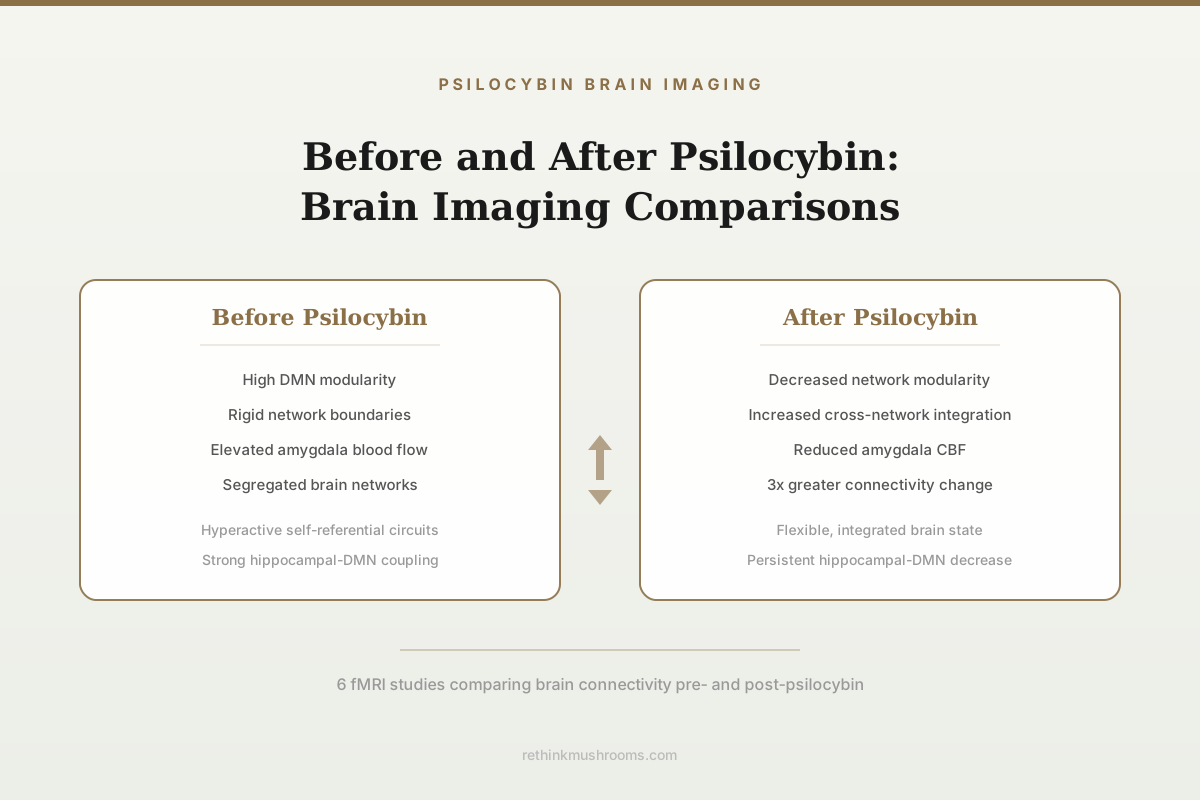
Psilocybin produced more than threefold greater functional connectivity change than methylphenidate (a stimulant control), with effects strongest in the default mode network.
Siegel et al., Nature, 2024
DMN
default mode network most affected
Weeks
persistent hippocampal-DMN changes post-dose
6 Studies
with pre/post fMRI brain scans
Siegel et al., Nature, 2024
 View as image ↗
Brain imaging has transformed the study of psilocybin from subjective experience reports into measurable neuroscience. Functional magnetic resonance imaging (fMRI) can capture brain activity before a psilocybin dose and compare it to activity after treatment, revealing which networks change, how they change, and whether those changes correlate with clinical improvement. The consistent finding across multiple research groups is that psilocybin reduces the rigidity of brain network organization, increases communication between networks that are normally segregated, and produces persistent changes in specific circuits relevant to depression. These are not subtle effects. In the most precisely measured study to date, psilocybin produced over three times more functional connectivity disruption than a stimulant control drug. For the broader context on fMRI during the acute psilocybin experience, see the companion article.
Key Takeaways
- Psilocybin caused more than threefold greater functional connectivity change than methylphenidate, with effects strongest in the default mode network and persisting for weeks in the hippocampal-DMN circuit (Siegel et al., Nature, 2024)
- In 19 patients with treatment-resistant depression, psilocybin decreased cerebral blood flow in the amygdala, and this decrease correlated with reduced depressive symptoms at 5 weeks (Carhart-Harris et al., 2017)
- Across two clinical trials, psilocybin's antidepressant response correlated with decreased brain network modularity, while the same network changes were absent after escitalopram treatment (Daws et al., Nature Medicine, 2022)
- Baseline fMRI functional connectivity predicted psilocybin treatment outcomes with 85% accuracy in a machine learning analysis, identifying potential biomarkers for patient selection (Copa et al., 2024)
- The first psilocybin fMRI study showed decreased activity in key brain hubs including the medial prefrontal cortex and posterior cingulate cortex, overturning the assumption that psychedelics increase brain activity (Carhart-Harris et al., 2012)
- A single psilocybin dose increased synaptic density by 12% in pig hippocampus while decreasing 5-HT2A receptor density by 15%, measured by PET imaging (Raval et al., 2021)
The First Psilocybin Brain Scan: Overturning Assumptions
Carhart-Harris et al. (2012) published the first fMRI study of psilocybin's acute effects in healthy volunteers, and the results contradicted the prevailing assumption that psychedelics work by increasing brain activity.[1]
Using a task-free fMRI protocol designed to capture the transition from normal waking consciousness to the psychedelic state, the researchers administered intravenous psilocybin to 30 healthy volunteers. Rather than the expected increases in brain activity, they observed pronounced decreases. Blood oxygen-level dependent (BOLD) signal dropped in key brain hubs: the medial prefrontal cortex (mPFC), the posterior cingulate cortex (PCC), and the anterior cingulate cortex. These regions form the core of the default mode network (DMN).
The magnitude of DMN deactivation correlated with the intensity of subjective psychedelic effects. Volunteers who experienced the strongest ego dissolution, visual phenomena, and altered sense of meaning showed the greatest reductions in DMN activity. This correlation established a direct link between measurable brain changes and the subjective psychedelic experience.[1]
This finding reframed the neuroscience of psychedelics. The DMN is associated with self-referential thinking, rumination, and the maintenance of a stable sense of self. In depression, the DMN tends to be hyperactive and rigidly connected, which correlates with the repetitive negative thought patterns characteristic of the disorder. Psilocybin's suppression of DMN activity offered a neurobiological explanation for both the acute psychedelic experience (dissolving the sense of self) and its potential therapeutic mechanism (breaking rigid patterns of self-referential thinking). For more on the receptor-level basis of these effects, see the article on 5-HT2A receptor density and psilocybin response.
Before and After in Treatment-Resistant Depression
Carhart-Harris et al. (2017) moved from healthy volunteers to patients with treatment-resistant depression, conducting fMRI scans before and after psilocybin treatment.[2]
Nineteen patients who had failed to respond to at least two antidepressant medications received two psilocybin doses (10 mg followed by 25 mg, one week apart) with psychological support. fMRI was performed at baseline and one day after the second (25 mg) dose.
Whole-brain analyses revealed post-treatment decreases in cerebral blood flow (CBF) in the temporal cortex, including the amygdala. This amygdala CBF decrease correlated with reduced depressive symptoms. The amygdala processes threat and negative emotional stimuli; its hyperactivity is one of the most replicated neuroimaging findings in depression. Psilocybin's acute suppression of amygdala blood flow may represent a resetting of threat-processing circuits.
Resting-state functional connectivity also changed. Post-treatment, the default mode network showed increased connectivity with other brain networks, a pattern opposite to what is typically seen in depression (where the DMN becomes increasingly isolated and self-referentially focused). At one week post-treatment, all 19 patients showed decreased depressive symptoms, and 47% met criteria for clinical response at five weeks.[2]
The limitation of this study was the open-label design: patients knew they were receiving psilocybin, and the absence of a placebo comparison makes it impossible to attribute brain changes exclusively to the drug rather than to expectation, therapeutic context, or time. The subsequent controlled trials addressed this gap.
Global Brain Integration: The Nature Medicine Finding
Daws et al. (2022) published the most methodologically rigorous before-and-after analysis, combining data from two clinical trials with different designs.[3]
Trial 1 was the open-label study in treatment-resistant depression (psilocybin 10 mg and 25 mg, seven days apart, fMRI at baseline and one day post-treatment). Trial 2 was a double-blind randomized controlled trial in major depressive disorder comparing psilocybin (two 25 mg doses, three weeks apart) plus daily placebo capsules against microdose psilocybin (two 1 mg doses) plus daily escitalopram (10-20 mg).
In both trials, the antidepressant response to psilocybin was rapid and sustained. The critical neuroimaging finding: psilocybin treatment correlated with decreased brain network modularity. This means the boundaries between functionally distinct brain networks became less rigid. Higher-order networks rich in 5-HT2A receptors became more functionally interconnected and flexible.[3]
The escitalopram comparison was revealing. Escitalopram produced a milder antidepressant response, and no changes in brain network organization were observed. The antidepressant that works through gradual serotonin reuptake inhibition did not produce the network-level reorganization that psilocybin induced. This dissociation suggests that psilocybin's therapeutic mechanism is fundamentally different from conventional antidepressants: it does not just alter serotonin levels but restructures the functional architecture of brain networks.
The correlation between decreased modularity and clinical improvement is consistent with the hypothesis that depression involves excessive network segregation (rigid patterns of connectivity that reinforce negative thought loops), and that psilocybin breaks these patterns by temporarily increasing cross-network communication.
Precision Mapping: Psilocybin Desynchronizes the Brain
Siegel et al. (2024) published the most technically advanced psilocybin neuroimaging study to date in Nature, using precision functional mapping with densely repeated fMRI sessions in individual participants.[4]
Six healthy adults underwent approximately 18 MRI visits each, providing individual-specific brain network maps rather than group-averaged data. Each participant received a single 25 mg psilocybin dose and 40 mg methylphenidate (Ritalin, as an active control), with follow-up imaging six to twelve months later.
Psilocybin produced more than threefold greater functional connectivity change than methylphenidate. The effect was characterized as "massive disruption" through brain desynchronization: within-network correlations decreased and between-network anticorrelations (the normal push-pull relationship between networks) diminished. The result was a dissolving of network distinctions, a brain temporarily operating without its normal organizational boundaries.
Effects were strongest in the default mode network, confirming and extending earlier findings. The DMN connects to the anterior hippocampus, a region involved in spatial memory, temporal processing, and self-referential thought. Psilocybin caused a persistent decrease in functional connectivity between the anterior hippocampus and the default mode network lasting for weeks after a single dose.[4]
Individual differences in functional connectivity changes were strongly linked to the subjective psychedelic experience. Participants who reported more intense experiences showed greater network disruption. An unexpected finding: performing a perceptual task during the psilocybin session reduced the drug-driven connectivity changes, indicating that focused cognitive engagement competes with psilocybin's desynchronizing effect.
The persistent hippocampal-DMN disconnection is a candidate mechanism for psilocybin's lasting antidepressant effects. The hippocampus-DMN circuit is implicated in autobiographical memory retrieval and rumination. Weakening this connection could reduce the automatic activation of negative self-referential memories that characterizes depression. For more on how PET imaging visualizes psilocybin receptor binding, see the dedicated analysis.
Predicting Who Will Respond
Copa et al. (2024) asked whether baseline brain scans before psilocybin treatment could predict who would respond and who would not.[5]
Using a machine learning pipeline applied to baseline resting-state fMRI data from patients with treatment-resistant and major depression who received psilocybin, the researchers achieved 85% prediction accuracy for treatment outcomes. Specific patterns of baseline functional connectivity in networks involving the prefrontal cortex, anterior cingulate, and default mode network differentiated eventual responders from non-responders before any drug was administered.
This finding has practical implications. If validated in larger samples, baseline fMRI could serve as a patient selection biomarker, identifying individuals most likely to benefit from psilocybin-assisted therapy and sparing others the cost and experience of a treatment unlikely to help them. Given that psilocybin-assisted therapy costs upward of AUD $20,000 per course in Australia's pioneering prescribing program, the ability to predict responders could dramatically improve the cost-effectiveness of the treatment model.
The 85% accuracy is high for psychiatric prediction models, which typically perform poorly with symptom-based measures. However, this was derived from a relatively small sample, and the specific connectivity features that predicted outcomes need replication across independent datasets before clinical use.
Synaptic Density Changes: PET Evidence
Raval et al. (2021) used positron emission tomography (PET) rather than fMRI to capture structural changes in the brain after psilocybin, providing a complementary perspective to the connectivity studies.[6]
In pigs given a single psychedelic dose of psilocybin, PET imaging with the synaptic vesicle glycoprotein 2A (SV2A) radioligand showed a 12% increase in hippocampal synaptic density one day post-dose. Simultaneously, 5-HT2A receptor density decreased by 15% in the hippocampus. Both changes were measured at the same timepoint.
The simultaneous increase in synaptic density and decrease in 5-HT2A receptor density suggests a dual mechanism: psilocybin promotes the growth of new synaptic connections (synaptogenesis) while downregulating the very receptors through which it acts. The synaptogenesis finding aligns with the "neural plasticity" hypothesis of psychedelic therapy: psilocybin does not just temporarily alter brain activity during the acute experience but promotes lasting structural changes in neural circuits. The receptor downregulation may explain the rapid tolerance to psilocybin's subjective effects with repeated dosing.
While pig brains are not human brains, the pig model offers closer anatomical and receptor distribution similarity to humans than rodent models. Whether the same magnitude of synaptic density increase occurs in human hippocampus after psilocybin remains unmeasured, as SV2A PET studies in humans receiving psychedelics have not yet been published. For how this relates to what brain scans reveal about psilocybin treating depression, see the companion analysis.
The Emerging Picture
The before-and-after brain imaging literature reveals a coherent sequence of psilocybin effects across temporal scales:
Acutely (during the dose): Brain networks desynchronize. The default mode network loses its normal coherence. Between-network boundaries dissolve. The magnitude of this disruption correlates with the intensity of the subjective psychedelic experience.
Subacutely (one day post-dose): Amygdala blood flow decreases. Brain network modularity drops. Higher-order networks become more interconnected. Synaptic density increases in the hippocampus.
Persistently (weeks to months): Hippocampal-DMN connectivity decreases. The degree of network integration change correlates with antidepressant response. Escitalopram produces clinical improvement without these network-level changes.
This temporal sequence suggests that psilocybin's therapeutic mechanism involves acute network disruption that enables a more flexible reorganization of brain connectivity during a window of enhanced neuroplasticity. The resulting connectivity patterns are measurably different from baseline and from the patterns produced by conventional antidepressants. Whether this reorganization is permanent or gradually reverts without repeated dosing is an open question, as most imaging studies have follow-up periods of weeks to months rather than years.
The ability to predict treatment outcomes from baseline scans suggests that specific patterns of brain connectivity predispose certain individuals to benefit from this network-level reorganization. Understanding these patterns could eventually enable personalized treatment selection in psychiatry, matching patients to the therapeutic mechanism most likely to address their specific neurocircuit dysfunction. For how these insights connect to the visual guide of fMRI during the psilocybin experience, see the dedicated article.
The Bottom Line
fMRI studies conducted before and after psilocybin treatment consistently show that the drug reduces brain network rigidity, increases cross-network communication, and produces persistent changes in default mode network connectivity. These changes correlate with antidepressant response and are absent after conventional SSRI treatment. The most precise study to date found that psilocybin causes over threefold greater connectivity disruption than a stimulant control, with default mode network effects persisting for weeks. Machine learning analysis of baseline brain scans can predict treatment response with 85% accuracy, and PET imaging shows psilocybin increases synaptic density while downregulating 5-HT2A receptors.
Frequently Asked Questions
Sources & References
- 1RTHM-00245·Carhart-Harris, Robin L et al. (2012). “Neural correlates of the psychedelic state as determined by fMRI studies with psilocybin..” Proceedings of the National Academy of Sciences of the United States of America.Study breakdown →PubMed →↩
- 2RTHM-00463·Carhart-Harris, Robin L et al. (2017). “Psilocybin for treatment-resistant depression: fMRI-measured brain mechanisms..” Scientific reports.Study breakdown →PubMed →↩
- 3RTHM-00752·Daws, Richard E et al. (2022). “Increased global integration in the brain after psilocybin therapy for depression..” Nature medicine.Study breakdown →PubMed →↩
- 4RTHM-00911·Siegel, Joshua S et al. (2024). “Psilocybin desynchronizes the human brain..” Nature.Study breakdown →PubMed →↩
- 5RTHM-00872·Copa, Débora et al. (2024). “Predicting the outcome of psilocybin treatment for depression from baseline fMRI functional connectivity..” Journal of affective disorders.Study breakdown →PubMed →↩
- 6RTHM-00714·Raval, Nakul Ravi et al. (2021). “A Single Dose of Psilocybin Increases Synaptic Density and Decreases 5-HT2A Receptor Density in the Pig Brain..” International journal of molecular sciences.Study breakdown →PubMed →↩