Psilocybin Neuroimaging
39%
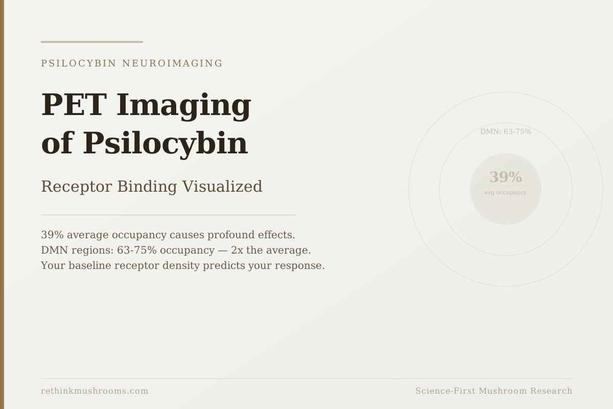
average 5-HT2A receptor occupancy is sufficient to produce profound psychedelic effects. Default mode network regions show 63-75% occupancy, far above the whole-brain average, explaining why these regions dominate the psilocybin experience.
Madsen et al., Neuropsychopharmacology, 2019
~20% threshold
minimum 5-HT2A occupancy where psychedelic effects begin to emerge
Baseline predicts
pre-dose receptor density predicts mystical experience intensity and time distortion
Pig PET 2021
single dose increased synaptic density AND decreased receptor density; both visible on scan
Madsen et al., Neuropsychopharmacology, 2019
 View as image ↗
Before PET imaging, psilocybin researchers could watch what the brain does during a psychedelic experience. What they could not directly measure was which receptors were responsible and how many were being occupied. PET imaging filled that gap. When Madsen and colleagues published the first human PET study of psilocybin in 2019, they found something that has not been widely explained in accessible language: profound psychedelic experiences occur when only about 39% of the brain's serotonin 2A receptors are occupied. That average conceals a distribution: in certain brain regions, particularly those in the default mode network, occupancy reaches 63-75%. This is a psilocybin PET imaging article about what those numbers mean and why they matter.
For the broader fMRI picture of what happens to the brain during a psilocybin experience, psilocybin and the default mode network: how it quiets the ego covers the complementary imaging technique.
Key Takeaways
- PET imaging directly measures receptor occupancy (the fraction of a given receptor type that is bound to a drug) using radioactive tracer molecules that compete with psilocin for the same binding sites
- The Madsen 2019 PET study (8 volunteers, doses 3-30mg) found psychedelic effects began emerging above approximately 20% 5-HT2A receptor occupancy, with subjective intensity correlating linearly with occupancy percentage[1]
- Average whole-brain 5-HT2A occupancy at pharmacologically active doses was approximately 39%, but default mode network regions (subgenual anterior cingulate and angular gyri) showed 63-75% occupancy, the highest regional concentrations[1]
- A 2021 PET study (Stenbæk) found that pre-dose 5-HT2A receptor density in the brain predicted the intensity of mystical experiences and temporal distortion under psilocybin; individual baseline receptor density explains part of why people respond differently[2]
- A 2021 pig PET study (Raval) found that a single psilocybin dose simultaneously increased synaptic density (measured by a different tracer targeting a synaptic protein) and decreased 5-HT2A receptor density in the prefrontal cortex; both changes visible on PET[3]
What PET Imaging Is and Why fMRI Couldn't Answer This Question
Functional MRI (fMRI) measures blood flow changes in the brain. When a region becomes more active, it needs more oxygen, and oxygenated blood rushes in; fMRI detects this as a signal change. fMRI tells you which regions of the brain are more or less active during an experience. It does not tell you which specific receptors are driving that activity.
PET imaging works through a different principle. Researchers first synthesize a radioactive molecule designed to bind to a specific receptor type, in this case the serotonin 2A receptor (5-HT2A). This radioactive molecule is called a radioligand. When you inject the radioligand into a research participant, it travels to the brain and binds to available 5-HT2A receptors. The PET scanner detects the radioactivity and produces an image showing where the radioligand is concentrated.
Now introduce psilocybin. Psilocin (what psilocybin converts into in the body) also binds to 5-HT2A receptors. If you give psilocybin before or alongside the radioligand, psilocin occupies some of the receptors first. When those receptors are already taken by psilocin, the radioligand has nowhere to bind and shows up at lower concentrations in that region. By measuring how much less radioligand binds compared to a baseline scan, researchers can calculate what percentage of receptors psilocin is occupying.
That percentage is receptor occupancy. It is a direct, quantitative measure of drug-receptor interaction that fMRI cannot provide.
The 5-HT2A receptor is the primary target for psilocin. Activating 5-HT2A receptors produces the changes in perception, consciousness, and sense of self associated with psychedelics. The receptor is expressed throughout the brain but is particularly concentrated in the cortex, especially in regions involved in self-referential processing and perception.
The Madsen 2019 Study: Mapping Occupancy to Experience
The first human PET study of psilocybin-receptor binding was published by Madsen and colleagues in 2019.[1] Eight healthy volunteers each completed multiple sessions at different psilocybin doses (3mg, 6mg, and up to 30mg). For each session, they received both PET scanning with a radioligand called [11C]Cimbi-36 (which targets 5-HT2A) and comprehensive ratings of their subjective experience.
The core finding was a direct dose-response relationship: the more psilocin was present in the blood, the more 5-HT2A receptors were occupied, and the more intense the psychedelic experience was. This sounds obvious in retrospect but had never been directly measured in humans. The PET scan made the receptor engagement quantifiable.
The threshold finding: psychedelic effects began emerging when occupancy exceeded approximately 20%. Below that threshold, most participants reported minimal subjective effects despite measurable drug levels in the blood. The 20% floor appears to represent the minimum receptor engagement needed to meaningfully alter perceptual and cognitive processing.
The average whole-brain occupancy at pharmacologically active doses settled around 39%. Across the entire brain, roughly 4 in 10 5-HT2A receptors were occupied by psilocin during the peak experience.
But the average masks the most important finding: the distribution was not uniform. When Madsen's team looked at specific brain regions, they found that regions within the default mode network (the brain circuit most active during self-reflection, rumination, and the sense of being a self) showed far higher occupancy. The subgenual anterior cingulate cortex and the angular gyri, both key nodes of the default mode network, showed occupancy of 63-75%.
The rest of the brain (sensory cortex, motor cortex, subcortical structures) showed occupancy much closer to the 39% average or below it.
This regional selectivity explains something that had been puzzling: why does psilocybin so consistently and powerfully affect the subjective sense of self while producing less dramatic changes in basic sensory or motor function? Because the self-generating circuit has 2-3 times more 5-HT2A occupancy than the rest of the brain.
For more on the default mode network and what its suppression produces experientially, seeing the psychedelic brain: fMRI studies on psilocybin covers that angle in detail. For the receptor pharmacology, psilocybin and 5-HT2A receptors: the gateway to psychedelic effects covers the receptor mechanism specifically.
Why Some People Are Affected More Than Others: The Baseline Prediction Finding
One of the most practically important PET findings came not from measuring receptor occupancy during a session, but from measuring receptor density before it.
Stenbæk and colleagues conducted a PET study that scanned participants before they received psilocybin, measuring their baseline 5-HT2A receptor density.[2] The radioligand [11C]Cimbi-36 (the same one used by Madsen) measures how many 5-HT2A receptors are present per unit of brain tissue. This varies from person to person.
The finding: people with higher baseline 5-HT2A receptor density (meaning more receptors available before receiving any drug) reported significantly more intense mystical experiences and stronger temporal distortion under psilocybin. Their brains had more binding sites available, so the same dose of psilocin produced more total receptor activation.
This is a clinically meaningful finding for therapeutic use. Two people given identical doses of psilocybin will have different experiences partly because of differences in how many 5-HT2A receptors their brains express. This individual variation is measurable on a pre-dose PET scan.
It also partially explains anecdotal reports of dramatic differences in response between people taking the same dose in the same setting. Some of that variation is psychological (set and setting). Some of it is pharmacokinetic (how quickly you absorb and metabolize psilocybin). But some is receptor-level biology: how densely you express the receptor that produces the experience.
The practical implication: in a future where precision dosing of psilocybin therapy might be possible, a pre-treatment PET scan could theoretically inform dose selection for a given therapeutic target; more receptors available means a lower dose might achieve the same occupancy and the same therapeutic threshold.
What a Single Dose Does to the Brain Durably: Evidence from PET Imaging in Pigs
One of the most striking PET findings comes from a 2021 study by Raval and colleagues, and it requires an important upfront note: this study was conducted in pigs, not humans.[3] The mechanisms may translate to humans, but the direct evidence is preclinical.
Raval's team gave pigs a single dose of psilocybin and then conducted two different PET scans: one measuring synaptic density using a tracer that binds to a synaptic vesicle protein (SV2A), and one measuring 5-HT2A receptor density using the same Cimbi-36 radioligand.
The finding was a paradox. After a single dose, two things happened simultaneously in the prefrontal cortex. Synaptic density increased (the SV2A tracer bound to more synaptic proteins, indicating more synaptic connections). And 5-HT2A receptor density decreased (the Cimbi-36 tracer bound to fewer receptors, indicating receptor downregulation).
More synapses forming. Fewer receptors available.
This combination is a plausible cellular mechanism for two well-documented observations about psilocybin: (1) a single dose produces neuroplastic effects that outlast the drug's presence in the body by weeks or months, suggesting lasting structural changes; and (2) tolerance develops rapidly after repeated doses, consistent with receptor downregulation reducing response to subsequent exposures.
Both of these observations had been hypothesized from indirect evidence. The Raval pig PET study made both visible simultaneously on a scan, in a single experiment.
The caveat is important: pig brain neuroanatomy and receptor pharmacology differ from human neuroanatomy in meaningful ways. Whether a single psilocybin dose produces the same synaptic density increase and 5-HT2A receptor downregulation simultaneously in humans at clinical doses has not been directly demonstrated by human PET data. The Raval study is a mechanistic proof of concept, not a clinical finding.
For the neuroplasticity evidence specifically, psilocybin and neuroplasticity: rewiring the brain covers the full synaptogenesis research including human imaging work.
What PET Cannot Yet Tell Us
PET imaging has produced the clearest direct evidence of the receptor mechanism underlying psilocybin's effects. It has also revealed individual variation in baseline receptor density as a predictor of response. But several important questions about psilocybin that clinicians and researchers care about remain beyond current PET capabilities.
Lasting receptor changes in humans. The Raval pig study suggests a single dose reduces 5-HT2A receptor density. Whether the same thing happens in human brains, and if so for how long, requires human PET scans conducted before and after a psilocybin session. These studies are technically feasible but require participants to undergo multiple scanner sessions and involve radiation exposure from the radioligand. As of 2025, this data is limited.
The relationship between receptor occupancy and therapeutic outcome. Madsen 2019 established the occupancy-experience link, but the therapeutic question is whether higher occupancy (more intense experience) is necessary for antidepressant effects, or whether sub-threshold doses could produce lasting changes through a different mechanism. PET studies in patient populations measuring occupancy alongside clinical outcomes would answer this.
The full receptor picture. Psilocin binds to 5-HT2A most potently, but it also interacts with other serotonin receptor subtypes (5-HT2C, 5-HT1A) at varying occupancies. The Madsen study measured only 5-HT2A. A complete receptor occupancy map across all psilocin-binding receptor types would give a more complete pharmacological picture of what is happening during the experience.
For what brain scans have revealed about psilocybin as a depression treatment, what brain scans reveal about how psilocybin treats depression covers the fMRI and PET evidence for therapeutic mechanisms. For the connectivity changes that fMRI has documented, psilocybin and brain connectivity: the hyperconnected brain covers that angle.
The Bottom Line
PET imaging directly measures 5-HT2A receptor occupancy during psilocybin sessions. Madsen 2019 found profound effects emerge above approximately 20% occupancy, with average whole-brain occupancy around 39% at pharmacologically active doses; default mode network regions show 63-75%. Stenbæk 2021 found pre-dose baseline receptor density predicts mystical experience intensity; part of individual variation in response is receptor-level biology. Raval 2021 showed a single psilocybin dose simultaneously increased synaptic density and decreased receptor density in pig prefrontal cortex, both visible on PET. Human PET data on lasting receptor changes after clinical doses remains limited.
Frequently Asked Questions
Sources & References
- 1RTHM-00578·Madsen, Martin K et al. (2019). “Psychedelic effects of psilocybin correlate with serotonin 2A receptor occupancy and plasma psilocin levels..” Neuropsychopharmacology : official publication of the American College of Neuropsychopharmacology.Study breakdown →PubMed →↩
- 2RTHM-00720·Stenbæk, Dea Siggaard et al. (2021). “Brain serotonin 2A receptor binding predicts subjective temporal and mystical effects of psilocybin in healthy humans..” Journal of psychopharmacology (Oxford.Study breakdown →PubMed →↩
- 3RTHM-00714·Raval, Nakul Ravi et al. (2021). “A Single Dose of Psilocybin Increases Synaptic Density and Decreases 5-HT2A Receptor Density in the Pig Brain..” International journal of molecular sciences.Study breakdown →PubMed →↩