Chaga for Skin Health
3 Models Preclinical Only
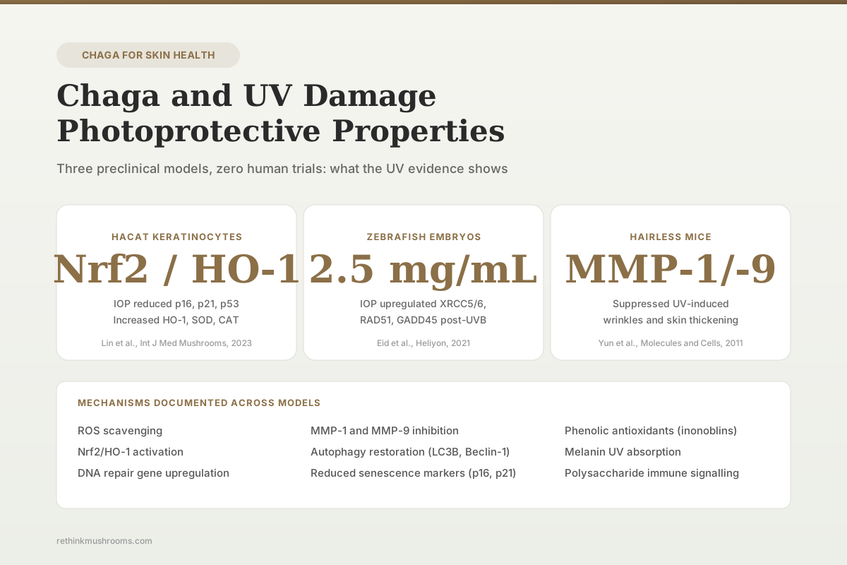
Chaga polysaccharides (IOP) reduced UVB-induced senescence and apoptosis in human HaCaT keratinocytes by modulating the Nrf2/HO-1 pathway and restoring autophagy. Supporting evidence exists in UVB-exposed zebrafish embryos and UV-exposed hairless mice. No human clinical trial has tested chaga for photoprotection.
Lin et al., Int J Med Mushrooms, 2023
HaCaT UVB model
IOP reduced p16, p21, p53 and increased HO-1, SOD, CAT via Nrf2/HO-1 (Lin et al., 2023)
Zebrafish genoprotection
IOP 2.5 mg/mL upregulated XRCC5, XRCC6, RAD51, GADD45 after UVB (Eid et al., 2021)
0 human trials
no RCT has tested oral or topical chaga for UV-induced skin damage in humans
Lin et al., Int J Med Mushrooms, 2023
 View as image ↗
Ultraviolet radiation damages skin through overlapping mechanisms: direct DNA photoadducts (cyclobutane pyrimidine dimers, 6-4 photoproducts) from UVB, reactive oxygen species (ROS) generation from UVA, matrix metalloproteinase activation that degrades dermal collagen and elastin, and inflammatory cytokine release that amplifies tissue damage. Over repeated exposures these processes produce photoaging (wrinkles, loss of elasticity, dyspigmentation) and contribute to non-melanoma and melanoma skin cancers. Against this biology, chaga (Inonotus obliquus) has a small but mechanistically consistent preclinical literature: one UVB-induced HaCaT keratinocyte study documenting Nrf2-mediated antioxidant activation and restored autophagy, one UVB-exposed zebrafish embryo study showing upregulated DNA repair genes, and one in vivo hairless mouse study documenting reduced UV-induced skin thickening and wrinkle formation.[1][2][3] No randomised clinical trial in humans has tested chaga for any UV-related endpoint. For the dedicated melanin-focused sibling, see chaga melanin and UV protection: can a mushroom protect your skin?.
Key Takeaways
- Lin et al. (2023) used a UVB-induced HaCaT keratinocyte photoaging model and showed that chaga polysaccharides (IOP) reduced p16, p21, and p53 protein expression, increased HO-1, SOD, and CAT via the Nrf2/HO-1 pathway, and restored autophagy markers p-AMPK, LC3B, and Beclin-1
- Eid et al. (2021) exposed zebrafish embryos to UVB (12 J/m²/s, 305–310 nm) and treated with 2.5 mg/mL IOP; treated embryos showed reduced DNA damage by comet assay, upregulation of XRCC5, XRCC6, RAD51, and GADD45, and downregulation of p53 and BAX
- Yun et al. (2011) reported that I. obliquus suppressed UV-induced skin thickening and wrinkle formation in hairless mice in vivo and inhibited MMP-1 and MMP-9 activity in hydrogen peroxide–treated human fibroblasts
- Park et al. (2004) documented that chaga mushroom extract inhibited oxidative DNA damage in human lymphocytes in a comet assay, providing parallel evidence that chaga can attenuate ROS-driven genotoxicity relevant to UV exposure
- Lee et al. (2007) isolated new antioxidant polyphenols (inonoblins A–C; phelligridins D, E, G) from chaga with scavenging activity against ABTS, DPPH, and superoxide radicals, establishing chemical classes with mechanistic plausibility for photoprotection
- Comprehensive chaga reviews (Glamočlija 2015; Fordjour 2023; Ern 2024) catalogue antioxidant and anti-inflammatory evidence relevant to UV damage while documenting the complete absence of clinical trials for any dermatological endpoint
How UV Damages Skin: The Targets a Photoprotective Agent Must Hit
UV radiation reaching skin is partitioned between UVA (315–400 nm, penetrates to dermis) and UVB (280–315 nm, absorbed mostly by epidermis). UVB is the primary driver of erythema, direct DNA damage, and keratinocyte photoaging; UVA contributes ROS-driven oxidative damage reaching dermal fibroblasts. The molecular consequences are well characterised.
Direct DNA damage from UVB produces cyclobutane pyrimidine dimers and 6-4 photoproducts at adjacent pyrimidines. These lesions are repaired by nucleotide excision repair involving XRCC family proteins; failure activates p53-dependent cell cycle arrest (via p21 and p16) or apoptosis (via BAX and downstream caspases). Premature senescence markers (p16, p21) accumulate in repeatedly irradiated keratinocytes and contribute to cellular dysfunction that manifests clinically as photoaging.
Indirect damage is mediated by ROS. Both UVA and UVB generate superoxide, hydrogen peroxide, and hydroxyl radicals in skin cells. ROS activate MAPK pathways (particularly JNK and p38), which in turn activate AP-1 transcription factor and drive expression of matrix metalloproteinases MMP-1 (collagenase) and MMP-9 (gelatinase B). MMP-1 cleaves type I and III collagen in dermal matrix; MMP-9 cleaves denatured collagen and elastin. Repeated MMP activation produces the wrinkled, inelastic appearance of photoaged skin.
Endogenous defences include enzymatic antioxidants (superoxide dismutase SOD, catalase CAT, glutathione peroxidase GPx), non-enzymatic antioxidants (glutathione, ascorbate, tocopherols), and the Nrf2/HO-1 transcriptional axis that upregulates these systems under oxidative stress. Autophagy also contributes to photodamage recovery by clearing damaged organelles and protein aggregates. A candidate photoprotective agent should plausibly do some combination of: scavenge ROS directly, activate Nrf2-driven antioxidant enzyme upregulation, inhibit MMP activity, enhance DNA repair gene expression, or support autophagic clearance of photodamaged components.
The Lin 2023 HaCaT Keratinocyte Study: Chaga Polysaccharides Against UVB Photoaging
Lin et al. (2023) used a UVB-induced human immortalised keratinocyte (HaCaT) cell photoaging model to characterise the mechanism of chaga polysaccharides (IOP) in relieving photoaging.[1] The HaCaT line is a standard in vitro model for human epidermal keratinocyte photobiology; UVB induces reproducible senescence (p16, p21, p53 elevation), apoptosis, ROS accumulation, and autophagy dysregulation.
IOP pretreatment produced three distinct mechanistic signatures. First, cell senescence and apoptosis markers were suppressed: p16 (a canonical senescence marker), p21 (cell cycle arrest mediator), and p53 (stress-response transcription factor driving apoptosis) all showed reduced protein expression compared with UVB-alone cells. Second, the Nrf2/HO-1 antioxidant axis was activated: HO-1, SOD, and CAT protein expression increased, consistent with Nrf2 nuclear translocation and antioxidant response element (ARE) engagement; the authors explicitly interpret this as reduced ROS generation. Third, autophagy markers were restored: p-AMPK (phosphorylated AMP-activated protein kinase), LC3B (autophagosome marker), and Beclin-1 (autophagy initiation) were all upregulated, consistent with IOP reversing UVB-induced autophagy inhibition.
The limitations are standard for cell-line photobiology. HaCaT cells are immortalised and carry p53 mutations that distinguish them from primary keratinocytes; endpoints are single-timepoint western blot rather than longitudinal tissue-level measures; one UVB dose was used; no in vivo validation was paired with the HaCaT experiments in the same publication. The positive signal is informative but establishes mechanism in a cell model, not clinical photoprotection.
The Eid 2021 Zebrafish Study: DNA Repair Gene Upregulation After UVB
Eid et al. (2021) tested the genoprotective effects of chaga polysaccharides in UVB-exposed zebrafish embryos.[2] Embryos (12 hours post fertilisation) were exposed to UVB at 12 J/m²/s (305–310 nm) for 10 seconds, then treated with IOP at 2.5 mg/mL from 24 hpf through 7 days post fertilisation. Genotoxicity was quantified by acridine orange staining, alkaline comet assay, and qRT-PCR of DNA repair genes; ELISA quantified XRCC5 and RAD51 protein.
IOP treatment significantly reduced DNA damage and rescued developmental deformities in UVB-exposed embryos. At the transcriptional level, DNA repair and survival gene expression was coordinated: XRCC5 and XRCC6 (non-homologous end joining), RAD51 (homologous recombination), and GADD45 (growth arrest and DNA damage-inducible) were upregulated in IOP-treated UVB embryos, while apoptosis drivers p53 and BAX were downregulated. Protein-level ELISA confirmed that XRCC5 and RAD51 were decreased in UVB-exposed embryos and restored in IOP co-treated embryos; p53 and BAX protein levels mirrored the transcriptional pattern, with elevated expression in UVB-only embryos indicating ongoing apoptotic signalling.
Zebrafish embryos are a validated vertebrate screening system for genotoxicity. They share with human skin many DNA repair pathways and oxidative stress responses, but embryonic development is not equivalent to adult epidermal photodamage, and the 2.5 mg/mL aqueous exposure does not translate directly to any human topical or oral dose.
The Yun 2011 Hairless Mouse Study: In Vivo Photoaging Endpoints
Yun et al. (2011) is the closest existing in vivo evidence for chaga photoprotection at the tissue level.[3] The investigators tested I. obliquus in both human fibroblast cultures and UV-exposed hairless mice. In hydrogen peroxide-treated human fibroblasts, I. obliquus pretreatment scavenged intracellular ROS, prevented lipid peroxidation, and suppressed hydrogen peroxide–induced apoptosis and premature senescence. At the ECM level, I. obliquus inhibited MMP-1 and MMP-9 activity in these cells, consistent with reduced collagen-degrading capacity.
In hairless mice exposed to UV in vivo, I. obliquus suppressed the characteristic morphological photoaging changes: skin thickening and wrinkle formation. Collagen synthesis increased. The mouse data integrate the mechanistic signals from fibroblasts (ROS scavenging, MMP-1/MMP-9 inhibition) into a tissue-level outcome that is directly relevant to human photoaging pathology.
The limitations are those of the hairless-mouse model in general: accelerated photoaging kinetics, different epidermal structure than human skin, and the route and formulation of I. obliquus delivery are not directly comparable to how chaga is used in human products. The positive result nevertheless strengthens the preclinical case beyond cell-model work.
Supporting Evidence from Broader Chaga Research
Chaga's photoprotective signal sits inside a substantially larger literature on antioxidant and anti-inflammatory activity. Park et al. (2004) used the comet assay to show that chaga mushroom extract inhibited oxidative DNA damage in human lymphocytes exposed to hydrogen peroxide.[4] Lymphocytes are not keratinocytes, but the comet-assay endpoint (reduced single- and double-strand breaks) parallels the DNA protection demonstrated in zebrafish embryos after UVB.
Lee et al. (2007) isolated new antioxidant polyphenols from chaga including inonoblins A, B, and C and phelligridins D, E, and G, with scavenging activity against ABTS, DPPH, and superoxide radicals.[5] These compound classes have structural and mechanistic plausibility for contributing to reduced ROS in UV-exposed skin models. Debnath et al. (2013) documented antioxidant and anti-inflammatory activity of chaga cultured on germinated brown rice in mouse tissue, reinforcing the cross-model antioxidant signal.[6]
Glamočlija et al. (2015) chemically characterised chaga samples from multiple geographic origins and catalogued phenolic acids, flavonoids, and melanin content alongside documented antioxidant activity.[7] This chemical profiling is important for photoprotection claims because compound content varies substantially between wild and cultivated material and between extraction methods. For the topical-product context, see chaga in skincare products: topical application research.
What the Chaga Reviews Say About Photoprotection
Fordjour et al. (2023) and Ern et al. (2024) both reference the UVB and photoaging studies within broader antioxidant and dermatological sections of their chaga reviews.[8][9] Both reviews frame the evidence as entirely preclinical and note the absence of any clinical trial in humans for UV-related endpoints. The reviews also converge on a consistent mechanistic narrative: chaga's antioxidant phenolics and polysaccharides, together with melanin content, drive the observed cellular and tissue-level protection.
Neither review recommends chaga for UV protection. Both treat the evidence as hypothesis-generating for formulation development, not as basis for use. The gap between in vitro/in vivo preclinical signals and validated human clinical endpoints (minimal erythema dose, sunburn cell quantification, DNA photoproduct measurement, photoaging scoring) is unchanged across the literature.
Topical Versus Oral: What the Evidence Does and Does Not Address
The three core photoprotection studies use different delivery routes: HaCaT keratinocytes receive direct exposure to IOP in culture; zebrafish embryos are bathed in 2.5 mg/mL IOP; hairless mice receive I. obliquus either orally or topically depending on the protocol detail. None of these routes maps cleanly onto typical human use patterns, which include oral tea, capsule supplements, and topical cosmetic formulations.
For topical delivery, the key unknowns are skin penetration of active phenolics, stability in a cosmetic matrix, concentration required at the target tissue, and UV stability of the phenolic components (some polyphenols are themselves photolabile). For oral delivery, systemic bioavailability of chaga polysaccharides and triterpenoids through human digestion is not characterised, and there is no pharmacokinetic basis for expecting that oral intake would deliver photoprotective concentrations to basal keratinocytes or dermal fibroblasts.
The reviewed chaga SPF-testing literature (e.g., reports of SPF >30 at 10 mg/mL aqueous or ethanolic extract in vitro) is based on in vitro optical absorbance assays, not validated in vivo SPF testing in humans following standard methods (ISO 24444). Chaga extract absorbs UV light in vitro, but that is not equivalent to clinical SPF.
What the Evidence Supports and What It Does Not
| Question | Evidence | Confidence |
|---|---|---|
| Does chaga reduce UV-induced cellular damage in keratinocytes? | Yes: IOP in HaCaT UVB model reduced senescence/apoptosis markers and activated Nrf2 (Lin et al., 2023) | Moderate (single cell line) |
| Does chaga protect DNA from UVB damage in a vertebrate model? | Yes: IOP in zebrafish embryos reduced DNA damage and upregulated repair genes (Eid et al., 2021) | Moderate (embryo model) |
| Does chaga reduce UV-induced skin changes in vivo? | Yes: I. obliquus suppressed skin thickening and wrinkle formation in hairless mice (Yun et al., 2011) | Moderate (single study) |
| Does chaga inhibit MMP-1 and MMP-9 in skin cells? | Yes in H₂O₂-treated fibroblasts (Yun et al., 2011) | Low to moderate (indirect UV relevance) |
| Do chaga topical products protect human skin from UV? | Not tested: no randomised clinical trial published | None |
| Is oral chaga effective for UV photoprotection in humans? | Not tested; no pharmacokinetic basis | None |
| Is chaga's SPF >30 claim clinically meaningful? | No: based on in vitro absorbance, not ISO 24444 SPF testing | Very low |
Safety Considerations for Use Adjacent to Sun Exposure
Three oxalate nephropathy case reports have been published for chaga (Kikuchi 2014; Kwon 2020; Lee 2022), all involving long-term high-dose oral use. These are relevant context for anyone considering chaga supplementation in the context of photoprotection, but they do not specifically implicate topical use. Chaga's documented antiplatelet activity adds bleeding risk for patients on aspirin or anticoagulants.
A separate concern specific to photoprotection framing: substituting chaga for evidence-based UV protection (mineral or chemical sunscreens validated to ISO standards, sun-protective clothing, shade, avoidance of peak UV hours) would increase UV exposure risk. No preclinical evidence for chaga currently justifies omitting validated photoprotection measures. For detailed safety analysis of topical formulations, see chaga in skincare products: topical application research and for the related skin-health sibling see chaga for skin health: melanin protection research.
Active Compounds Most Plausibly Responsible
The compound classes with mechanistic plausibility for the observed UV effects are:
Polysaccharides (IOP): The photoaging study (Lin 2023) and zebrafish study (Eid 2021) both used polysaccharide fractions. Fungal β-glucans can engage innate immune and antioxidant signalling at the cellular level; the Nrf2/HO-1 activation signal is mechanistically plausible for polysaccharide-driven stress response activation.
Polyphenols (inonoblins, phelligridins, caffeic acid derivatives): Lee et al. (2007) established direct ROS scavenging for chaga polyphenols. These compounds are the most direct mechanistic candidate for reduced ROS in UV-exposed cells.
Melanin: Chaga is among the most melanin-rich fungi, with the black sclerotium containing substantial melanin that absorbs UV across the spectrum. Isolated chaga melanin shows strong free radical scavenging in vitro. For the dedicated sibling on melanin and UV, see chaga melanin and UV protection: can a mushroom protect your skin?.
Triterpenoids (inotodiol, trametenolic acid, betulin derivatives): Anti-inflammatory mechanisms (NF-κB suppression, MAPK modulation) are relevant to UV-induced inflammatory signalling, though direct photoprotection studies of isolated chaga triterpenoids have not been published.
The Bottom Line
Chaga (Inonotus obliquus) shows preclinical photoprotective activity across three species models. Chaga polysaccharides reduced UVB-induced senescence, apoptosis, and ROS in human HaCaT keratinocytes through Nrf2/HO-1 activation and restored autophagy (Lin et al., 2023); the same compound class reduced UVB-induced DNA damage and upregulated repair genes in zebrafish embryos (Eid et al., 2021); and I. obliquus suppressed skin thickening, wrinkle formation, and MMP-1/-9 activity in UV-exposed hairless mice and H₂O₂-treated fibroblasts (Yun et al., 2011). Supporting evidence comes from chaga's documented antioxidant polyphenols (Lee et al., 2007) and parallel DNA-damage-protection data in human lymphocytes (Park et al., 2004). No randomised clinical trial has tested chaga for any UV endpoint in humans, and in vitro SPF-absorbance results do not meet ISO 24444 clinical SPF standards.
Frequently Asked Questions
Sources & References
- 1RTHM-00840·Lin, Jun et al. (2023). “Chaga Medicinal Mushroom, Inonotus obliquus (Agaricomycetes), Polysaccharides Alleviate Photoaging by Regulating Nrf2 Pathway and Autophagy..” International journal of medicinal mushrooms.Study breakdown →PubMed →↩
- 2RTHM-00683·Eid, Jehane Ibrahim et al. (2021). “Chaga mushroom (Inonotus obliquus) polysaccharides exhibit genoprotective effects in UVB-exposed embryonic zebrafish (Danio rerio) through coordinated expression of DNA repair genes..” Heliyon.Study breakdown →PubMed →↩
- 3RTHM-00241·Yun, Jong Seok et al. (2011). “Inonotus obliquus protects against oxidative stress-induced apoptosis and premature senescence..” Molecules and cells.Study breakdown →PubMed →↩
- 4RTHM-00076·Park, Yoo Kyoung et al. (2004). “Chaga mushroom extract inhibits oxidative DNA damage in human lymphocytes as assessed by comet assay..” BioFactors (Oxford.Study breakdown →PubMed →↩
- 5RTHM-00118·Lee, In-Kyoung et al. (2007). “New antioxidant polyphenols from the medicinal mushroom Inonotus obliquus..” Bioorganic & medicinal chemistry letters.Study breakdown →PubMed →↩
- 6RTHM-00283·Debnath, Trishna et al. (2013). “Anti-oxidant and anti-inflammatory activities of Inonotus obliquus and germinated brown rice extracts..” Molecules (Basel.Study breakdown →PubMed →↩
- 7RTHM-00373·Glamočlija, Jasmina et al. (2015). “Chemical characterization and biological activity of Chaga (Inonotus obliquus), a medicinal "mushroom"..” Journal of ethnopharmacology.Study breakdown →PubMed →↩
- 8RTHM-01694·Fordjour, Eric et al. (2023). “Chaga mushroom: a super-fungus with countless facets and untapped potential..” Frontiers in pharmacology.Study breakdown →PubMed →↩
- 9RTHM-01886·Ern, Phoebe Tee Yon et al. (2024). “Chaga Therapeutic Properties: What This 2024 Review Actually Shows.” Mycology.Study breakdown →PubMed →↩