Mushroom Cultivation
25,000 Conidia/m³
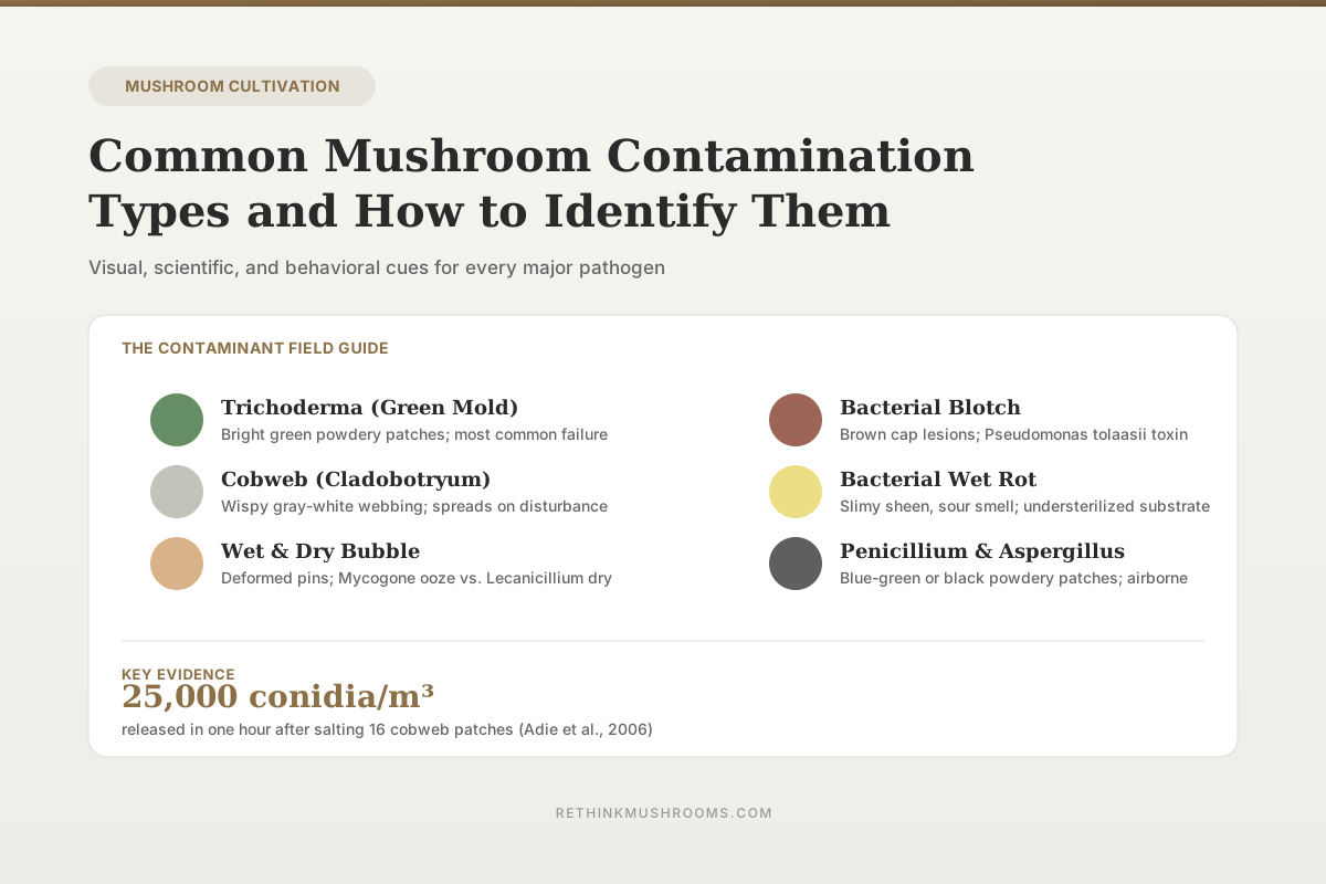
A single salting of 16 cobweb patches (0.55 m²) released roughly 25,000 Cladobotryum conidia per cubic meter of growing room air within one hour, demonstrating how mechanical disturbance of contamination drives its spread.
Adie et al., Applied and Environmental Microbiology, 2006
Green Mold
most common cultivation failure
4+ Fungal
major pathogen groups
2 Bacterial
primary blotch pathogens
Adie et al., Applied and Environmental Microbiology, 2006
 View as image ↗
Every mushroom grower meets contamination eventually. A jar goes green. A fruiting block sprouts white fuzz that is not the species you inoculated. Pins turn brown and slimy overnight. The question is not whether contamination will happen but whether you can name it when it does, because the name drives every decision that follows: salvage or toss, which room to seal off, which step in your process to change. This guide catalogs the contaminants that show up most often in mushroom cultivation, the science behind each, and the visual and behavioral cues that separate them. For the workspace that prevents most of these problems, see building a still air box for mushroom work. For the clean-culture techniques that catch contamination early, see agar work for mushroom growers. For broader workflow, see sterile technique for mushroom growing.
Key Takeaways
- Trichoderma (green mold) is the single most common contamination failure in mushroom cultivation; a 24-month monitoring study identified 248 airborne Trichoderma isolates across four species in cultivation houses, with T. harzianum dominant (Kim et al., 2019)
- Six distinct Trichoderma species have been documented on shiitake, including T. harzianum, T. atroviride, T. viride, T. pleuroticola, T. longibrachiatum, and T. oblongisporum, and pH and temperature adjustments do not control them (Wang et al., 2016)
- Cladobotryum mycophilum is now the dominant cobweb pathogen on Agaricus bisporus, not C. dendroides as historically reported, and many isolates are resistant to methylbenzimidazole carbamate (MBC) fungicides (McKay et al., 1999)
- Salting cobweb patches releases up to 25,000 conidia per cubic meter into growing-room air, and 85-97% of conidia settle within 15 minutes of air movement stopping (Adie et al., 2006)
- The bacterial blotch toxin tolaasin is produced by Pseudomonas tolaasii via a 65 kbp gene cluster encoding three proteins of 465, 440, and 435 kDa, which together direct toxin synthesis (Rainey et al., 1993)
- A screen of 97 Pleurotus strains identified five strains that were highly resistant to Pseudomonas tolaasii brown blotch disease, all within P. pulmonarius and P. cf. floridanus (Azu et al., 2019)
Field identification at a glance
One Discriminating Cue Per Contaminant
The fastest way to name what you are looking at is the single most telling visual cue, plus where it typically appears. Match the row, then act. Detail and citations for each organism follow below.
White at first, then bright green / yellow-green powdery patches within 24–72 h
Agar, grain, fruiting substrate
Toss — conidia cross a plate in days and disperse readily
Fine, wispy, pale gray webbing spreading flat across the casing; later pink / orange patches
Casing surface, fruiting bodies
Wet and cover before removing — never brush or dry-salt
White woolly, tumor-like masses that ooze fetid amber liquid
Pins / primordia (casing-borne)
Remove without agitation; track and drop the casing batch
Dry, leathery, deformed caps and split stems — no ooze
Pins and early fruiting (casing-borne)
Remove before sporulation; check for water splash
Sunken brown / yellow-brown lesions on the cap surface only
Developing and mature caps
Dry the caps, cut misting; switch strain for repeat problems
Wet, shiny, gummy patches with a sour, fermented smell; free liquid in the jar
Agar and grain spawn
Discard; review sterilization and grain moisture
Soft blue-gray (Penicillium) or black / olive (Aspergillus) powdery patches
Grain and substrate
Toss; bag and remove heavy Aspergillus without disturbance
Color is the fastest cue on a plate; smell and texture separate the bacterial blooms; substrate location narrows the casing-borne bubble diseases. When two candidates remain, the salvage-or-toss reflex is the same for every fungal and bacterial contaminant here — contain and discard rather than disturb.
Sources: Kim et al. 2019; Wang et al. 2016; McKay et al. 1999 (RTHM-00067); Adie et al. 2006 (RTHM-00174); Largeteau & Savoie 2010; Rainey et al. 1993 (RTHM-00034); Azu et al. 2019; Shi et al. 2019 — each detailed and cited in the article body.
How Contamination Presents at Each Stage
Contamination looks different depending on where in the process you find it. On agar, you see isolated colonies with distinct colors and textures. On grain spawn, you see patches breaking through the colonizing mycelium. On fruiting substrate, contamination competes visually with the mushroom mycelium and eventually with the fruiting bodies themselves. On mature mushrooms, certain pathogens cause discoloration, deformation, or decay that the grower only notices at harvest.
Largeteau and Savoie (2010) reviewed the range of microbially induced diseases that affect Agaricus bisporus, the most studied cultivated mushroom, and identified Pseudomonas tolaasii and Pseudomonas reactans as the dominant bacterial pathogens, Trichoderma aggressivum as the dominant fungal competitor in compost, and Lecanicillium fungicola as the cause of dry bubble disease.[5] Each of these organisms has a signature appearance and a signature impact on the crop. Recognizing them is the first step in deciding whether a given flush, jar, or plate can be salvaged.
Trichoderma (Green Mold)
What it looks like. White or translucent mycelium initially, then rapidly shifting to bright green, yellow-green, or forest green patches as spores form. The spore-forming surface has a powdery, dusty texture. On agar it outcompetes mushroom mycelium and can cover a plate in 48-72 hours. On grain spawn it emerges as a vivid green patch that expands radially. On fruiting substrate it can appear at any point, often where moisture pools.
Why it is so common. Kim et al. (2019) conducted 24 months of continuous air monitoring in oak wood mushroom cultivation houses and obtained 1,063 fungal isolates from indoor air alone. Of these, 248 were identified as Trichoderma across four species: T. harzianum, T. atroviride, T. citrinoviride, and T. pseudokoningii, with T. harzianum dominant.[1] All four species produced cellulase, beta-glucosidase, amylase, xylanase, and protease, the exact enzymes needed to degrade sawdust and other mushroom substrates. Trichoderma is not a rare intruder. It is a ubiquitous part of the indoor microbial community in any space where mushrooms are grown, and it is adapted to consume the same materials mushrooms consume.
Species diversity on host crops. Wang et al. (2016) characterized Trichoderma isolates from diseased Lentinula edodes (shiitake) cultivation across the main shiitake regions of China and identified six species: T. harzianum, T. atroviride, T. viride, T. pleuroticola, T. longibrachiatum, and T. oblongisporum.[2] In confrontation cultures, all Trichoderma isolates distorted, swelled, and inhibited shiitake mycelium. The authors noted that adjusting pH or temperature did not control green disease, meaning environmental manipulation of the substrate is not a reliable fix once Trichoderma is established.
Substrate-level control. Jaramillo et al. (2024) evaluated ozone as a substrate treatment for Pleurotus ostreatus production against Trichoderma contamination.[3] Ozone bubbled through water containing wheat straw substrate suppressed Trichoderma conidia and reduced subsequent contamination rates compared to untreated substrate. This is additional evidence that Trichoderma pressure is primarily a substrate problem, not a technique problem, and that substrate sterilization or decontamination intensity matters.
What to do. On agar, toss the plate; Trichoderma will cross a plate in days and contaminate neighbors. On a grain jar, toss the jar and clean the incubation area before returning. On a fruiting block, the block is generally lost once green mold is visible in quantity; contain and discard rather than attempting to cut out infected areas, since conidia disperse readily.
Cobweb Disease (Cladobotryum)
What it looks like. Fine, wispy, pale gray or white aerial mycelium that spreads rapidly across the casing layer or fruiting bodies. Named because the growth pattern resembles spider webbing. Unlike mushroom mycelium, which is typically denser and more localized around the base of pins, cobweb mycelium spreads horizontally across the casing surface in a thin, loose layer. Within days, the mycelium darkens and produces pink, orange, or tan patches as conidia mature.
Pathogen identity. McKay et al. (1999) sequenced internal transcribed spacer (ITS) regions of Cladobotryum isolates causing cobweb disease and found that the organism historically blamed for cobweb, C. dendroides (formerly Dactylium dendroides), is not the common pathogen in modern commercial production.[6] The most frequent isolate was C. mycophilum, split into three ITS subgroups: two sensitive to methylbenzimidazole carbamate (MBC) fungicides and one resistant. The resistant group, recovered from Ireland and Great Britain, appeared clonal by RAPD analysis. This matters because growers and advisors sometimes recommend benzimidazole fungicides for cobweb control, but the resistant lineage has been spreading in commercial production for over two decades.
Fact check
Will a benzimidazole fungicide clear cobweb?
The claim
“Reach for a methylbenzimidazole carbamate (MBC) fungicide and cobweb disease is under control.”
Older commercial guidance from when C. dendroides was assumed to be the cobweb pathogen and MBC fungicides still worked against it.
What research shows
Context DependentThe dominant modern cobweb pathogen on Agaricus bisporus is Cladobotryum mycophilum, not C. dendroides, and an MBC-resistant subgroup has been circulating in commercial production for over two decades.
McKay et al. found three ITS subgroups of C. mycophilum — two MBC-sensitive and one resistant. The resistant lineage recovered from Ireland and Great Britain was clonal by RAPD analysis. Resistance is real but region-specific; it should not be assumed for every cobweb isolate worldwide.
McKay et al., Applied and Environmental Microbiology, 1999
Why disturbance matters. Adie et al. (2006) measured airborne Cladobotryum conidia in experimental and commercial mushroom growing rooms during standard disease treatment (covering cobweb patches with salt).[8] Salting released conidia into the air in direct response to physical disturbance. In a 75 m³ experimental growing room, salting 16 cobweb patches (0.55 m² total) produced a peak of approximately 25,000 conidia per cubic meter in the following hour. Once air conditioning fans stopped, 85% of conidia settled within a 0.5 m radius of the source, and 91-97% settled within 15 minutes. Covering patches with damp paper tissue before salting, or holding a dust extractor above the patch during treatment, reduced airborne conidia to undetectable levels.
Adie et al. (2006) · Applied and Environmental Microbiology
- Population
- Experimental (75 m³) and commercial Agaricus bisporus growing rooms
- Intervention
- Standard cobweb treatment — covering 16 patches (0.55 m² total) with salt
- Comparator
- Damp-tissue cover or a dust extractor held above the patch during treatment
- Finding
- Salting produced a peak of roughly 25,000 Cladobotryum conidia per cubic meter in the following hour. Once air conditioning stopped, 85% of conidia settled within a 0.5 m radius and 91–97% settled within 15 minutes. Pre-covering with damp tissue or extracting dust during treatment dropped airborne conidia to undetectable levels.
positive Direct dispersal measurements, not modeling — the numbers are the reason the reflex is 'wet and cover, never brush.'
Adie et al., Applied and Environmental Microbiology, 2006
What to do. Do not brush, wipe, or dry-salt a cobweb patch; every disturbance multiplies the conidia in the air. Wet the patch first with damp tissue or fine mist, then cover it before removing it. Isolate affected trays and reduce airflow during treatment.
Wet Bubble Disease (Mycogone perniciosa)
What it looks like. Pins fail to develop into normal mushrooms and instead form irregular, tumor-like masses covered in white woolly mycelium. As the masses mature, they ooze an amber liquid with a fetid smell. The affected fruiting bodies are unrecognizable as mushrooms.
Biology and impact. Wet bubble is caused by the mycoparasite Mycogone perniciosa, which infects Agaricus bisporus primordia early in development and blocks normal differentiation. Largeteau and Savoie (2010) note that wet bubble, alongside dry bubble and cobweb, is one of the three major fungal diseases that cause economic loss in commercial A. bisporus production.[5] The pathogen is primarily soilborne and enters growing rooms through contaminated casing material, making casing source and handling a critical control point.
What to do. Remove affected mushrooms carefully without agitation, since the amber ooze contains infective spores. Disinfect tools and hands between handling diseased and clean areas. The casing layer is the likely source, so track the casing batch that produced the outbreak and avoid reusing it.
Dry Bubble Disease (Lecanicillium fungicola)
What it looks like. Distorted, leathery, dry-looking deformations of mushroom stems and caps. Affected pins may form a small dry mass rather than developing into a mushroom, or mature mushrooms may show gray-brown lesions on the cap or a split, stubbed stem. Unlike wet bubble, there is no amber ooze; the affected tissue is dry and firm.
Biology. Lecanicillium fungicola (formerly Verticillium fungicola) is a mycoparasite that attacks A. bisporus during pinning and early fruiting. Largeteau and Savoie (2010) identified it as one of the three dominant fungal pathogens in commercial mushroom production and noted that the host-pathogen interaction is only partially characterized at the biochemical level.[5] The pathogen produces spores that survive in soil and casing material and are dispersed by water splashing, tools, and insects such as sciarid flies.
What to do. Identify affected mushrooms by their dry, deformed appearance. Remove them before sporulation surfaces develop. Check watering practices; overhead watering can splash spores across a tray. Review casing source, since Lecanicillium, like Mycogone, is typically introduced through casing.
Bacterial Blotch (Pseudomonas tolaasii)
What it looks like. Brown or yellow-brown lesions on the caps of developing or mature mushrooms, sometimes with a sunken, leathery texture. The lesions remain localized to the cap surface rather than producing aerial mycelium. Caps may look normal when harvested and develop blotches within hours as the bacteria proliferate.
The toxin. Rainey et al. (1993) identified the genetic basis for brown blotch disease. Transposon mutagenesis of P. tolaasii NCPPB 1116 generated 5,000 chromosomal insertions; 35 (0.7%) produced no tolaasin, and 12 (0.25%) produced reduced amounts.[7] The mutations mapped to a 65 kbp genetic locus encoding three high-molecular-weight proteins: TL1 (465 kDa), TL2 (440 kDa), and TL3 (435 kDa). These proteins together direct the synthesis of tolaasin, the extracellular lipodepsipeptide toxin that is the primary disease determinant of pathogenicity on A. bisporus. The gene cluster sits at one end of a 640 kbp PacI chromosomal fragment in the 6.7 Mbp P. tolaasii genome.
Pleurotus blotch. P. tolaasii also infects oyster mushrooms. Azu et al. (2019) screened 97 Pleurotus strains (21 P. cf. floridanus, 20 P. ostreatus, 56 P. pulmonarius) for brown blotch resistance using two inoculation methods: inoculation on the pileus (IMP) and inoculation on the spawned substrate (IMSS).[9] Using IMP, 22 P. pulmonarius strains and 4 P. cf. floridanus strains were moderately resistant. Using IMSS, 11 P. pulmonarius, 6 P. cf. floridanus, and 1 P. ostreatus strain were highly resistant; eight strains were completely resistant under IMSS conditions. Combining both methods, five strains were highly resistant (four P. pulmonarius and one P. cf. floridanus) and 11 were moderately resistant. The authors concluded that P. pulmonarius and P. cf. floridanus are promising sources for breeding resistant cultivars, and that cultivar choice meaningfully affects disease susceptibility.
What to do. Reduce surface moisture on fruiting mushrooms; bacterial blotch thrives where water sits on caps. Improve air circulation and shorten misting durations. For repeat problems, switch strains; the 97-strain screen shows that substantial resistance variation exists within the same species.
Bacterial Contamination on Agar and Grain
What it looks like. Wet, shiny, sometimes gummy patches that may be white, cream, yellow, pink, or iridescent. The patches often emit a sour, fermented, or rotting smell distinct from the earthy smell of healthy mycelium. On grain, bacterial contamination appears as a cloudy wet layer or slimy sheen, and shaking the jar may reveal free liquid at the bottom.
Why it happens. Understerilized substrate is the most common source. Bacterial endospores survive standard pressure cooking if times, pressures, or grain moisture are wrong, and germinate once mycelium lowers the environmental competitive pressure. Bacterial blooms can also follow overhydration of grain, which creates the free-water conditions bacteria need.
The Cabin-Sequestering method. Shi et al. (2019) developed the Cabin-Sequestering (CS) technique to remove bacterial contamination from fungal cultures without antibiotics.[4] A 3-5 mm round or square hole (the cabin) is excavated in a solid agar plate. The contaminated fungal inoculum is placed in the cabin and covered with a sterile coverslip. Fungi, which grow hyphally and expand under the coverslip into surrounding agar, escape the cabin within 7-10 days. Bacteria, which grow colonially in bulk liquid conditions, remain trapped. The method avoids the drawbacks of antibiotic treatment (incomplete removal, limited spectra, recontamination risk, effects on the fungal strain, environmental residues) and produces clean fungal cultures without chemical intervention.
What to do. On agar, attempt the CS method if the culture is genetically valuable; otherwise discard. On a grain jar with bacterial contamination, discard the jar and review sterilization. Check pressure-cooker gauge accuracy, grain moisture (28-30% is standard), and hold time at full pressure (typically 90 minutes minimum for grain at 15 PSI).
Penicillium and Aspergillus
Penicillium (blue-green mold). Appears as blue-green, powdery patches. Unlike Trichoderma's vivid kelly green, Penicillium is a softer, grayer, blue-tinted shade. Colonies often develop a white perimeter with the blue-green center expanding outward. Common in grain and substrate.
Aspergillus (black mold). Appears as black, dark brown, or olive-green powdery patches depending on species. A. niger produces the most recognizable black form. Some Aspergillus species produce mycotoxins and pose a direct human health risk at high concentrations.
What to do. Both are common airborne contaminants controlled by still-air technique, properly sterilized substrate, and sealed fruiting environments. Toss contaminated material. Do not attempt to work around heavy Aspergillus growth; use a sealed bag for removal and dispose of it outside.
Grower safety
ModerateDisturbing contaminated material puts spores in the air you breathe
Concern
The same physical disturbance that spreads contamination to fresh substrate also aerosolizes spores into the room's breathing air. Adie et al. measured roughly 25,000 Cladobotryum conidia per cubic meter within an hour of salting cobweb patches, and molds such as Aspergillus can additionally produce mycotoxins and provoke respiratory hypersensitivity in cultivators exposed repeatedly to spore-laden air.
What the research says
Treat heavily contaminated jars and blocks as a respiratory hazard, not just a lost crop. Wet and cover patches before handling so spores stay put, bag and seal contaminated material rather than opening it in the grow space, and remove it without brushing, shaking, or dry-salting.
Particularly relevant for: Home and commercial mushroom growers, especially those working repeatedly around contamination in enclosed rooms
What to do
Wear a fitted particulate respirator (N95 or better) when handling visibly contaminated material, keep the material sealed, and ventilate or leave the space after disturbance. This article is educational and not medical advice.
Adie et al., Applied and Environmental Microbiology, 2006; general mycology
When to Salvage, When to Toss
Some contamination can be cut around and some cannot. Cultures on agar with a single small bacterial colony or an isolated mold spot can sometimes be salvaged by clean-sector transfer to a fresh plate, especially for valuable genetics. The CS method (Shi et al., 2019) extends this option to bacterial contamination without antibiotics.[4]
Grain spawn generally cannot be salvaged. The substrate is homogeneous and contamination spreads throughout before it is visible. A single green patch on a jar means the jar is lost. Fruiting blocks with active contamination are similarly lost, and the conidia-dispersal work of Adie et al. (2006) is the reason why: disturbing a contaminated block multiplies the spore load in the room by orders of magnitude.[8] The conservative response is to bag, seal, and remove without disturbance.
What the Evidence Does Not Say
Many published treatment recommendations are species-specific to commercial A. bisporus and do not necessarily transfer to other cultivated species. Fungicide data, in particular, is sparse for Pleurotus, Lentinula, and specialty species. The MBC-resistance finding of McKay et al. (1999) applies to Cladobotryum isolates in specific geographic regions and should not be generalized to assume all cobweb pathogens worldwide are MBC-resistant.[6] Published sterilization protocols also reflect commercial compost production and may be conservative or aggressive for small-scale work. Where published evidence is thin, what is available is pathogen biology, dispersal physics, and general microbiology; these are reliable guides even where species-specific trials are limited.
The Bottom Line
Mushroom contamination falls into a small number of recurring categories: green mold (Trichoderma), cobweb (Cladobotryum), wet bubble (Mycogone), dry bubble (Lecanicillium), bacterial blotch (Pseudomonas tolaasii), bacterial contamination in agar and grain, and the generalist molds Penicillium and Aspergillus. Each has a signature appearance, a characteristic substrate preference, and a distinct dispersal pattern, and published research on air monitoring, genetic characterization, and resistance screening provides practical guidance for identification and response. Prevention through sterile technique and appropriate substrate treatment is far more effective than attempting to cure an established contamination.
Frequently Asked Questions
Sources & References
- 1RTHM-00564·Kim, Jun Young et al. (2019). “Isolation and Characterization of Airborne Mushroom Damaging Trichoderma spp. from Indoor Air of Cultivation Houses Used for Oak Wood Mushroom Production Using Sawdust Media..” The plant pathology journal.Study breakdown →PubMed →↩
- 2RTHM-00455·Wang, Gangzheng et al. (2016). “Diversity and effect of Trichoderma spp. associated with green mold disease on Lentinula edodes in China..” MicrobiologyOpen.Study breakdown →PubMed →↩
- 3RTHM-00881·Jaramillo Mejía, Santiago et al. (2024). “Use of Ozone as a Substrate Treatment for the Control of Trichoderma in the Production of Pleurotus ostreatus..” Current microbiology.Study breakdown →PubMed →↩
- 4RTHM-00587·Shi, Xiao-Xiao et al. (2019). “A handy method to remove bacterial contamination from fungal cultures..” PloS one.Study breakdown →PubMed →↩
- 5RTHM-00189·Largeteau, Michèle L et al. (2010). “Microbially induced diseases of Agaricus bisporus: biochemical mechanisms and impact on commercial mushroom production..” Applied microbiology and biotechnology.Study breakdown →PubMed →↩
- 6RTHM-00030·McKay, G J et al. (1999). “Genetic and morphological characterization of Cladobotryum species causing cobweb disease of mushrooms..” Applied and environmental microbiology.Study breakdown →PubMed →↩
- 7RTHM-00013·Rainey, P B et al. (1993). “Identification of a gene cluster encoding three high-molecular-weight proteins, which is required for synthesis of tolaasin by the mushroom pathogen Pseudomonas tolaasii..” Molecular microbiology.Study breakdown →PubMed →↩
- 8RTHM-00090·Adie, Bruce et al. (2006). “Temporal and spatial dispersal of cladobotryum conidia in the controlled environment of a mushroom growing room..” Applied and environmental microbiology.Study breakdown →PubMed →↩
- 9RTHM-00544·Azu Okorley, Benjamin et al. (2019). “Resistance Sources to Brown Blotch Disease (Pseudomonas tolaasii) in a Diverse Collection of Pleurotus Mushroom Strains..” Pathogens (Basel.Study breakdown →PubMed →↩