Cordycepin Anti-Inflammatory
5 routes To One Cytokine
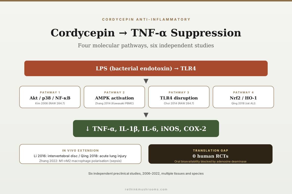
Cordycepin suppresses LPS-induced tumor necrosis factor-alpha (TNF-α) through NF-κB inhibition at Akt and p38 (Kim 2006), TLR4 and MyD88 downregulation (Choi 2014), AMPK activation via its own monophosphate metabolite (Zhang 2014; Hawley 2020), Nrf2/HO-1 engagement (Qing 2018), and inhibition of polyadenylation itself (Ashraf 2019). Six preclinical studies anchor the story — five of which actually measured TNF-α — alongside a single human trial in which TNF-α also fell in the placebo group.
Kim et al., European J Pharmacology, 2006; Zhang et al., IJMS, 2014; Ontawong et al., Scientific Reports, 2024
5 of 6 studies
measured TNF-α directly; Li 2016 is widely counted in this literature but reported matrix enzymes, nitric oxide and PGE2 in disc cells and no TNF-α at all
AMPK via a metabolite
Hawley 2020 showed cordycepin is a pro-drug here — it is phosphorylated into cordycepin monophosphate, an AMP mimic, and that metabolite is what activates AMPK
1 human trial
Ontawong 2024 measured TNF-α in people given a fermented Cordyceps beverage; TNF-α fell in the treatment arm and in the placebo arm, with no significant difference between them
Kim et al., European J Pharmacology, 2006; Zhang et al., IJMS, 2014; Ontawong et al., Scientific Reports, 2024
 View as image ↗
TNF-α is one of the dominant effector cytokines in innate immunity. It drives fever, leukocyte recruitment, and vascular endothelial activation during acute inflammation, and it sits at the centre of the cytokine network driving chronic inflammatory disease from rheumatoid arthritis to inflammatory bowel disease. Every major biologic therapy in rheumatology (infliximab, adalimumab, etanercept, certolizumab, golimumab) either neutralises TNF-α directly or blocks its receptor. Anything that measurably suppresses TNF-α in a well-controlled model has pharmacological relevance, which is why the cordycepin TNF-α literature has accumulated six independent preclinical studies between 2006 and 2022.[1][2] For the NF-κB-specific mechanism deep-dive, see cordycepin and nf-kb: how it suppresses inflammatory pathways, and for the broader cordyceps inflammation framing see cordyceps and inflammation: anti-inflammatory mechanisms.
Key Takeaways
- Kim et al. (2006) established the foundational mechanism in RAW 264.7 macrophages: suppression of LPS-induced NF-κB activation through inhibition of Akt and p38 MAPK signalling, with downstream reduction in TNF-α, iNOS, and COX-2 expression — the study did not measure IL-1β or IL-6
- Zhang et al. (2014) in International Journal of Molecular Sciences demonstrated that cordycepin suppresses LPS-induced TNF-α in mouse macrophages and in ex vivo PBMCs from Kawasaki disease patients, with AMPK confirmed as the mediating node by both pharmacological inhibition and siRNA knockdown
- Hawley et al. (2020) in Cell Chemical Biology resolved how that AMPK step works: cordycepin is imported by adenosine transporters and phosphorylated into cordycepin monophosphate, an AMP mimic that is the actual activating species — cordycepin is a pro-drug at this node
- Choi et al. (2014) showed receptor-level intervention in LPS-stimulated RAW 264.7: cordycepin reduced LPS binding to the macrophage surface and lowered TLR4 and MyD88 expression, blunting the cascade before it entered the cell
- Li et al. (2016) extended cordycepin's anti-inflammatory activity to intervertebral disc nucleus pulposus cells, suppressing matrix-degrading enzymes, nitric oxide and PGE2 via NF-κB — this study is frequently cited in TNF-α discussions but reported no TNF-α measurement
- Qing et al. (2018) showed cordycepin attenuates LPS-induced acute lung injury in Wistar rats via Nrf2/HO-1 activation, lowering plasma TNF-α and IL-6 — at intravenous doses of 1–30 mg/kg that oral dosing cannot reach
- Ashraf et al. (2019) in Scientific Reports connected cordycepin's defining molecular action — polyadenylation inhibition — to inflammation directly, reducing pain and joint pathology in two rodent arthritis models while blocking nuclear entry of NF-κB and of the polyadenylation factors WDR33 and CPSF4
- One randomised human trial exists: Ontawong et al. (2024) gave healthy adults a fermented Cordyceps militaris beverage carrying 2.85 mg cordycepin for eight weeks and measured TNF-α, IL-1β and IL-6. TNF-α declined within the treatment arm — and within the placebo arm, more steeply in men. No cytokine differed significantly between groups
Why TNF-α Is the Anchor Outcome
Inflammation is not a single phenomenon and inflammatory cytokines are not interchangeable. TNF-α occupies a privileged position because it is produced early in the inflammatory cascade, drives downstream IL-1β and IL-6 production, activates endothelial adhesion molecules that recruit neutrophils, and directly causes tissue damage at sustained elevated concentrations. TNF-α-driven inflammation is the disease process in rheumatoid arthritis, the target of the most commercially successful biologic drug category in history, and the upstream driver of the cytokine storms seen in severe sepsis, COVID-19 acute respiratory distress, and chimeric antigen receptor T-cell (CAR-T) therapy toxicity.
For a natural product or investigational compound, demonstrating TNF-α suppression at physiologically plausible concentrations in a well-controlled inflammatory stimulation model is a meaningful mechanism-level signal. The cordycepin literature has done this across multiple tissue models, inflammatory stimuli, and molecular pathway framings, which is the strongest form of preclinical mechanism evidence available before human trials.
It is also a literature that gets summarised carelessly. The phrase "six studies show cordycepin suppresses TNF-α" is repeated widely, including in places that should know better. Five of the six put a number on TNF-α. The sixth measured something else entirely and is worth reading for what it did find rather than for what it is cited for.
Auditing the Six-Study Claim
5 of the 6 Studies Actually Measured TNF-α
“Six preclinical studies” is the number usually attached to cordycepin and TNF-α. Five of them put a number on TNF-α. One — Li 2016 — measured matrix-degrading enzymes and oxidative markers in disc cells and never reported TNF-α at all. It is a real anti-inflammatory result. It is not TNF-α evidence.
Kim et al. (2006)
[1]Kangwon Nat’l University, South Korea
Cell cultureRAW 264.7 murine macrophages + LPS
TNF-α
MeasuredPathway framing
NF-κB, blocked upstream at Akt and p38
What was measured
TNF-α expression, NO, iNOS, COX-2, Akt + p38 phosphorylation, IκB-α phosphorylation, NF-κB translocation
Limit: IL-1β and IL-6 were not reported
Zhang et al. (2014)
[2]Zhejiang University, China
Human cells, ex vivoRAW 264.7 + bone-marrow macrophages + ex vivo PBMCs from Kawasaki disease patients
TNF-α
MeasuredPathway framing
AMPK — confirmed as the mediator by compound C and siRNA knockdown
What was measured
TNF-α as the primary outcome in all three systems; ROS; NF-κB activation
Limit: No in vivo arm; PBMCs are ex vivo, not dosed patients
Choi et al. (2014)
[3]Dongeui University, Busan, South Korea
Cell cultureRAW 264.7 murine macrophages + LPS
TNF-α
MeasuredPathway framing
TLR4 / MyD88 at the receptor, upstream of MAPK and NF-κB
What was measured
TNF-α and IL-1β release + mRNA, NO, PGE2, iNOS, COX-2, MAPK phosphorylation, NF-κB translocation, LPS binding to macrophages, TLR4 and MyD88 expression
Limit: No in vivo arm
Li et al. (2016)
[4]Shanghai Jiao Tong University, China
Cell cultureRat nucleus pulposus cells + intervertebral disc organ culture + LPS
TNF-α
Not measuredPathway framing
NF-κB — inferred from matrix and oxidative endpoints, not from TNF-α
What was measured
MMP-3, MMP-13, ADAMTS-4, ADAMTS-5, nitric oxide, PGE2, macrophage infiltration, NF-κB activation
Limit: No TNF-α measurement is reported anywhere in this study
Qing et al. (2018)
[5]Shaoyang University, China
Whole animalWistar rats, IV LPS 30 mg/kg then IV cordycepin 1 / 10 / 30 mg/kg at +1 h
TNF-α
MeasuredPathway framing
Nrf2 / HO-1 — a redox route, distinct from the NF-κB axis
What was measured
TNF-α, IL-6 and IL-10 in plasma by ELISA; lung wet-to-dry ratio, myeloperoxidase, nitrite/nitrate and superoxide in lung tissue; HO-1 activity; Nrf2 translocation
Limit: Cytokines were plasma, not lung tissue; IV dosing bypasses the oral bioavailability problem entirely
Zhang et al. (2022)
[6]Xi’an Children’s Hospital, China
Whole animalCecal ligation and puncture sepsis in immature mice
TNF-α
MeasuredPathway framing
NF-κB / p65, reframed as a macrophage polarisation shift
What was measured
TNF-α, iNOS, Arg-1, MCP-1, IL-4, IL-10; M1 (F4/80⁺iNOS⁺) and M2 (F4/80⁺CD206⁺) populations by flow cytometry; NF-κB/p65; liver and lung histology
Limit: The abstract reports the polarisation direction inconsistently — see the note below
Two things the table makes visible
Geography. All six studies come from two countries — South Korea and China. Independent replication in a different research culture, with different reagents and different incentives, has not happened. That is a meaningful gap, not a detail. Directional reporting. The Zhang 2022 abstract states that cordycepin increased both M1 and M2 populations, while its own conclusion states that cordycepin reduced M1/M2 polarisation. Those cannot both be true as written. The finding that survives the inconsistency is that cordycepin altered macrophage phenotype and lowered TNF-α through NF-κB/p65 — not a clean, confirmed M1-to-M2 switch.
Source: Kim 2006; Zhang 2014; Choi 2014; Li 2016; Qing 2018; Zhang 2022 (refs 1–6)
The Foundational Mechanism: NF-κB via Akt and p38 (Kim 2006)
Kim et al. (2006) produced the first definitive cordycepin anti-inflammatory mechanism paper in European Journal of Pharmacology.[1] The group stimulated RAW 264.7 murine macrophages with bacterial lipopolysaccharide (LPS, the classic TLR4 agonist that drives innate inflammatory activation) in the presence or absence of cordycepin.
Cordycepin suppressed NF-κB nuclear translocation, the regulatory step that controls transcription of TNF-α, iNOS, and COX-2 among many other inflammatory genes. Mechanistically, Kim et al. localised the cordycepin effect upstream of NF-κB: inhibition of Akt (protein kinase B) and p38 MAPK phosphorylation, both of which feed into IKK-mediated IκB-α phosphorylation and the release of NF-κB for nuclear translocation. The result is a transcription-level intervention that reduces NF-κB-driven inflammatory gene expression in parallel rather than targeting a single downstream cytokine.
Two details are worth carrying forward. The measured outputs in this study were TNF-α expression, nitric oxide, iNOS, and COX-2 — IL-1β and IL-6 were not among them, despite frequently appearing in summaries of this paper. And the author list includes an affiliation with Mushtech Co. Ltd, a commercial mushroom company. That does not invalidate the work, which has been broadly consistent with everything that followed, but a foundational paper with an industry tie is a fact a careful reader should hold.
AMPK Activation, and the Metabolite That Does the Work (Zhang 2014, Hawley 2020)
Zhang et al. (2014) in International Journal of Molecular Sciences extended the mechanism upstream by implicating AMP-activated protein kinase (AMPK) as a principal cordycepin target in the TNF-α suppression pathway.[2] AMPK is the cellular energy sensor activated by high AMP-to-ATP ratios; its activation shifts metabolism toward catabolism and away from anabolism, and it has well-documented anti-inflammatory effects through multiple downstream targets including direct NF-κB inhibition.
The Zhang study tested cordycepin in parallel systems: LPS-stimulated RAW 264.7 cells and bone marrow-derived macrophages on the mouse side, and ex vivo peripheral blood mononuclear cells (PBMCs) from Kawasaki disease patients — a childhood vasculitis driven by TNF-α-mediated endothelial damage during the acute phase. Cordycepin suppressed TNF-α in both. Critically, the causal role of AMPK was established rather than inferred: blocking AMPK with compound C or depleting it with siRNA abolished cordycepin's effect on TNF-α production. Cordycepin also reduced ROS production and NF-κB activation, and PBMCs from Kawasaki patients showed higher basal ROS and NF-κB activity than healthy controls.
The Kawasaki PBMC data are distinctive because they used cells from real disease-state patients rather than from healthy donors or a pure cell line. That translational proximity is a step closer to clinical relevance, though the cells were treated in a dish rather than in a patient, and nothing about human cordycepin pharmacokinetics is tested by this design.
What Zhang could not answer was how cordycepin activates AMPK. Hawley et al. (2020), working in D. Grahame Hardie's laboratory — the group that has defined most of what is known about AMPK — answered it in Cell Chemical Biology.[8] Cordycepin enters cells through adenosine transporters and is then converted by ordinary cellular metabolism into cordycepin mono-, di-, and triphosphates. AMPK activation tracks the concentration of cordycepin monophosphate specifically, not of cordycepin itself or of the other phosphorylated forms. In cell-free assays, cordycepin monophosphate reproduces all three of the effects that AMP normally has on AMPK, and in intact cells the activation is abolished by a γ-subunit mutation that blocks AMP sensing.
TLR4 Engagement: The Receptor-Level Intervention (Choi 2014)
Choi et al. (2014), publishing in Drug Design, Development and Therapy, tested cordycepin's effects on the TLR4 pathway itself.[3] TLR4 is the receptor that recognises bacterial LPS and initiates the entire downstream inflammatory cascade. Most anti-inflammatory interventions target downstream signalling nodes (NF-κB, p38, JAK/STAT, inflammasome assembly) because those sit inside the cell and are easier to drug. Receptor-level intervention is less common but mechanistically cleaner when it works, because it prevents the signal from entering the cell rather than blunting it after entry.
The Choi data showed cordycepin reduced production of nitric oxide and prostaglandin E2 by suppressing iNOS and COX-2 gene expression, and inhibited release of TNF-α and IL-1β through downregulation of their respective mRNAs. Pretreatment blocked LPS-induced MAPK phosphorylation and attenuated NF-κB nuclear translocation, with IκB-α degradation prevented.
The receptor-level finding is more concrete than it is usually reported. Choi et al. did not merely speculate about TLR4 involvement: they showed that cordycepin potently inhibited the binding of LPS to macrophages, and reduced LPS-induced expression of both TLR4 and its adaptor protein MyD88. What remains undissected is the physical basis of that reduced binding — whether cordycepin competes at the receptor, displaces a co-receptor such as MD-2 or CD14, or interferes with TLR4 dimerisation. The observation is solid; the molecular explanation for the observation is not yet established.
Tissue-Specific Models: Intervertebral Disc (Li 2016) and Acute Lung Injury (Qing 2018)
Two studies moved cordycepin anti-inflammatory testing out of the generic RAW 264.7 macrophage model into tissue-specific disease-relevant systems.
Li et al. (2016), in PeerJ, tested cordycepin in rat nucleus pulposus cells — the cartilage-like cells in intervertebral discs that degenerate in degenerative disc disease — and in intervertebral disc organ culture.[4] LPS stimulation in this model drives matrix-degrading enzymes that break down disc tissue. Cordycepin suppressed MMP-3, MMP-13, ADAMTS-4 and ADAMTS-5, reduced nitric oxide and PGE2, limited macrophage infiltration into the disc, and suppressed LPS-induced NF-κB activation on both western blot and luciferase reporter assays.
This is a genuine and useful anti-inflammatory result, and it is the study most often mis-cited in this literature. Li et al. did not measure TNF-α. Their inflammatory readouts were oxidative and matrix-degradative, and their pathway evidence is NF-κB activation rather than cytokine output. The study supports a therapeutic rationale for testing cordycepin in disc degeneration; it does not add a data point to the count of studies showing TNF-α suppression.
Qing et al. (2018) tested cordycepin in a Wistar rat model of LPS-induced acute lung injury (ALI), a mechanistic surrogate for acute respiratory distress syndrome (ARDS).[5] The group administered intravenous LPS (30 mg/kg) to induce lung injury, then intravenous cordycepin (1, 10, or 30 mg/kg) one hour later. In lung tissue, cordycepin reduced the wet-to-dry weight ratio (a standard pulmonary edema readout), myeloperoxidase activity (a neutrophil infiltration marker), nitrite/nitrate content, and superoxide. Cytokines were measured separately, in plasma rather than lung tissue: TNF-α, IL-6 and IL-10 were all regulated through HO-1. Mechanistically, cordycepin induced Nrf2 translocation from cytoplasm to nucleus and raised both HO-1 expression and its enzymatic activity — a redox-driven route that is molecularly distinct from, and complementary to, the NF-κB axis.
The significance of these two studies lies in the move from generic cell-culture activity to measurable effects in disease-relevant models, one of them in a whole animal. The Qing study also sets an important boundary: a dose range of 1 to 30 mg/kg given intravenously is unreachable by oral dosing given cordycepin's adenosine deaminase problem.[9] These findings establish a mechanistic ceiling, not a clinically translatable protocol.
The Mechanism the Four-Pathway Framing Leaves Out (Ashraf 2019)
Cordycepin is 3'-deoxyadenosine. The missing 3'-hydroxyl is the whole molecule: RNA polymerase extends a growing RNA chain by bonding to the 3'-hydroxyl of the previous nucleotide, so when cordycepin is incorporated, elongation stops. Its best-characterised consequence is inhibition of polyadenylation — the addition of the poly(A) tail that mRNA needs for stability and translation. This is cordycepin's defining biochemical action, and it is routinely omitted from anti-inflammatory mechanism summaries that list NF-κB, AMPK, TLR4 and Nrf2.
Ashraf et al. (2019), in Scientific Reports, connected it directly to inflammation.[12] The group first examined joint tissue from people with osteoarthritis, comparing those with high-grade and low-grade synovial inflammation against non-arthritic post-mortem controls, and found that the polyadenylation factor CPSF4 and NF-κB were both elevated in the synovia of patients with high-grade inflammation. They then tested cordycepin in two independent rodent osteoarthritis models — monosodium iodoacetate injection in rats and surgical destabilisation of the medial meniscus in mice — where it reduced pain behaviour, synovial inflammation, and joint pathology. In human monocyte-derived macrophages and a mouse macrophage line, cordycepin inhibited the nuclear localisation not only of NF-κB but of the polyadenylation factors WDR33 and CPSF4.
That last result is the interesting one, because it suggests the pathways described in this article may be less separate than they look. If cordycepin restricts nuclear entry of both NF-κB and the polyadenylation machinery, then several of the "distinct mechanisms" reported across these studies may be different observational windows onto one upstream event.
Arthritis is also where the most translationally proximate cell model in this literature sits. Noh et al. (2009), in Rheumatology, isolated synovial fibroblasts from twelve rheumatoid arthritis patients and stimulated them with IL-1β rather than LPS.[13] Cordycepin inhibited IL-1β-induced MMP-1 and MMP-3 expression dose-dependently, and among the chemokines tested — MCP-1, GRO-α, RANTES and ENA-78 — it specifically blocked ENA-78 production. The pathway result is the one worth pausing on. Noh et al. ran electrophoretic mobility shift assays for the DNA binding of both NF-κB and AP-1, and cordycepin inhibited AP-1 but not NF-κB; the kinases it blocked were p38 and JNK, with ERK unaffected. In the tissue that sits closest to human rheumatoid disease anywhere in this literature, the NF-κB node that carries almost every other study in this article is not the node that moved. These are human cells from diagnosed patients, which is as close to the target tissue as this literature gets. They are still cells in a dish, treated at concentrations chosen by the experimenter.
Mechanism · Five Entry Points, One Endpoint
Where Cordycepin Actually Interrupts the TNF-α Signal
Cordycepin is usually described as having four anti-inflammatory pathways. There is a fifth, and it is the one that defines the molecule: it jams the machinery that finishes messenger RNA. Convergence on a single endpoint from five structurally different directions is what makes the target credible — and it is also why no single “the mechanism” answer exists.
Polyadenylation inhibition
Inside the nucleus
Cordycepin lacks the 3′ hydroxyl that RNA chains need to extend, so poly(A) tails are cut short. Blocking the polyadenylation factors WDR33 and CPSF4 also blocks NF-κB from entering the nucleus.
Human macrophages + rat and mouse arthritis models
Ref 12Akt and p38
Kinases above NF-κB
Dose-dependent loss of Akt and p38 phosphorylation stops IκB-α from being tagged for destruction, so NF-κB never releases.
RAW 264.7 macrophages
Ref 1AMPK
The cell’s energy sensor
Cordycepin is imported by adenosine transporters and phosphorylated into cordycepin monophosphate, an AMP look-alike that switches AMPK on. AMPK then restrains NF-κB.
Mouse macrophages + Kawasaki patient PBMCs
Refs 2, 8TLR4 and MyD88
At the receptor
Cordycepin reduces LPS binding to the macrophage surface and lowers TLR4 and MyD88 expression, cutting the signal before it enters the cell.
RAW 264.7 macrophages
Ref 3Nrf2 and HO-1
The redox branch
Nrf2 moves into the nucleus and drives HO-1. The anti-inflammatory effect runs through HO-1 rather than through NF-κB — a parallel route, not the same one.
LPS-injured Wistar rats
Ref 5Less TNF-α transcribed and released
Four of the five routes end at the NF-κB switch. The Nrf2/HO-1 branch reaches the same outcome without it.
Where the diagram stops
Every route on this map was drawn in cell culture or in animals, at concentrations reached by bathing cells or by intravenous injection. None of it has been demonstrated in a person taking cordycepin by mouth. The arrows are real; the distance between the last arrow and a clinical effect is the unsolved part.
Source: Kim 2006; Zhang 2014; Choi 2014; Qing 2018; Hawley 2020; Ashraf 2019 (refs 1, 2, 3, 5, 8, 12)
Macrophage Polarisation: The M1/M2 Framing (Zhang 2022)
Zhang et al. (2022) in Journal of Pharmacy and Pharmacology reframed cordycepin's anti-inflammatory effect as a shift in macrophage polarisation rather than simple cytokine suppression.[6] M1 macrophages are the classically activated pro-inflammatory state driven by TLR engagement and IFN-γ exposure; M2 macrophages are alternatively activated tissue-repair macrophages driven by IL-4/IL-13 and regulatory signals. Chronic inflammatory disease typically features M1-skewed macrophage populations; resolution requires a shift toward M2.
Using cecal ligation and puncture (CLP) to induce sepsis in immature mice, the group reported improved liver and lung injury on histology and TUNEL staining, altered distribution of M1 (F4/80⁺iNOS⁺) and M2 (F4/80⁺CD206⁺) macrophages on flow cytometry, changes in iNOS, Arg-1, TNF-α, MCP-1, IL-4 and IL-10, and suppression of the CLP-induced rise in NF-κB/p65.
The polarisation direction, however, should be reported cautiously. The paper's own abstract states that cordycepin increased both M1-like and M2-like populations, while its concluding sentence states that cordycepin reduced M1/M2 polarisation. Those are not compatible as written. What survives the inconsistency is that cordycepin altered macrophage phenotype, lowered TNF-α, and acted through NF-κB/p65 in a whole-animal sepsis model — not a cleanly demonstrated M1-to-M2 conversion. The model is also worth noting: immature mice with surgically induced polymicrobial sepsis is a specific and severe context, and one whose relevance to chronic inflammatory disease in adults is indirect.
The Comprehensive Review Framing (Ashraf 2020)
Ashraf et al. (2020) reviewed cordycepin as a nutraceutical and therapeutic candidate in Molecules, covering the anti-inflammatory literature alongside anti-cancer, anti-viral, and metabolic effects.[7] The review frames cordycepin as an adenosine structural analogue distinguished only by the missing hydroxyl at the 3' position of the ribose moiety — the single structural difference that drives both the biological activity and the bioavailability problem.
That bioavailability problem has its own dedicated literature. Chen et al. (2023), in Phytotherapy Research, reviewed it specifically: cordycepin undergoes rapid deamination by adenosine deaminase (ADA) in vivo, which shortens its half-life and decreases bioavailability, and the three viable strategies identified are co-administration of an ADA inhibitor, structural modification to produce derivatives that are not ADA substrates, and new drug delivery systems.[9] Molecular dynamics work confirms cordycepin binds the human ADA1 active site, with binding affinity between that of adenosine and 2'-deoxyadenosine.[11]
What the Human Data Actually Say
The most commonly repeated claim about cordycepin and inflammation in humans is that no randomised trial has ever measured an inflammatory biomarker after cordycepin dosing. That claim is no longer true, and the trial that makes it untrue is worth reading closely.
Ontawong et al. (2024), in Scientific Reports, ran a randomised, placebo-controlled trial at the University of Phayao in Thailand.[10] Healthy adult volunteers, ten per sex per arm, received either a functional beverage produced by submerged fermentation of Cordyceps militaris carrying 2.85 mg of cordycepin, or a placebo, for eight weeks. TNF-α, IL-1β and IL-6 were measured in blood by ELISA at baseline, four weeks, and eight weeks, alongside NK cell activity, immunoglobulins, and safety parameters.
TNF-α did decline. In men on the beverage it fell by week eight (p = 0.047); in women it fell at both four and eight weeks (p = 0.008 for each). IL-1β fell in men at four weeks (p = 0.049) and IL-6 fell in women (p = 0.047).
TNF-α also declined in the placebo group. In men, the placebo arm's decline was both earlier and stronger than the treatment arm's (p = 0.018 at four weeks, p = 0.009 at eight). In women, placebo TNF-α also decreased by week eight. Every one of these comparisons is within-group against the participant's own baseline, and no cytokine reached statistical significance between the treatment and placebo groups.
The One Human Trial
TNF-α Fell in the Placebo Arm Too
It is often said that no randomised trial has measured TNF-α in humans given cordycepin. One has — Ontawong and colleagues, 2024. It is worth knowing about, and it is worth reading carefully, because what it found is not what a headline would make of it.
Trial design
- Design — Randomised, placebo-controlled; blinding not described
- Participants — 10 per sex per arm, healthy adults — no inflammatory disease
- What was given — Fermented Cordyceps militaris beverage, not isolated cordycepin
- Cordycepin dose — 2.85 mg per serving, 8 weeks
- Cytokine assay — TNF-α, IL-1β, IL-6 by ELISA at 0 / 4 / 8 weeks
- Comparison used — Within-group versus baseline — no between-group cytokine significance reported
Reported TNF-α movement, by arm
Men — cordycepin beverage
TreatmentDown at 8 weeks (p = 0.047)
IL-1β down at 4 weeks (p = 0.049); IL-6 not significant
Men — placebo
PlaceboDown at 4 weeks (p = 0.018) and 8 weeks (p = 0.009)
Stronger and earlier TNF-α decline than the treatment arm
Women — cordycepin beverage
TreatmentDown at 4 weeks (p = 0.008) and 8 weeks (p = 0.008)
IL-6 down (p = 0.047); IL-1β not significant
Women — placebo
PlaceboAlso decreased by 8 weeks
Same direction as treatment
Why this is a null result
Every comparison reported is within-group against that participant’s own baseline. In men, TNF-α fell further and sooner on placebo than on the beverage. No cytokine reached significance between the treatment and placebo groups. A biomarker that drifts down over eight weeks in both arms is telling you about the study conditions — season, diet, repeated blood draws, regression to the mean — not about cordycepin.
And the dose is not in the same universe
Qing 2018, rat, top dose
30 mg/kg IV
≈ 292 mg for a 60 kg adult by standard allometric conversion — and delivered intravenously
Ontawong 2024, human
2.85 mg oral
Roughly 100-fold below the converted animal dose, before accounting for the enzyme that clears it
The human trial did not fail to show a cordycepin effect at a fair dose. It was never dosed anywhere near the range where the animal work produced its results, and it gave the compound by the one route where adenosine deaminase gets to it first.
Source: Ontawong 2024 (ref 10); dose comparison versus Qing 2018 (ref 5)
This is a null result for cordycepin-attributable TNF-α suppression, and it is a more useful piece of information than the absence of a trial would have been. It also comes with three structural caveats that limit what could have been detected: participants were healthy, so there was little inflammation to suppress; the intervention was a whole fermented mushroom beverage rather than isolated cordycepin, so any effect could belong to polysaccharides, adenosine, or other constituents; and the cordycepin dose was roughly two orders of magnitude below the human equivalent of the doses that produced effects in the rat lung injury work — by the oral route, where adenosine deaminase acts first.
What the Preclinical Coherence Means and What It Does Not
The coherence across six independent studies spanning 2006 to 2022, multiple cell and animal models, and five distinct molecular pathway framings is a real preclinical signal. TNF-α suppression by cordycepin in LPS-stimulated inflammatory models is reproducible and mechanistically well-characterised. That is the strongest basis currently available for expecting that cordycepin has genuine anti-inflammatory pharmacology.
Two qualifications sit alongside it. The first is geographic, and it needs stating precisely. All six of the TNF-α studies originate from research groups in South Korea and China. No group outside that region has run the core experiment — stimulate with LPS, add cordycepin, measure TNF-α — and published it. That matters in a literature whose central claim rests on convergence, because agreement among groups drawing on shared reagent suppliers, animal facilities and publication incentives is weaker evidence than agreement across them.
It is not, however, a blanket absence of outside work, and the article would be overstating the gap if it said so. Two of the mechanism studies leaned on here came from British laboratories: Hawley et al. (2020) at the University of Dundee, who took the AMPK claim from Zhang 2014 and established what the activating species actually is,[8] and Ashraf et al. (2019) at the University of Nottingham, who established the polyadenylation route and its effect on joint pathology.[12] Neither measured LPS-induced TNF-α, so neither replicates the six. But the upstream biology those six depend on — cordycepin activates AMPK, cordycepin restricts nuclear entry of NF-κB — has been examined outside the region and held up. What remains unreplicated is the specific cytokine endpoint, not the mechanism underneath it.
The second qualification is the mis-citation problem already described — a six-study count that includes a study which measured no TNF-α is a count that has not been checked.
What the coherence does not demonstrate is clinical efficacy in any human inflammatory condition. The preclinical studies use LPS stimulation, a specific and standardised inflammatory stimulus, rather than the heterogeneous drivers of actual clinical disease. The in vivo studies use intravenous or intraperitoneal routes that bypass the oral bioavailability problem. The one human trial that measured TNF-α used a whole-mushroom beverage at a dose far below the preclinical range, in healthy volunteers, and found no between-group difference.
The clinical translation gap is not a shortcoming of the mechanism research; it is a statement about how far preclinical pharmacology has actually been taken. For the pharmaceutical development status and translation strategies see cordycepin as a pharmaceutical: current development status.
Nothing in this article is medical advice, and the distance between a mechanism in a dish and a treatment in a person is the entire subject of the article.
Before Drawing Conclusions
ModerateCordycepin is not a treatment for any inflammatory condition
Concern
Every result in this article is preclinical except one, and that one — a randomised trial of a fermented Cordyceps militaris beverage in healthy adults — found no significant between-group difference in TNF-α, IL-1β or IL-6 against placebo. TNF-α-driven diseases such as rheumatoid arthritis and inflammatory bowel disease are diagnosed and managed by physicians, and the drugs that target this cytokine are immunosuppressive biologics with monitoring requirements. A compound that modulates the same pathway is not a reason to change any of that.
What the research says
Read this literature as mechanism research, not as dosing guidance. There is no established human anti-inflammatory dose of cordycepin, no approved indication, and no trial of isolated cordycepin against an inflammatory endpoint. The animal doses that produced effects were given intravenously and sit roughly a hundredfold above the only human dose ever tested for cytokines.
Particularly relevant for: Anyone with a diagnosed inflammatory or autoimmune condition, anyone taking immunosuppressants, biologics or anticoagulants, and anyone on prescription medication of any kind
What to do
Do not stop, reduce or substitute prescribed anti-inflammatory therapy — including corticosteroids or anti-TNF biologics — on the basis of preclinical data. Discuss any Cordyceps supplement with your prescriber first.
Ontawong et al., Scientific Reports, 2024 (ref 10); Chen et al., Phytotherapy Research, 2023 (ref 9)
The Bottom Line
Cordycepin (3'-deoxyadenosine) suppresses LPS-induced TNF-α through five molecularly distinct routes documented across six independent preclinical studies: NF-κB inhibition via Akt and p38 MAPK suppression (Kim et al., 2006), AMPK activation — shown by Hawley et al. (2020) to be driven by the metabolite cordycepin monophosphate rather than cordycepin itself (Zhang et al., 2014) — TLR4 and MyD88 downregulation with reduced LPS binding at the receptor (Choi et al., 2014), Nrf2/HO-1 antioxidant engagement (Qing et al., 2018), and inhibition of polyadenylation itself, which Ashraf et al. (2019) tied to reduced pain and joint pathology in two rodent arthritis models. Five of the six preclinical studies measured TNF-α directly; Li et al. (2016) is widely cited in this context but reported matrix-degrading enzymes, nitric oxide and PGE2 rather than any cytokine. All six originate from research groups in South Korea and China, and no group outside that region has run the LPS-plus-cordycepin-plus-TNF-α experiment — though the upstream mechanism has been examined independently in the UK by Hawley et al. (2020) at Dundee and Ashraf et al. (2019) at Nottingham, neither of which measured TNF-α. One randomised human trial has measured TNF-α after cordycepin dosing: Ontawong et al. (2024) gave healthy adults a fermented Cordyceps militaris beverage carrying 2.85 mg cordycepin for eight weeks and observed TNF-α declines within the treatment arm and within the placebo arm, with no significant between-group difference for any cytokine. The translation gap is driven primarily by rapid adenosine deaminase inactivation of cordycepin after oral absorption, and the human dose tested sits roughly a hundredfold below the allometric equivalent of the effective animal doses.
Frequently Asked Questions
Sources & References
- 1RTHM-00103·Kim, Ho Gyoung et al. (2006). “Cordycepin inhibits lipopolysaccharide-induced inflammation by the suppression of NF-kappaB through Akt and p38 inhibition in RAW 264.7 macrophage cells..” European journal of pharmacology.Study breakdown →PubMed →↩
- 2RTHM-00361·Zhang, Jian-Li et al. (2014). “Cordycepin inhibits lipopolysaccharide (LPS)-induced tumor necrosis factor (TNF)-α production via activating amp-activated protein kinase (AMPK) signaling..” International journal of molecular sciences.Study breakdown →PubMed →↩
- 3RTHM-00317·Choi, Yung Hyun et al. (2014). “Anti-inflammatory effects of cordycepin in lipopolysaccharide-stimulated RAW 264.7 macrophages through Toll-like receptor 4-mediated suppression of mitogen-activated protein kinases and NF-κB signaling pathways..” Drug design.Study breakdown →PubMed →↩
- 4RTHM-00433·Li, Yan et al. (2016). “Cordycepin inhibits LPS-induced inflammatory and matrix degradation in the intervertebral disc..” PeerJ.Study breakdown →PubMed →↩
- 5RTHM-00526·Qing, Rui et al. (2018). “Cordycepin alleviates lipopolysaccharide-induced acute lung injury via Nrf2/HO-1 pathway..” International immunopharmacology.Study breakdown →PubMed →↩
- 6RTHM-00808·Zhang, Yudan et al. (2022). “Cordycepin induces M1/M2 macrophage polarization to attenuate the liver and lung damage and immunodeficiency in immature mice with sepsis via NF-κB/p65 inhibition..” The Journal of pharmacy and pharmacology.Study breakdown →PubMed →↩
- 7RTHM-00605·Ashraf, Syed Amir et al. (2020). “Cordycepin for Health and Wellbeing: A Potent Bioactive Metabolite of an Entomopathogenic Cordyceps Medicinal Fungus and Its Nutraceutical and Therapeutic Potential..” Molecules (Basel.Study breakdown →PubMed →↩
- 8RTHM-00618·Hawley, Simon A et al. (2020). “Mechanism of Activation of AMPK by Cordycepin..” Cell chemical biology.Study breakdown →PubMed →↩
- 9RTHM-00817·Chen, Min et al. (2023). “Cordycepin: A review of strategies to improve the bioavailability and efficacy..” Phytotherapy research : PTR.Study breakdown →PubMed →↩
- 10RTHM-00899·Ontawong, Atcharaporn et al. (2024). “A randomized controlled clinical trial examining the effects of Cordyceps militaris beverage on the immune response in healthy adults..” Scientific reports.Study breakdown →PubMed →↩
- 11RTHM-00647·Niramitranon, Jitti et al. (2020). “Exploring the binding modes of cordycepin to human adenosine deaminase 1 (ADA1) compared to adenosine and 2'-deoxyadenosine..” Journal of molecular modeling.Study breakdown →PubMed →↩
- 12RTHM-00543·Ashraf, Sadaf et al. (2019). “The polyadenylation inhibitor cordycepin reduces pain, inflammation and joint pathology in rodent models of osteoarthritis..” Scientific reports.Study breakdown →PubMed →↩
- 13RTHM-00167·Noh, E-M et al. (2009). “Cordycepin inhibits IL-1beta-induced MMP-1 and MMP-3 expression in rheumatoid arthritis synovial fibroblasts..” Rheumatology (Oxford.Study breakdown →PubMed →↩