Cordyceps & Anti-Inflammatory
NF-κB pathway suppressed
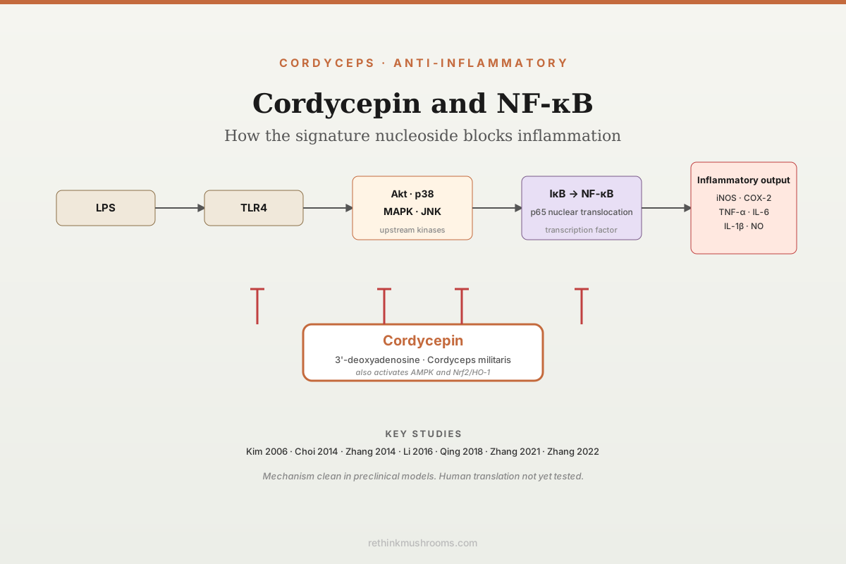
Cordycepin blocks NF-kB nuclear translocation in LPS-stimulated macrophages, down-regulates iNOS and COX-2, and attenuates TNF-α and IL-6 production. Multiple studies converge on the same upstream kinase targets: Akt, p38, MAPK and AMPK.
Kim et al., 2006; Choi et al., 2014; Zhang et al., 2014
NF-κB blocked
nuclear translocation suppressed
iNOS + COX-2 down
downstream gene expression reduced
Akt + p38 + AMPK
upstream kinase targets identified
Kim et al., 2006; Choi et al., 2014; Zhang et al., 2014
 View as image ↗
Cordycepin (3'-deoxyadenosine) is the signature nucleoside isolated from Cordyceps militaris and, in smaller amounts, from Ophiocordyceps sinensis. Its anti-inflammatory profile is one of the most mechanistically studied features of the molecule, and the evidence converges on a consistent picture: cordycepin interrupts NF-κB (nuclear factor kappa B) activation in immune cells by inhibiting the upstream kinases that drive it, with measurable reductions in downstream inflammatory gene expression (iNOS, COX-2) and cytokine secretion (TNF-α, IL-1β, IL-6).[1] This article lays out the specific studies, the specific kinases, and what the evidence does and does not yet establish in human physiology.
For broader context on this family of findings see cordyceps and inflammation: anti-inflammatory mechanisms, cordycepin and TNF-alpha suppression: molecular anti-inflammatory evidence, cordyceps for chronic inflammatory conditions: research review, and cordyceps and joint inflammation: arthritis research.
Key Takeaways
- Kim et al. (2006) established the foundational NF-κB finding: cordycepin suppressed LPS-induced NF-κB activation in RAW 264.7 macrophages by inhibiting Akt and p38 phosphorylation, with downstream reductions in iNOS, COX-2, TNF-α and NO production
- Choi et al. (2014) showed cordycepin attenuates NF-κB nuclear translocation by suppressing Toll-like receptor 4 (TLR4) signaling and MAPK phosphorylation in LPS-stimulated macrophages
- Zhang et al. (2014) identified AMPK activation as a parallel mechanism: cordycepin blocks LPS-induced TNF-α expression via AMPK signaling, independent of the Akt/p38 axis
- Li et al. (2016) confirmed cordycepin inhibits NF-κB signaling and MAPK phosphorylation in LPS-treated nucleus pulposus cells, extending the mechanism beyond macrophages
- Qing et al. (2018) demonstrated cordycepin alleviates LPS-induced acute lung injury via Nrf2/HO-1 up-regulation alongside NF-κB suppression, linking anti-inflammatory and antioxidant pathways
- Zhang et al. (2021) extended the finding to neuroinflammation: cordycepin attenuated microglial NF-κB activation and reduced dopaminergic neuron loss in a rotenone Parkinson's disease model
- Zhang et al. (2022) showed cordycepin drives M1-to-M2 macrophage polarisation via NF-κB/p65 inhibition in a sepsis model, providing a physiological context for the in vitro mechanism
What NF-κB Does In Inflammation
NF-κB is a family of transcription factors that translate pathogen and damage signals into transcriptional programs controlling the acute inflammatory response. In a resting cell, NF-κB dimers (typically p65/p50) are sequestered in the cytoplasm by the inhibitor IκB. On receipt of an inflammatory signal, typically via Toll-like receptor (TLR), tumour necrosis factor receptor, or IL-1 receptor engagement, IκB is phosphorylated by IκB kinase (IKK) and degraded. Freed p65/p50 translocate to the nucleus and activate transcription of pro-inflammatory genes: cytokines (TNF-α, IL-1β, IL-6), enzymes (iNOS, COX-2) and chemokines (CCL2, CXCL8). Pharmacological inhibitors of NF-κB are a major class of anti-inflammatory candidates because the pathway is central, ancient, and clinically relevant across autoimmune, metabolic and neurodegenerative disease states.
A compound that blocks NF-κB activation can do so at several points: at the receptor (TLR4), at adapter molecules (MyD88), at upstream kinases (Akt, p38 MAPK, ERK, JNK, IKK), at IκB phosphorylation or degradation, at nuclear translocation, or at DNA binding. Cordycepin has been shown to operate at multiple levels of this cascade, which is both scientifically interesting and a source of mechanistic complexity.
The Foundational Study: Kim et al. 2006
Kim and colleagues (2006) published the first paper establishing the cordycepin-NF-κB connection in a defined cell system. Using RAW 264.7 murine macrophages stimulated with lipopolysaccharide (LPS), the standard innate-immunity activator, the investigators measured NF-κB activation, kinase phosphorylation and downstream gene expression with and without cordycepin pretreatment.[1]
Their findings were unusually clean. Cordycepin inhibited LPS-induced phosphorylation of both Akt and p38 MAPK in a dose-dependent manner. It suppressed IκB-α phosphorylation and subsequent degradation, blocking NF-κB p65 nuclear translocation. Downstream, iNOS and COX-2 mRNA and protein were reduced, nitric oxide (NO) production was lowered, and TNF-α secretion was attenuated. The pattern is a canonical anti-inflammatory signature: upstream kinase inhibition, blocked NF-κB translocation, and proportional down-regulation of classical pro-inflammatory effectors. Kim 2006 is cited as the primary reference in almost every subsequent cordycepin anti-inflammatory paper.
Confirmation And Extension: Choi 2014 And Zhang 2014
Choi and colleagues (2014) extended the mechanism upward to TLR4 itself.[2] Using the same RAW 264.7 model, Choi demonstrated that cordycepin suppressed TLR4-mediated activation, attenuated MAPK phosphorylation (ERK, JNK and p38), and blocked NF-κB nuclear translocation. The practical implication is that cordycepin's anti-inflammatory effect is not restricted to events downstream of TLR4; it partially operates by dampening the receptor signal itself.
Zhang and colleagues (2014) added a parallel mechanism.[3] In mouse macrophages, cordycepin activated AMP-activated protein kinase (AMPK) and suppressed LPS-induced TNF-α expression in an AMPK-dependent manner. AMPK activation is known to cross-talk with NF-κB suppression, and the Zhang finding places cordycepin among a growing group of natural products whose anti-inflammatory effect is at least partially mediated by metabolic-stress-sensor activation rather than by direct NF-κB inhibition alone. The Akt/p38/NF-κB pathway (Kim 2006) and the AMPK pathway (Zhang 2014) are not mutually exclusive; cordycepin appears to act on both.
Beyond Macrophages: Nucleus Pulposus, Lung And Microglia
Li and colleagues (2016) tested cordycepin in LPS-stimulated nucleus pulposus cells, the tissue of the intervertebral disc, a system relevant to disc degeneration and chronic low-back inflammation.[4] Cordycepin inhibited NF-κB signaling and MAPK phosphorylation, reduced matrix metalloproteinase expression (MMP-3, MMP-13), and preserved aggrecan and collagen II, suggesting that the macrophage-derived mechanism translates to structural cells in an inflammatory disease-relevant context.
Qing and colleagues (2018) examined cordycepin in LPS-induced acute lung injury in mice.[5] Cordycepin reduced lung oedema, neutrophil infiltration and BALF protein, with concurrent activation of the Nrf2/HO-1 antioxidant pathway and suppression of NF-κB in lung tissue. The Nrf2/HO-1 arm matters because oxidative stress and NF-κB activation are tightly coupled at the cellular level, and compounds that engage both pathways are mechanistically more robust than those engaging either alone.
Zhang and colleagues (2021) brought the mechanism into neuroinflammation.[6] In a rotenone-induced Parkinson's disease model, cordycepin attenuated microglial NF-κB activation, reduced dopaminergic neuron loss in the substantia nigra, and improved motor performance. Microglia are resident CNS macrophages and show the same NF-κB-dependent inflammatory programme as peripheral macrophages, so the Kim 2006 mechanism extending into microglia is biologically predictable.
Zhang and colleagues (2022), a separate group, tested cordycepin in a sepsis model in immature mice.[7] Cordycepin attenuated liver and lung damage, improved survival metrics, and drove M1-to-M2 macrophage polarisation, with NF-κB/p65 inhibition identified as the mechanistic driver. M1/M2 macrophage polarisation is the macroscopic expression of the NF-κB axis in tissue: M1 macrophages carry the pro-inflammatory programme, M2 the resolving programme. A pharmacological shift toward M2 via NF-κB suppression converts the in vitro signal into a plausible in vivo mechanism of action.
The Signaling Map, Integrated
Putting the papers together, the pharmacological map looks like this:
| Upstream target | Effect on NF-κB | Key study |
|---|---|---|
| TLR4 signaling | Suppressed | Choi 2014 |
| Akt phosphorylation | Inhibited | Kim 2006 |
| p38 MAPK | Inhibited | Kim 2006, Choi 2014 |
| ERK / JNK | Inhibited | Choi 2014 |
| AMPK activation | Activated (parallel) | Zhang 2014 |
| Nrf2 / HO-1 | Activated (parallel) | Qing 2018 |
| NF-κB p65 nuclear translocation | Blocked | Kim 2006, Zhang 2022 |
| iNOS, COX-2 transcription | Reduced | Kim 2006 |
| TNF-α, IL-1β, IL-6 output | Reduced | Kim 2006, Zhang 2014, Zhang 2022 |
The picture is of a molecule that touches the NF-κB pathway at multiple levels rather than at a single high-specificity node. This is common for small-molecule natural products and is part of why cordycepin is mechanistically interesting but pharmacologically complex.
How Does Cordycepin Do This At The Molecular Level?
Cordycepin is an adenosine analogue with a missing 3'-hydroxyl on the ribose sugar. Its most-characterised primary biochemical activity is chain termination of RNA synthesis, through incorporation into growing RNA transcripts by polymerases that cannot then add the next nucleotide. That mechanism is central to its antiviral and anticancer effects, but it does not cleanly explain the kinase-level effects on NF-κB.
Several complementary mechanisms have been proposed. Cordycepin can be phosphorylated by cellular kinases to cordycepin triphosphate (3'-dATP), which interferes with polyadenylation and with some ATP-binding kinases. It is also a substrate for adenosine deaminase, and its deaminated product 3'-deoxyinosine has its own receptor activity. Adenosine receptor engagement (A1, A2A, A2B, A3) has documented anti-inflammatory effects that include NF-κB suppression. Sharma and colleagues (2022) reviewed these candidate mechanisms and noted that no single molecular target has been definitively established as the basis of the anti-inflammatory signal.[8] Ashraf and colleagues (2020) and Phull and colleagues (2022) reached similar conclusions in their reviews of the cordycepin therapeutic literature.[9][10]
The honest summary is that the empirical evidence for NF-κB suppression by cordycepin is strong across multiple cell types and disease models, while the proximal molecular target responsible for that suppression is still not definitively identified.
What The Human Evidence Shows
The NF-κB evidence for cordycepin is almost entirely preclinical: murine macrophages, murine disease models, murine tissue systems. Phull and colleagues (2022) surveyed the cordycepin clinical and preclinical literature and found no completed randomised controlled trial specifically testing the NF-κB anti-inflammatory mechanism in humans.[10] Clinical trials of Cordyceps militaris or Ophiocordyceps sinensis extracts in humans, where they exist, test mushroom products containing cordycepin alongside polysaccharides, ergosterol, adenosine, cordyglycoside and many other bioactive constituents. Attribution of any human clinical effect specifically to cordycepin-mediated NF-κB suppression is not possible with current evidence.
This is the central translational gap in the cordycepin anti-inflammatory story. The mechanism is well-characterised in the laboratory. The clinical endpoints linked mechanistically to that laboratory evidence have not been measured rigorously in humans.
Limitations Of The Evidence
Several limitations warrant explicit statement.
The published anti-inflammatory studies are overwhelmingly in murine cell lines (RAW 264.7 macrophages) and mouse disease models. Translation to human myeloid cells and human disease states is assumed, not directly demonstrated.
Cordycepin is rapidly deaminated to 3'-deoxyinosine by adenosine deaminase in vivo, which substantially limits oral bioavailability and complicates dose-response interpretation. Chen and colleagues (2023) reviewed formulation and delivery strategies for cordycepin and noted that achieving macrophage-relevant concentrations in humans at tolerable doses is a non-trivial pharmacological problem.
The molecular target responsible for cordycepin's NF-κB suppression has not been definitively identified. The kinase-level effects may be downstream of adenosine-receptor engagement, 3'-dATP accumulation, or other mechanisms.
Most in vivo studies use intraperitoneal or intravenous cordycepin administration, not oral delivery, which is the only practical route for a dietary supplement or food product. Oral bioavailability of isolated cordycepin in humans at doses approaching those used in mouse studies has not been robustly characterised.
The NF-κB pathway is physiologically essential for normal immune function. A compound that durably suppresses NF-κB could, in principle, also impair host defence against infection. This risk has not been systematically assessed for cordycepin at supplement-equivalent exposures.
Finally, most whole-Cordyceps products contain cordycepin at modest concentrations, and the mechanism-level findings in this article apply directly to purified cordycepin at experimentally defined concentrations. Extrapolation to a daily capsule of Cordyceps militaris extract is not straightforward.
What The Evidence Actually Establishes
- Cordycepin blocks LPS-induced NF-κB nuclear translocation in macrophages and in several other cell types through inhibition of upstream kinases (Akt, p38, MAPK) and via TLR4 signal suppression
- The mechanism is paralleled by AMPK activation (Zhang 2014) and Nrf2/HO-1 engagement (Qing 2018), giving cordycepin multiple levers on the inflammatory-oxidative axis
- Downstream pro-inflammatory outputs (iNOS, COX-2, TNF-α, IL-1β, IL-6) are reduced in proportion to the NF-κB suppression
- The mechanism has been demonstrated in macrophages (Kim 2006, Choi 2014, Zhang 2014), nucleus pulposus cells (Li 2016), lung tissue (Qing 2018), microglia (Zhang 2021) and an in vivo sepsis model (Zhang 2022)
- The proximal molecular target has not been definitively identified; the effect may operate through adenosine-receptor engagement, 3'-dATP accumulation, or other mechanisms
- No human randomised controlled trial has tested the NF-κB-specific anti-inflammatory mechanism, and isolated-cordycepin oral bioavailability in humans remains a translational constraint
The Bottom Line
Cordycepin suppresses NF-κB activation in LPS-stimulated macrophages by inhibiting Akt and p38 phosphorylation (Kim 2006), attenuating TLR4 signaling (Choi 2014) and activating AMPK (Zhang 2014). The mechanism extends into nucleus pulposus cells, lung tissue, microglia and sepsis models, with downstream reductions in iNOS, COX-2, TNF-α and IL-6. Translation to human clinical anti-inflammatory endpoints has not been formally tested.
Frequently Asked Questions
Sources & References
- 1RTHM-00103·Kim, Ho Gyoung et al. (2006). “Cordycepin inhibits lipopolysaccharide-induced inflammation by the suppression of NF-kappaB through Akt and p38 inhibition in RAW 264.7 macrophage cells..” European journal of pharmacology.Study breakdown →PubMed →↩
- 2RTHM-00317·Choi, Yung Hyun et al. (2014). “Anti-inflammatory effects of cordycepin in lipopolysaccharide-stimulated RAW 264.7 macrophages through Toll-like receptor 4-mediated suppression of mitogen-activated protein kinases and NF-κB signaling pathways..” Drug design.Study breakdown →PubMed →↩
- 3RTHM-00361·Zhang, Jian-Li et al. (2014). “Cordycepin inhibits lipopolysaccharide (LPS)-induced tumor necrosis factor (TNF)-α production via activating amp-activated protein kinase (AMPK) signaling..” International journal of molecular sciences.Study breakdown →PubMed →↩
- 4RTHM-00433·Li, Yan et al. (2016). “Cordycepin inhibits LPS-induced inflammatory and matrix degradation in the intervertebral disc..” PeerJ.Study breakdown →PubMed →↩
- 5RTHM-00526·Qing, Rui et al. (2018). “Cordycepin alleviates lipopolysaccharide-induced acute lung injury via Nrf2/HO-1 pathway..” International immunopharmacology.Study breakdown →PubMed →↩
- 6RTHM-00735·Zhang, Xiao-Ling et al. (2021). “Anti-inflammatory and neuroprotective effects of natural cordycepin in rotenone-induced PD models through inhibiting Drp1-mediated mitochondrial fission..” Neurotoxicology.Study breakdown →PubMed →↩
- 7RTHM-00808·Zhang, Yudan et al. (2022). “Cordycepin induces M1/M2 macrophage polarization to attenuate the liver and lung damage and immunodeficiency in immature mice with sepsis via NF-κB/p65 inhibition..” The Journal of pharmacy and pharmacology.Study breakdown →PubMed →↩
- 8RTHM-00795·Sharma, Shivani et al. (2022). “Cordycepin as a Metabolite with Pharmacological Potential: A Review..” International journal of medicinal mushrooms.Study breakdown →PubMed →↩
- 9RTHM-00605·Ashraf, Syed Amir et al. (2020). “Cordycepin for Health and Wellbeing: A Potent Bioactive Metabolite of an Entomopathogenic Cordyceps Medicinal Fungus and Its Nutraceutical and Therapeutic Potential..” Molecules (Basel.Study breakdown →PubMed →↩
- 10RTHM-00790·Phull, Abdul-Rehman et al. (2022). “Cordyceps militaris as a Bio Functional Food Source: Pharmacological Potential, Anti-Inflammatory Actions and Related Molecular Mechanisms..” Microorganisms.Study breakdown →PubMed →↩