Cordyceps and Inflammation
100 µM Effective Dose
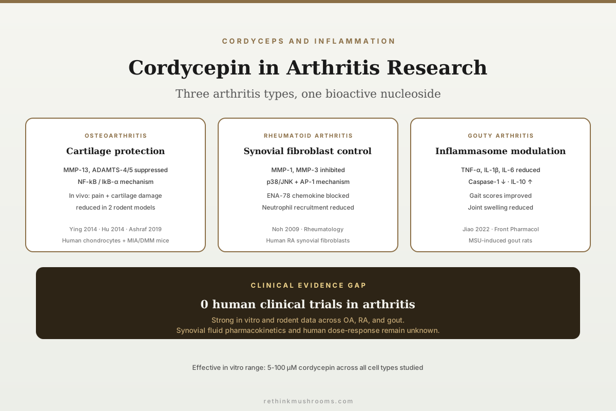
Cordycepin at 10-100 µM dose-dependently suppressed IL-1β-induced MMP-13, IL-6, iNOS, and COX-2 expression in human osteoarthritis chondrocytes while blocking NF-kB activation through IkB-α stabilisation (Ying et al., International Orthopaedics, 2014).
Ying et al., International Orthopaedics, 2014
Pain + pathology
Cordycepin reduced pain behaviour and cartilage damage in both MIA rat and DMM mouse models of osteoarthritis in vivo (Ashraf et al., 2019)
MMP-1/3 in RA
Cordycepin inhibited IL-1β-induced MMP-1 and MMP-3 expression in rheumatoid arthritis synovial fibroblasts via p38/JNK/AP-1 blockade (Noh et al., 2009)
Gouty arthritis
Cordyceps militaris extract reduced TNF-α, IL-1β, IL-6, and Caspase-1 while increasing IL-10 in MSU-induced gout model rats (Jiao et al., 2022)
Ying et al., International Orthopaedics, 2014
 View as image ↗
Cordycepin, the primary bioactive nucleoside from Cordyceps militaris, has accumulated a focused body of evidence in joint inflammation research spanning osteoarthritis (OA), rheumatoid arthritis (RA), and gouty arthritis. The data cover human chondrocyte cultures, synovial fibroblast assays, macrophage models, and two in vivo OA models in rodents. The consistent finding across these systems is that cordycepin suppresses inflammatory mediators and matrix-degrading enzymes through NF-kB pathway inhibition and related transcription factor blockade.[1][2] For the broader NF-kB mechanism, see cordycepin and NF-kB: how it suppresses inflammatory pathways; for the TNF-alpha angle, see cordycepin and TNF-alpha suppression: molecular anti-inflammatory evidence.
Key Takeaways
- Ying et al. (2014) in International Orthopaedics showed cordycepin (10-100 µM) dose-dependently inhibited IL-1β-induced PGE2, NO, MMP-13, IL-6, iNOS, and COX-2 in human OA chondrocytes by suppressing IkB-α degradation and blocking NF-kB nuclear translocation
- Hu et al. (2014) in International Journal of Clinical and Experimental Pathology demonstrated cordycepin (5-100 µM) suppressed IL-1β-induced glycosaminoglycan release and gene expression of six catabolic enzymes (MMP-1, MMP-13, ADAMTS-4, ADAMTS-5, cathepsin K, cathepsin S) in advanced-stage OA chondrocytes
- Ashraf et al. (2019) in Scientific Reports provided the first in vivo evidence that oral cordycepin reduces pain behaviour, synovial inflammation, and cartilage damage in both MIA rat and DMM mouse models of OA, acting through polyadenylation factor inhibition in macrophages
- Noh et al. (2009) in Rheumatology showed cordycepin inhibited IL-1β-induced MMP-1 and MMP-3 expression in RA synovial fibroblasts by blocking p38/JNK and AP-1 activation, without affecting ERK or NF-kB in that cell type
- Jiao et al. (2022) in Frontiers in Pharmacology demonstrated Cordyceps militaris extract reduced TNF-α, IL-1β, IL-6, and Caspase-1 while increasing IL-10 in MSU-induced acute gouty arthritis rats, with transcriptomics identifying CCL7, CSF2RB, and IL-1β as key regulated genes
- Kim et al. (2006) in European Journal of Pharmacology established that cordycepin suppresses LPS-induced inflammation in macrophages through NF-kB inhibition via Akt and p38 MAPK pathways, providing the mechanistic foundation for the joint-specific studies
Osteoarthritis: The Chondrocyte Evidence
OA is characterised by progressive cartilage degradation driven by an imbalance between matrix synthesis and matrix-degrading enzyme activity. The key destructive enzymes are matrix metalloproteinases (MMPs) and aggrecanases (ADAMTS family), both upregulated by the inflammatory cytokine IL-1β through NF-kB-dependent transcription. Two independent studies tested cordycepin directly on human OA chondrocytes.
Ying et al. (2014) cultured human OA chondrocytes with cordycepin at 10, 50, and 100 µM, then stimulated them with IL-1β (5 ng/ml). Cordycepin dose-dependently inhibited the production of prostaglandin E2 (PGE2) and nitric oxide (NO), both direct mediators of pain and inflammation in OA joints. Gene expression and protein production of MMP-13 (the primary collagen-degrading enzyme in cartilage), IL-6, iNOS, and COX-2 were all significantly suppressed. The mechanism mapped to NF-kB: cordycepin prevented the degradation of IkB-α (the cytoplasmic inhibitor that holds NF-kB inactive), thereby blocking NF-kB nuclear translocation and downstream inflammatory gene transcription.[1]
Hu et al. (2014) took this further using chondrocytes from advanced-stage OA patients (Kellgren-Lawrence grade III-IV). They measured a broader panel of catabolic targets and found cordycepin (5-100 µM) dose-dependently suppressed IL-1β-induced glycosaminoglycan (GAG) release from cartilage explants, confirming reduced matrix degradation. At the gene level, cordycepin inhibited MMP-1, MMP-13, ADAMTS-4, ADAMTS-5, cathepsin K, and cathepsin S, covering the major collagenase, aggrecanase, and cysteine protease families responsible for cartilage destruction. The simultaneous suppression of COX-2 and iNOS at the transcript level, along with blocked NO production, replicated Ying et al.'s findings in a more severely diseased cell population.[2]
These two in vitro studies establish that cordycepin has chondroprotective activity in human OA cells through a broad-spectrum suppression of catabolic and inflammatory mediators. The convergent NF-kB mechanism across both studies, using different OA patient populations and different outcome panels, strengthens the mechanistic claim. The limitation is that both studies are in vitro. Whether cordycepin reaches joint tissue at effective concentrations after oral administration is a separate question, addressed by the in vivo work below.
In Vivo Evidence: Pain, Inflammation, and Joint Structure
Ashraf et al. (2019) published the most comprehensive in vivo study of cordycepin in OA, testing it in two complementary rodent models. In the monosodium iodoacetate (MIA) rat model (which produces rapid joint pain and inflammation), cordycepin reduced pain behaviour and synovial inflammation. In the destabilisation of the medial meniscus (DMM) mouse model (which mimics the slow mechanical progression of human OA), cordycepin reduced cartilage damage measured as proteoglycan loss and decreased expression of MMP-13 and ADAMTS-5.[3]
The mechanistic contribution of this study extends beyond replicating in vitro findings in vivo. Ashraf et al. identified a novel mechanism: cordycepin acts as a polyadenylation inhibitor. In OA synovial tissue from human patients, the polyadenylation factor CPSF4 was elevated alongside NF-kB in samples with high-grade inflammation. In macrophage cultures, LPS stimulation induced nuclear localisation of both NF-kB and polyadenylation factors (WDR33 and CPSF4), and cordycepin inhibited both. Critically, siRNA knockdown of polyadenylation factors alone prevented NF-kB nuclear localisation, establishing that polyadenylation is upstream of NF-kB activation in this inflammatory context.
This mechanism distinguishes cordycepin from conventional anti-inflammatory drugs. Rather than blocking a single cytokine or enzyme, cordycepin may interfere with the post-transcriptional processing machinery that enables inflammatory gene expression. The authors characterised cordycepin as a potential lead compound for a novel class of orally available anti-inflammatory and analgesic drugs for OA patients with high synovial inflammation. No clinical trial has yet tested this proposition.
Ashraf et al. (2019) · Scientific Reports
- Population
- MIA-injected rats (rapid pain model) and DMM-operated mice (slow mechanical OA model), plus human OA synovial tissue
- Intervention
- Oral cordycepin (3'-deoxyadenosine)
- Comparator
- Untreated / vehicle model controls
- Finding
- Cordycepin reduced pain behaviour and synovial inflammation in the MIA rat and lowered cartilage proteoglycan loss with reduced MMP-13 and ADAMTS-5 in the DMM mouse. The novel mechanism: cordycepin inhibits polyadenylation factors (CPSF4/WDR33) that sit upstream of NF-kB activation in inflamed macrophages.
preliminary This is the only in vivo demonstration and the only study to identify the polyadenylation mechanism, but it remains an animal model — no human OA outcomes exist.
Ashraf et al., Scientific Reports, 2019
Rheumatoid Arthritis: Synovial Fibroblast Data
RA differs from OA in that it is an autoimmune disease where the synovial lining itself becomes the primary site of destructive inflammation. RA synovial fibroblasts (RASFs) produce MMPs that degrade cartilage and bone, driven by cytokine stimulation. Noh et al. (2009) tested cordycepin directly on human RASFs.
Cordycepin inhibited IL-1β-induced MMP-1 and MMP-3 expression in RASFs in a dose-dependent manner. It also specifically blocked IL-1β-induced ENA-78 (CXCL5) production, a chemokine that recruits neutrophils into the inflamed joint. The signalling mechanism in RASFs differed from the chondrocyte studies: cordycepin inhibited p38/JNK and AP-1 activation but did not affect ERK or NF-kB activation in this cell type.[4]
This cell-type-specific signalling difference is informative. In macrophages and chondrocytes, cordycepin's primary mechanism is NF-kB inhibition. In RASFs, the same functional outcome (MMP suppression) occurs through AP-1 rather than NF-kB. This suggests cordycepin engages multiple signalling nodes depending on cellular context, which is consistent with its broad-spectrum anti-inflammatory profile but complicates predictions about which pathway will dominate in intact joints where multiple cell types interact. For how cordycepin's anti-inflammatory effects extend beyond joints, see cordyceps for chronic inflammatory conditions: research review.
Gouty Arthritis: Cordyceps militaris Extract
Gout is driven by monosodium urate (MSU) crystal deposition in joints, triggering acute NLRP3 inflammasome activation, Caspase-1 processing, and IL-1β release. Unlike OA and RA, gout involves a crystal-driven innate immune response rather than chronic autoimmune or mechanical joint degradation.
Jiao et al. (2022) tested Cordyceps militaris extract (CME, whole extract rather than isolated cordycepin) in an MSU-induced acute gouty arthritis rat model. CME significantly improved gait scores and reduced joint swelling. Inflammatory markers showed a clear pattern: the pro-inflammatory cytokines TNF-α, IL-1β, IL-6, and Caspase-1 were reduced, while the anti-inflammatory cytokine IL-10 was increased. Histological analysis confirmed reduced inflammatory cell infiltration in joint tissue.[5]
The transcriptomic analysis identified four key regulated genes: CCL7 (a monocyte-recruiting chemokine), CSF2RB (the common beta chain of the GM-CSF/IL-3/IL-5 receptor), LIF (leukaemia inhibitory factor), and IL-1β. The enriched pathway was cytokine-cytokine receptor interaction, connecting CME's effect to inflammasome regulation and immune cell signalling. This is the only arthritis study using whole C. militaris extract rather than purified cordycepin, making it more directly relevant to commercially available cordyceps supplements, which are typically C. militaris-derived.
The Mechanistic Foundation: NF-kB Suppression
The joint-specific studies build on Kim et al.'s (2006) foundational work showing that cordycepin suppresses LPS-induced inflammatory mediator production in RAW 264.7 macrophages. Kim et al. demonstrated that cordycepin inhibits NF-kB by suppressing both Akt and p38 MAPK phosphorylation, preventing IkB kinase activation and subsequent NF-kB nuclear translocation. This reduces the transcription of iNOS, COX-2, TNF-α, and IL-1β.[6]
Choi et al. (2014) extended this mechanism to the Toll-like receptor 4 (TLR4) pathway, showing cordycepin inhibits LPS-induced inflammatory gene expression in macrophages through TLR4-mediated suppression of both MAPK and NF-kB signalling cascades.[7]
These macrophage studies are relevant to arthritis because macrophages are the dominant inflammatory cell type in both OA synovium and RA pannus tissue. The macrophage NF-kB mechanism is consistent with what Ying et al. found in chondrocytes and what Ashraf et al. confirmed in vivo. The exception is Noh et al.'s RA synovial fibroblast data showing AP-1 rather than NF-kB as the primary cordycepin target in that cell type.
How the Evidence Connects Across Arthritis Types
The studies above address three distinct forms of arthritis: OA (mechanical and inflammatory cartilage degradation), RA (autoimmune synovial inflammation), and gout (crystal-triggered innate immune activation). Despite different disease drivers, cordycepin shows consistent effects on the downstream inflammatory cascade. In OA chondrocytes, it suppresses MMP-13 and ADAMTS enzymes that directly degrade cartilage matrix. In RA synovial fibroblasts, it suppresses MMP-1 and MMP-3 that drive pannus-mediated joint destruction. In gouty arthritis, it modulates Caspase-1 and the inflammasome pathway that converts pro-IL-1β to its active form.
Arthritis Type · Signalling Node · Evidence Tier
Same Endpoint, Different Upstream Switch
Across three forms of arthritis cordycepin converges on one outcome — matrix-metalloproteinase suppression and cytokine reduction — but the pathway it works through changes with the cell type. That divergence is why no single “mechanism of action” captures it.
Osteoarthritis
In vitroHuman OA chondrocytes
Targets suppressed
MMP-13, IL-6, iNOS, COX-2, ADAMTS-4/5, cathepsin K/S
Dominant pathway
NF-κB
Blocks IκB-α degradation, stopping NF-κB nuclear translocation
Anchor: Cordycepin 10–100 µM dose-dependently suppressed catabolic enzymes in IL-1β-stimulated chondrocytes
Osteoarthritis
In vivoMIA rat + DMM mouse (in vivo)
Targets suppressed
Pain behaviour, cartilage loss, MMP-13, ADAMTS-5
Dominant pathway
Polyadenylation → NF-κB
Inhibits CPSF4/WDR33 upstream of NF-κB activation
Anchor: Oral cordycepin reduced pain, synovial inflammation, and structural cartilage damage in two rodent OA models
Rheumatoid arthritis
In vitroHuman RA synovial fibroblasts
Targets suppressed
MMP-1, MMP-3, ENA-78 (CXCL5)
Dominant pathway
p38/JNK → AP-1
Acts through AP-1, NOT NF-κB or ERK, in this cell type
Anchor: Cordycepin dose-dependently inhibited IL-1β-induced MMP-1/MMP-3 and neutrophil-recruiting ENA-78
Gouty arthritis
In vivoMSU-induced rat model (in vivo)
Targets suppressed
TNF-α, IL-1β, IL-6, Caspase-1 (↑ IL-10)
Dominant pathway
NLRP3 / Caspase-1
Whole C. militaris extract modulates the crystal-driven inflammasome
Anchor: CME improved gait and joint swelling while shifting the cytokine balance anti-inflammatory
Only the two in-vivo columns cross beyond cell culture, and both are rodent models. No human randomized controlled trial has tested cordycepin or cordyceps for any form of arthritis.
Source: Ying (2014); Hu (2014); Ashraf (2019); Noh (2009); Jiao (2022)
View as image ↗The shared thread is matrix metalloproteinase suppression and cytokine reduction, but the upstream signalling varies by cell type. This suggests cordycepin is not a single-pathway drug candidate but rather a compound that interfaces with multiple inflammatory signalling nodes, with the dominant node depending on the cellular and disease context. Whether this multi-target profile would translate to efficacy across arthritis subtypes in humans, or whether cell-type-specific pharmacokinetics would limit its reach, remains untested. The lack of synovial fluid pharmacokinetic data after oral cordycepin administration is the most critical missing piece for all three arthritis types.
Limitations
All human cell studies used in vitro models. Joint tissue penetration of cordycepin after oral administration has not been directly measured, and only Ashraf et al. (2019) demonstrated in vivo efficacy. Effective in vitro concentrations (10-100 µM) may not be achievable in synovial fluid with standard oral supplementation. Cordycepin's half-life in plasma is short due to rapid deamination by adenosine deaminase, and no study has measured whether biologically active concentrations persist long enough to affect joint tissue remodelling over weeks or months.
No randomised controlled trial has tested cordycepin or cordyceps preparations in human arthritis patients. The animal OA models (MIA and DMM) replicate some features of human OA but not the full multi-decade disease progression with its mechanical, metabolic, and age-related drivers. The gouty arthritis study used whole C. militaris extract, making it difficult to attribute effects specifically to cordycepin versus polysaccharides, adenosine, or other bioactive components present in whole-extract preparations.
The NF-kB mechanism has been demonstrated across multiple cell types, but the cell-type specificity of signalling (NF-kB in macrophages and chondrocytes, AP-1 in synovial fibroblasts) means the dominant pathway in an intact arthritic joint with mixed cell populations remains unclear. Cross-talk between these pathways has not been characterised for cordycepin. The dose ranges used in vitro (5-100 µM) and in animal models have not been validated against achievable human tissue concentrations at standard oral supplement doses. For a broader perspective on how mushroom anti-inflammatory compounds work beyond cordyceps, see anti-inflammatory mushrooms for metabolic inflammation.
- Cell & Lab Studiescompleted
Human OA chondrocytes, RA synovial fibroblasts, and macrophage models map the NF-kB, AP-1, and inflammasome mechanisms (Ying 2014; Hu 2014; Noh 2009; Kim 2006; Choi 2014)
- Animal Studiescompleted
Two rodent OA models (MIA rat, DMM mouse) and an MSU-induced gout rat model show pain, inflammation, and structural benefit in vivo (Ashraf 2019; Jiao 2022)
- Case Reports & Early Seriesmissing
No published human case reports of cordycepin or cordyceps used for arthritis
- Small Human Trialsmissing
No randomized controlled trial has tested cordycepin or a cordyceps preparation in arthritis patients
- Large Randomized Trialsmissing
None
- Meta-Analyses & Systematic Reviewsmissing
None for arthritis endpoints
The mechanism is well-characterized in human cells and confirmed in animal models across three arthritis types, but the evidence stops at rodents. There is no human trial, no case series, and no synovial-fluid pharmacokinetic data — so cordycepin for arthritis is a well-supported preclinical hypothesis, not an established human treatment.
Aggregate of the seven cited studies (Ying 2014; Hu 2014; Ashraf 2019; Noh 2009; Jiao 2022; Kim 2006; Choi 2014)
Safety
ModerateThis is preclinical research, not a treatment protocol
Concern
Every finding in this article comes from cell cultures and animal models. Effective concentrations in the lab (10-100 µM) have not been shown to be reachable in human joint tissue after swallowing a cordyceps supplement, and cordycepin is rapidly broken down in the bloodstream by adenosine deaminase. Cordyceps supplements are also not standardized for cordycepin content, so what is in a capsule rarely matches what was used in these studies.
What the research says
The evidence supports cordycepin as a promising anti-inflammatory candidate worth studying in humans. It does not support using cordyceps as a treatment for osteoarthritis, rheumatoid arthritis, or gout, and it says nothing about safe or effective doses in people.
Particularly relevant for: Anyone with arthritis or joint pain considering cordyceps in place of, or alongside, prescribed treatment
What to do
This is not medical advice. Do not stop or replace prescribed arthritis medication with a cordyceps supplement; discuss any supplement with a rheumatologist or physician, especially if you take immunosuppressants, anticoagulants, or urate-lowering drugs.
Based on the limitations of Ying (2014), Hu (2014), Ashraf (2019), Noh (2009), and Jiao (2022), none of which included human arthritis patients.
The Bottom Line
Cordycepin demonstrates consistent anti-inflammatory and chondroprotective effects across osteoarthritis chondrocytes, rheumatoid arthritis synovial fibroblasts, gouty arthritis animal models, and two in vivo OA rodent models. The primary mechanism is NF-kB pathway inhibition in macrophages and chondrocytes, with AP-1 as the dominant target in synovial fibroblasts. Ashraf et al.'s 2019 study provided the strongest evidence by showing oral cordycepin reduced pain, inflammation, and structural joint damage in vivo. No human clinical trial in arthritis has been conducted.
Frequently Asked Questions
Sources & References
- 1RTHM-00357·Ying, Xiaozhou et al. (2014). “Cordycepin prevented IL-β-induced expression of inflammatory mediators in human osteoarthritis chondrocytes..” International orthopaedics.Study breakdown →PubMed →↩
- 2RTHM-00326·Hu, Pengfei et al. (2014). “Cordycepin modulates inflammatory and catabolic gene expression in interleukin-1beta-induced human chondrocytes from advanced-stage osteoarthritis: an in vitro study..” International journal of clinical and experimental pathology.Study breakdown →PubMed →↩
- 3RTHM-00543·Ashraf, Sadaf et al. (2019). “The polyadenylation inhibitor cordycepin reduces pain, inflammation and joint pathology in rodent models of osteoarthritis..” Scientific reports.Study breakdown →PubMed →↩
- 4RTHM-00167·Noh, E-M et al. (2009). “Cordycepin inhibits IL-1beta-induced MMP-1 and MMP-3 expression in rheumatoid arthritis synovial fibroblasts..” Rheumatology (Oxford.Study breakdown →PubMed →↩
- 5RTHM-00769·Jiao, Chunwei et al. (2022). “Transcriptomic analysis of the anti-inflammatory effect of Cordyceps militaris extract on acute gouty arthritis..” Frontiers in pharmacology.Study breakdown →PubMed →↩
- 6RTHM-00103·Kim, Ho Gyoung et al. (2006). “Cordycepin inhibits lipopolysaccharide-induced inflammation by the suppression of NF-kappaB through Akt and p38 inhibition in RAW 264.7 macrophage cells..” European journal of pharmacology.Study breakdown →PubMed →↩
- 7RTHM-00317·Choi, Yung Hyun et al. (2014). “Anti-inflammatory effects of cordycepin in lipopolysaccharide-stimulated RAW 264.7 macrophages through Toll-like receptor 4-mediated suppression of mitogen-activated protein kinases and NF-κB signaling pathways..” Drug design.Study breakdown →PubMed →↩