Cordyceps & Inflammation
0 Trials in Patients
Cordycepin suppresses inflammatory signaling through NF-kB, Akt, p38 MAPK, TLR4, and AMPK across cell and animal models. Not one randomized trial has enrolled a patient with arthritis, asthma, colitis, or any other chronic inflammatory disease.
Kim et al. 2006 through Ontawong et al. 2024
17 Studies
spanning macrophage biology, arthritis, gout, asthma, disc degeneration, and pharmacokinetics
2.85 mg
cordycepin per day in the only human randomized trial, in healthy volunteers
Lost Head-to-Head
the one study with active comparators found cordyceps weaker than prednisolone and montelukast
Kim et al. 2006 through Ontawong et al. 2024
 View as image ↗
Chronic inflammation drives a constellation of conditions: osteoarthritis, rheumatoid arthritis, gout, inflammatory bowel disease, asthma, and age-related tissue degeneration. These diseases share a molecular feature: sustained activation of inflammatory signaling that perpetuates tissue damage long after the initial trigger has resolved. Cordycepin, the principal bioactive nucleoside analog from Cordyceps species, suppresses these pathways at multiple points in preclinical studies. For a detailed look at the NF-kB mechanism, see cordycepin and NF-kB: how it suppresses inflammatory pathways. For TNF-alpha specifically, see cordycepin and TNF-alpha suppression: molecular anti-inflammatory evidence.
This article reviews that evidence across conditions, and is organized around a question the mechanistic literature rarely asks: how far does each finding actually reach? The mechanisms are real and, in human cartilage cells, independently replicated. The clinical evidence does not exist.
Key Takeaways
- Cordycepin suppressed LPS-induced NF-kB activation in RAW 264.7 macrophages by inhibiting Akt and p38 MAPK phosphorylation, reducing nitric oxide, iNOS, COX-2, and TNF-alpha (Kim et al., 2006)
- In the same cell line, cordycepin blocked LPS binding and TLR4/MyD88 expression at the receptor level, and reduced PGE2, TNF-alpha, and IL-1beta (Choi et al., 2014)
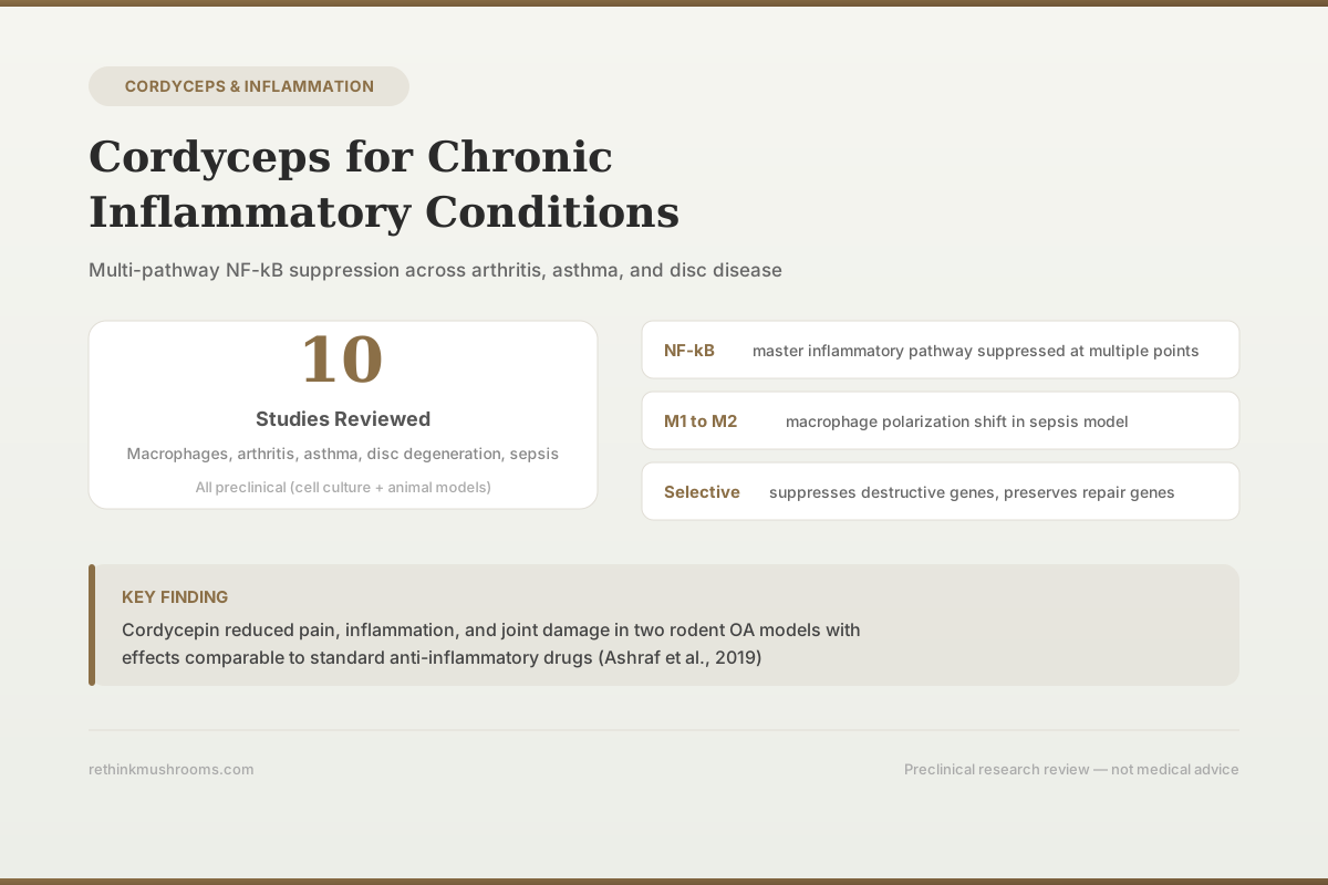
- Cordycepin reduced pain behavior, synovial inflammation, and joint pathology in two rodent osteoarthritis models, and the same study found polyadenylation factor CPSF4 elevated in synovia from OA patients (Ashraf et al., 2019)
- Two independent 2014 teams tested cordycepin on human osteoarthritis chondrocytes and agreed on every overlapping endpoint — nitric oxide, iNOS, COX-2, and MMP-13 all fell (Hu et al., 2014; Ying et al., 2014)
- The only mouse asthma study that included active drug comparators found C. militaris less effective than both prednisolone and montelukast, with the authors concluding it "is unable to adequately block the potent mediators of asthmatic airway inflammation" (Hsu et al., 2008)
- The single human randomized trial gave 2.85 mg of cordycepin daily to 40 healthy adults — roughly 20 to 80 times below the animal-scaled dose — and found within-group cytokine changes only (Ontawong et al., 2024)
How Far the Evidence Reaches
Deep in the Dish, Absent in the Clinic
The anti-inflammatory case for cordycepin is not weak — it is narrow in reach. Each rung below asks a different question, and the answers get thinner as you climb. Human cells and animal disease models both deliver. The single human randomized trial enrolled healthy volunteers, not patients. The top rung — people who actually have a chronic inflammatory disease — is empty.
Human patients with a chronic inflammatory disease
Does cordyceps improve the disease?
- Studies
- No study at this level
- Refs
- —
- What limits it
- Not a single randomized trial has enrolled patients with arthritis, asthma, colitis, or any other chronic inflammatory condition.
Human randomized trial — healthy volunteers
Does it move inflammatory markers in people?
- Studies
- Ontawong 2024
- Refs
- [12]
- What limits it
- 40 healthy adults, 8 weeks, 2.85 mg cordycepin/day. IL-1β fell in the male treatment group and IL-6 fell from baseline in the female treatment group, but these were within-group changes in 10-person cells, and the trial studied immune function, not disease.
Human cells and tissue
Does it work in human biology?
- Studies
- Hu 2014 · Ying 2014 · Ashraf 2019 · Zhang 2014 · Noh 2009
- Refs
- [5] [6] [4] [3] [17]
- What limits it
- Osteoarthritis cartilage and chondrocytes, OA synovial tissue, human monocyte-derived macrophages, PBMCs from Kawasaki disease patients, and synovial fibroblasts from 12 rheumatoid arthritis patients. Real human cells, but no clinical outcome, and cordycepin was applied directly to the cells rather than having to survive a bloodstream first.
Animal disease models
Does it change whole-organism outcomes?
- Studies
- Ashraf 2019 · Yang 2015 · Hsu 2008 · Jiao 2022 · Li 2016 · Zhang 2022
- Refs
- [4] [9] [8] [7] [10] [11]
- What limits it
- Osteoarthritis, asthma, gout, disc degeneration, and sepsis models show pain, inflammation, and tissue benefits — but the one study that included active drug comparators found cordyceps weaker than both (Hsu 2008).
Immortalized cell lines
What is the molecular mechanism?
- Studies
- Kim 2006 · Choi 2014
- Refs
- [1] [2]
- What limits it
- RAW 264.7 mouse macrophages stimulated with LPS. This is where the NF-κB, Akt, p38, and TLR4 story comes from — foundational, but the furthest removed from a patient.
Narrative review
What does the field believe?
- Studies
- Phull 2022
- Refs
- [13]
- What limits it
- Consolidates the preclinical literature. A narrative review inherits the limits of everything below it and adds no independent evidence.
Reading the ladder top-down rather than bottom-up is the point. The mechanism work is genuinely strong and has been reproduced by independent groups in human cells. What has never been done is the study that would matter to someone with osteoarthritis or asthma: give cordyceps to people who have the disease and measure whether they get better.
Evidence synthesis; rungs from refs [1]–[13] (see references)
The Core Mechanism: How Cordycepin Suppresses NF-kB
NF-kB is the master transcription factor for inflammatory gene expression. When activated by bacterial products (LPS), cytokines (TNF-alpha, IL-1beta), or damage signals, it translocates to the nucleus and drives production of inflammatory mediators that amplify and sustain tissue damage. In chronic inflammatory conditions, that activity becomes self-perpetuating.
Kim et al. (2006) established the foundational mechanism. In LPS-stimulated RAW 264.7 macrophages, cordycepin inhibited phosphorylation of Akt and p38 in a dose-dependent manner, suppressed IkB-alpha phosphorylation and NF-kB translocation, and reduced TNF-alpha expression. Downstream, iNOS and COX-2 expression fell significantly and nitric oxide production dropped. The dual inhibition of Akt and p38 matters because these are two independent upstream pathways converging on NF-kB, meaning cordycepin blocks the signal at more than one entry point.[1]
Choi et al. (2014) extended this upstream, to the receptor itself. In the same cell line, cordycepin inhibited the binding of LPS to macrophages and reduced LPS-induced Toll-like receptor 4 (TLR4) and MyD88 expression. It suppressed MAPK phosphorylation and attenuated NF-kB nuclear translocation by preventing IkB-alpha degradation. Functionally, it lowered nitric oxide and prostaglandin E2 by downregulating iNOS and COX-2, and reduced TNF-alpha and IL-1beta release through decreased mRNA expression. By acting where the signal enters the cell, cordycepin prevented the cascade rather than mopping up individual mediators.[2]
Zhang et al. (2014) identified a different mechanism, and did it partly in human cells. Cordycepin suppressed LPS-induced TNF-alpha in mouse macrophages and in peripheral blood mononuclear cells taken from patients with Kawasaki disease, a systemic vasculitis driven by TNF-alpha. The effect ran through AMP-activated protein kinase (AMPK): blocking AMPK pharmacologically or by siRNA depletion removed cordycepin's benefit, which is the kind of causal test most mechanism papers skip. Cordycepin also reduced reactive oxygen species and NF-kB activation.[3]
One caution about treating "NF-kB suppression" as the universal explanation. Noh et al. (2009) tested cordycepin on synovial fibroblasts isolated from 12 patients with rheumatoid arthritis and found it inhibited IL-1beta-induced MMP-1 and MMP-3 dose-dependently and specifically blocked the chemokine ENA-78 — but through p38/JNK and AP-1. In those human cells, cordycepin did not inhibit ERK or NF-kB activation.[17] The pathway cordycepin uses appears to depend on the cell type, which means mechanism findings from mouse macrophage lines should not be assumed to transfer to human joint tissue.
Osteoarthritis: The Strongest Disease-Specific Evidence
Osteoarthritis is driven by chronic low-grade inflammation in joint tissues that degrades cartilage through matrix metalloproteinase production and inflammatory cytokine release. The OA research on cordycepin is the most developed body of work in this article, and the only place where an independent replication exists.
Ashraf et al. (2019) · Scientific Reports
- Population
- Monosodium iodoacetate-injected rats and mice with surgical destabilisation of the medial meniscus, plus joint tissue from people with OA and non-arthritic post-mortem controls
- Intervention
- Cordycepin, a polyadenylation inhibitor
- Comparator
- Vehicle control — no active anti-inflammatory drug comparator was included
- Finding
- Cordycepin reduced pain behaviour, synovial inflammation, and joint pathology in both models. In human tissue, the polyadenylation factor CPSF4 and NF-kB were elevated in synovia from OA patients with high-grade inflammation, and knockdown of polyadenylation factors prevented NF-kB nuclear localisation in macrophages.
positive Strong for a preclinical study — two independent disease models plus a human tissue arm — but no active comparator and no patient outcomes.
Ashraf et al., 2019 [4]
Ashraf et al. (2019) tested cordycepin in two established rodent OA models: monosodium iodoacetate injection in rats and surgical destabilisation of the medial meniscus in mice. In both, cordycepin reduced pain behaviour, synovial inflammation, and joint pathology. The study's most interesting contribution is mechanistic and human: the researchers analysed joint tissues from people with OA and found the polyadenylation factor CPSF4 and NF-kB elevated in synovia from patients with high-grade inflammation. In macrophages, stimulation drove nuclear localisation of both NF-kB and polyadenylation factors, cordycepin inhibited that, and knocking down the polyadenylation factors independently prevented NF-kB from entering the nucleus.[4] That last result is what makes the mechanism credible rather than correlational.
Two separate groups published in 2014 on the same question in human cells. Hu et al. cultured human cartilage explants and chondrocytes with IL-1beta and cordycepin at 5–100 µM. Cordycepin suppressed IL-1beta-stimulated glycosaminoglycan release — meaning less matrix was lost — and dose-dependently decreased MMP-1, MMP-13, cathepsin K, cathepsin S, ADAMTS-4, and ADAMTS-5. It also inhibited COX-2 and iNOS expression and blocked nitric oxide production.[5] Ying et al. pretreated chondrocytes taken from human OA patients with cordycepin at 10, 50, or 100 µM before IL-1beta stimulation, and reported reductions in PGE2, nitric oxide, MMP-13, IL-6, iNOS, and COX-2, with NF-kB activation attenuated through preserved IkB-alpha.[6]
Independent Replication, and One Gap
Two 2014 Teams, the Same Human Cartilage Cells, Overlapping Results
Replication is what separates a finding from a result. Hu and Ying independently tested cordycepin on chondrocytes taken from people with advanced osteoarthritis, using different IL-1β doses and different measurement panels. Where the panels overlap, the two agree. The final row is the one that matters for a claim you will see repeated online but that neither study actually tested.
| Endpoint | Hu 2014 [5] | Ying 2014 [6] | Status |
|---|---|---|---|
| Nitric oxide production | Blocked | Inhibited | Replicated |
| iNOS expression | Decreased | Decreased | Replicated |
| COX-2 expression | Decreased | Decreased | Replicated |
| MMP-13 | Decreased, dose-dependent | Decreased (gene and protein) | Replicated |
| Glycosaminoglycan release | Suppressed | Not measured | One study |
| MMP-1, cathepsin K, cathepsin S | Decreased, dose-dependent | Not measured | One study |
| ADAMTS-4 and ADAMTS-5 | Decreased, dose-dependent | Not measured | One study |
| PGE2 production | Not measured | Inhibited | One study |
| IL-6 | Not measured | Decreased (gene and protein) | One study |
| NF-κB via IκB-α degradation | Not measured | Attenuated | One study |
| Aggrecan and type II collagen | Not measured | Not measured | Never measured |
Both studies applied cordycepin directly to cells in culture at 5–100 µM. That is the concentration question the rest of the article has to answer: whether anything close to those levels can reach cartilage in a living person taking a supplement by mouth.
Hu et al., 2014 [5]; Ying et al., 2014 [6]
Different teams, different IL-1beta concentrations, different readout panels, same direction on every shared endpoint. Independent replication in human cells is the strongest single fact in this article's evidence base, and it is usually collapsed into a single citation.
It also has a specific limit worth naming, because a claim circulates that cordycepin "preserves cartilage repair genes" such as aggrecan and type II collagen while suppressing destructive ones. Neither study measured those genes. Hu measured glycosaminoglycan release, which is matrix loss at the functional level, not anabolic gene expression. The honest version of the finding is that cordycepin reduced catabolic enzyme expression and matrix loss; whether it supports repair is untested.
The remaining limitation is structural. Isolated chondrocytes in monolayer culture behave differently from chondrocytes embedded in intact cartilage, where cell-matrix interactions, mechanical loading, and nutrient diffusion all shape the response. Hu's use of cartilage explants partly addresses this; nothing addresses the delivery question.
For context on how cordycepin interacts with joint inflammation through MMP suppression pathways, see cordyceps and joint inflammation: arthritis research.
Rheumatoid Arthritis and Gout
Osteoarthritis is a degenerative disease with an inflammatory component. Rheumatoid arthritis and gout are primarily inflammatory, and each has one study.
In rheumatoid arthritis, Noh et al. (2009) worked with synovial fibroblasts from 12 RA patients — the cells that drive joint destruction in RA. Cordycepin dose-dependently inhibited IL-1beta-induced MMP-1 and MMP-3 expression and selectively blocked production of the neutrophil chemoattractant ENA-78, while leaving MCP-1, GRO-alpha, and RANTES unaffected. The mechanism was p38/JNK/AP-1, not NF-kB.[17] The selectivity cuts both ways: a compound that blocks one chemokine out of four is more targeted than a steroid, and also less comprehensive.
In gout, Jiao et al. (2022) used monosodium urate crystal-induced acute gouty arthritis in rats and tested whole C. militaris extract rather than purified cordycepin. The extract improved gait scores and joint swelling, reduced inflammatory cell infiltration, lowered TNF-alpha, IL-1beta, IL-6, and Caspase-1, and raised the anti-inflammatory cytokine IL-10, with transcriptomic analysis confirming altered inflammatory gene expression.[7] This is one of the few studies in the corpus using an extract rather than an isolated compound, which makes it more representative of what a supplement contains and less informative about which constituent is responsible.
Airway Inflammation: Where Cordyceps Was Directly Compared
Asthma involves chronic airway inflammation characterised by eosinophil infiltration, Th2 cytokine overproduction, and airway hyperresponsiveness. Two mouse studies exist, and one of them is the most decision-relevant study in this entire article — for reasons that are usually left out of summaries.
Hsu et al. (2008) sensitised BALB/c mice to ovalbumin and then randomised them to C. militaris, prednisolone, montelukast, or placebo by gavage on days 15–27, after sensitisation was complete. Prednisolone and montelukast both inhibited the airway inflammatory response and reversed airway hyperresponsiveness to methacholine. C. militaris reduced airway inflammation less effectively than either drug, and although it initially reversed hyperreactivity, that effect disappeared at higher methacholine doses. The authors' stated conclusion is that C. militaris "is unable to adequately block the potent mediators of asthmatic airway inflammation."[8]
The One Head-to-Head Test
When Cordyceps Was Measured Against Real Asthma Drugs, It Lost
Almost every anti-inflammatory study of cordyceps compares it to an untreated control, which can only answer “is it better than nothing?” Hsu and colleagues asked the harder question and put Cordyceps militaris in the same experiment as prednisolone and montelukast. Both drugs inhibited airway inflammation and reversed airway hyperresponsiveness. Cordyceps did neither reliably.
Prednisolone
Corticosteroid — standard anti-inflammatory comparator
- Inflammation
- Inhibited the airway inflammatory response
- Hyperresponsiveness
- Reversed airway hyperresponsiveness to methacholine
Montelukast
Leukotriene receptor antagonist — standard controller comparator
- Inflammation
- Inhibited the airway inflammatory response
- Hyperresponsiveness
- Reversed airway hyperresponsiveness to methacholine
Cordyceps militaris
Cultivated C. militaris by gavage, days 15–27 after sensitization
- Inflammation
- Reduced airway inflammation less effectively than either drug
- Hyperresponsiveness
- Initially reversed hyperreactivity, but the effect disappeared at higher methacholine doses
Placebo
Vehicle control in ovalbumin-sensitized BALB/c mice
- Inflammation
- Significant airway inflammatory response developed
- Hyperresponsiveness
- Airway hyperresponsiveness present
A separate study using purified cordycepin rather than whole C. militaris reported stronger asthma results — reduced hyperreactivity, fewer eosinophils, and lower IL-4, IL-5, and IL-13 (Yang 2015 [9]) — but it included no active drug comparator, so it cannot be read as evidence that cordycepin matches a controller medication. The two studies also tested different things: one a whole mushroom preparation, the other an isolated compound.
Hsu et al., 2008 [8]; cordycepin comparison from Yang et al., 2015 [9]
This is the only experiment anywhere in the corpus that ran cordyceps against established anti-inflammatory drugs in the same protocol, and cordyceps lost. It is frequently cited as evidence that cordyceps "modulates airway inflammation," which is true and badly incomplete.
Yang et al. (2015) tested purified cordycepin at 10, 20, and 40 mg/kg in the same ovalbumin model, without drug comparators. Cordycepin attenuated airway hyperresponsiveness and mucus hypersecretion, reduced ovalbumin-specific IgE, decreased eosinophils and macrophages in bronchoalveolar lavage fluid, and lowered eotaxin, ICAM-1, IL-4, IL-5, and IL-13. The mechanism was suppression of p38-MAPK and NF-kB signalling.[9]
The asthma data are immunologically informative because they show cordycepin's activity extends past the macrophage/NF-kB axis into Th2-driven, eosinophilic inflammation, which is a distinct process. They are also the clearest illustration of the gap between "measurably active" and "clinically adequate."
Safety
CriticalCordyceps is not a substitute for prescribed anti-inflammatory or controller therapy
Concern
In the only head-to-head experiment, C. militaris controlled asthmatic airway inflammation less effectively than prednisolone and montelukast, and its effect on airway hyperreactivity failed at higher challenge doses. No human trial has tested cordyceps in anyone with asthma, arthritis, or any other inflammatory disease.
What the research says
Treat cordyceps as an unproven adjunct at best. Reducing or stopping a controller inhaler, corticosteroid, DMARD, or other prescribed anti-inflammatory therapy in favour of a supplement is not supported by any evidence in this literature and can be dangerous in asthma, where loss of control can be life-threatening.
Particularly relevant for: Anyone with diagnosed asthma, rheumatoid arthritis, inflammatory bowel disease, gout, or another inflammatory condition
What to do
Discuss any supplement with the clinician managing your condition before changing anything about your treatment. This article is research education, not medical advice.
Hsu et al., 2008 [8]
Disc Degeneration and Spinal Inflammation
Intervertebral disc degeneration involves chronic inflammatory mediator release from disc cells that drives progressive matrix breakdown and pain. Li et al. (2016) used rat nucleus pulposus cell culture together with an intervertebral disc organ culture model. Cordycepin inhibited LPS-induced expression and production of the matrix-degrading enzymes MMP-3, MMP-13, ADAMTS-4, and ADAMTS-5, and reduced the oxidative-stress-associated mediators nitric oxide and PGE2. It also protected discs against LPS-induced macrophage infiltration, and western blot and luciferase assays confirmed suppression of NF-kB pathway activation.[10]
The pattern mirrors the cartilage data: catabolic enzyme expression falls and NF-kB activation is suppressed. As with the chondrocyte work, the frequently repeated claim that cordycepin preserves type II collagen and aggrecan synthesis in disc tissue does not come from this study, which did not measure them.
Clinical relevance depends on whether cordycepin can reach nucleus pulposus cells in an intact human disc. The intervertebral disc is the largest avascular structure in the body, fed by diffusion from adjacent vertebral endplates. Drug delivery to the disc interior is a recognised problem in orthopaedic pharmacology, and many compounds that protect disc cells in culture never reach therapeutic concentrations in vivo. Li et al. demonstrate cellular efficacy and do not address delivery.
Macrophage Polarization in Sepsis
Zhang et al. (2022) induced sepsis in immature mice by cecal ligation and puncture and treated them with escalating cordycepin concentrations. Cordycepin improved liver and lung injury on histology and TUNEL assay, altered the distribution of F4/80+ CD206+ M2-like and F4/80+ iNOS+ M1-like macrophages, and shifted the protein expression of iNOS, Arg-1, TNF-alpha, MCP-1, IL-4, and IL-10. Elevated p65 induced by cecal ligation and puncture was repressed, and the authors attribute the effect to modulation of NF-kB/p65 signalling.[11]
Macrophage polarization is central to chronic inflammatory disease. In atherosclerosis, inflammatory bowel disease, and chronic wounds, the balance between M1 and M2 macrophages helps determine whether tissue damage progresses or resolves. A compound that rebalances that ratio could in principle help resolve inflammation rather than merely suppress mediators.
Two limits apply. First, the study reports cordycepin altering the distribution of both M1-like and M2-like populations, and the authors describe the result as modulating M1/M2 polarization rather than producing a clean one-directional M1-to-M2 conversion — the tidier version of this finding that circulates is a simplification. Second, cecal ligation and puncture models acute, overwhelming sepsis. Extrapolating from acute sepsis in immature mice to the low-grade, decades-long inflammation of osteoarthritis or IBD is a substantial leap that no study in this corpus has tested.
The Human Evidence, and the Dose Nobody Reconciles
There is exactly one randomized controlled trial. Ontawong et al. (2024) gave 40 healthy adults in Phayao Province, Thailand either a fermented C. militaris beverage containing 2.85 mg of cordycepin or placebo for eight weeks, with 10 men and 10 women per arm. NK cell activity rose in the male treatment group from baseline at four weeks, and in women receiving the beverage NK activity increased significantly versus placebo at eight weeks. Within-group analysis found IL-1beta reduced in the male treatment group and IL-6 reduced from baseline in the female treatment group. Blood sugar, lipid, and safety indices did not differ between groups, and the authors report no liver, kidney, or blood toxicity.[12]
Ontawong et al. (2024) · Scientific Reports
- Population
- 40 healthy adult men and women in Phayao Province, Thailand — 10 per sex per arm. No participant had an inflammatory disease.
- Intervention
- Fermented Cordyceps militaris beverage containing 2.85 mg cordycepin daily for 8 weeks
- Comparator
- Placebo beverage, randomized
- Finding
- NK cell activity rose in the male treatment group from baseline at 4 weeks and in the female treatment group versus placebo at 8 weeks. Within-group analysis found IL-1beta reduced in treated men and IL-6 reduced from baseline in treated women. Blood sugar, lipid, and safety indices did not differ between groups, with no liver, kidney, or blood toxicity reported.
narrow-scope Randomized and placebo-controlled, but the cytokine results are within-group changes in 10-person cells, the participants were healthy, and the trial was designed to assess immune stimulation rather than inflammatory disease.
Ontawong et al., 2024 [12]
That trial is genuinely the first human randomized evidence that a cordyceps product can move inflammatory cytokines. It is also the weakest possible version of that evidence: the participants were healthy, the cytokine findings were within-group rather than between-group, the sex-split results came from 10-person cells, and the study was designed to assess immune stimulation rather than inflammatory disease.
The Dose Nobody Reconciles
From 100 µM in a Dish to 2.85 mg in a Bottle
Every mechanism in this article was demonstrated at a concentration someone chose and applied directly. The question that decides whether any of it matters to a person is whether an oral dose can produce comparable exposure in inflamed tissue. Laid out on one scale, the numbers are not close — and an enzyme sits between them.
Concentration that worked on human cartilage cells
Measured5–100 µM
Cordycepin added directly to the culture medium, with no bloodstream, no liver, and no enzyme clearing it.
[5] [6]
Dose that worked in asthmatic mice
Measured10–40 mg/kg
Administered to ovalbumin-sensitized mice; reduced hyperreactivity, eosinophils, and Th2 cytokines.
[9]
Human-equivalent of that mouse dose
Derived estimate≈ 57–228 mg/day
Derived, not measured: standard body-surface-area scaling divides a mouse mg/kg dose by 12.3, then multiplies by 70 kg.
derivation
Cordycepin actually delivered in the only human trial
Measured2.85 mg/day
A fermented C. militaris beverage given to healthy adults for 8 weeks — roughly 20 to 80 times below the scaled animal dose.
[12]
What stands in between
BarrierAdenosine deaminase
Cordycepin is an adenosine analogue and a substrate for ADA1, which strips it to an inactive product. In rats, blood levels fall sharply within about 30 minutes with a short elimination half-life and high clearance, and blocking ADA prevents that breakdown.
[14] [15] [16]
None of this proves an oral dose cannot work — tissue concentrations after oral cordycepin have not been measured in humans, so the gap is unquantified rather than disproven. It does mean that citing a cell-culture result as evidence for a supplement skips the one step that has never been demonstrated. The published routes around the problem — co-dosing an ADA inhibitor, chemically modifying the molecule, or using a new delivery system [16] — all describe something other than a capsule of mushroom powder.
Values from refs [5] [6] [9] [12] [14] [15] [16]; scaled dose derived as noted
The dose is the part that deserves the most attention. Mechanistic work on human cartilage cells used 5–100 µM applied directly to culture medium.[5][6] The asthma work used 10–40 mg/kg in mice.[9] Standard body-surface-area scaling divides a mouse mg/kg dose by 12.3, which puts the human equivalent at roughly 57–228 mg per day for a 70 kg adult. The only human trial delivered 2.85 mg per day.[12] That is a gap of roughly 20 to 80-fold, and it is not addressed anywhere in the literature.
An enzyme sits in the middle of that gap. Cordycepin is an adenosine analogue and a substrate for adenosine deaminase. Tsai et al. (2010) measured cordycepin in rat blood and liver by microdialysis and found levels falling rapidly within about 30 minutes of administration, with inhibition of adenosine deaminase by EHNA preventing the breakdown.[14] Niramitranon et al. (2020) modelled cordycepin binding to human ADA1 directly and describe its short half-life in the human body as a consequence of ADA1 metabolism.[15] Chen et al. (2023) review the problem and conclude there are three viable routes around it: co-administering an ADA inhibitor, structurally modifying cordycepin into a more stable derivative, or using a new delivery system.[16] All three describe engineering that has not been applied to any marketed supplement.
Fact Check
Does cordyceps work as well as anti-inflammatory drugs?
The claim
“Cordycepin's anti-inflammatory effects are comparable to standard anti-inflammatory drugs.”
Rodent osteoarthritis results are strong enough on their own terms that summaries often add a drug comparison the underlying studies never made.
What research shows
MisleadingThe rodent OA study most often cited for this claim used vehicle controls, not an active drug comparator, so it produced no comparison to any anti-inflammatory drug. The one study in this literature that did include active comparators found the opposite: C. militaris controlled asthmatic airway inflammation less effectively than prednisolone and montelukast, and the authors concluded it could not adequately block the mediators of that inflammation.
Cordycepin's mechanisms are real, multi-target, and replicated in human cartilage cells. But 'measurably active' and 'as good as an established drug' are different claims, and only the first is supported.
Ashraf et al., 2019 [4]; Hsu et al., 2008 [8]
Review Evidence and What It Does Not Add
Phull et al. (2022) reviewed C. militaris as a bio-functional food source, consolidating its anti-inflammatory pharmacology and noting its potential to inhibit inflammation-related events in both in vitro and in vivo experiments, including regulation of pro-inflammatory cytokines across diseases such as cancer, colitis, and allergy.[13] The review positions itself as a foundation for eventual clinical use and a roadmap for future studies — which is an accurate description of where the field is.
It is worth being explicit that a narrative review adds no independent evidence. It inherits the limitations of every study beneath it, and reviews of a preclinical literature can create an impression of accumulated weight that the underlying studies do not individually support.
Evidence Boundaries and Clinical Gaps
The anti-inflammatory evidence for cordyceps is mechanistically robust and clinically untested, but "preclinical" is too blunt a summary. A meaningful share of this work used human biology: synovial fibroblasts from 12 RA patients, chondrocytes and cartilage explants from OA patients in two independent studies, OA synovial tissue, human monocyte-derived macrophages, and PBMCs from Kawasaki disease patients. That is real human evidence, and it is why the mechanism is credible.
What does not exist is any interventional trial in a patient with an inflammatory disease. The single randomized trial enrolled healthy volunteers and measured immune function. No trial has enrolled anyone with arthritis, asthma, colitis, gout, or disc degeneration, so no efficacy claim for any of those conditions is supportable.
Three specific gaps stand between the current evidence and a clinical answer:
Exposure. The concentrations that produce these effects in cells and the doses that produce them in rodents are both far above what the only human product studied delivered, and adenosine deaminase actively works against closing that gap.[14][16]
Comparator. Only one study compared cordyceps against active drugs, and cordyceps underperformed.[8] Until that is repeated in other models, the honest position is that the comparison has been made once and did not favour cordyceps.
Composition. Most supplements are whole extracts containing polysaccharides, adenosine, and other nucleosides alongside cordycepin, whereas most of the mechanistic work used purified cordycepin. The gout study is one of the few that tested an extract.[7] Whether effects observed with the purified compound are replicated, enhanced, or diluted in a commercial extract is unknown.
For the broader picture of cordyceps and inflammation, see cordyceps and inflammation: anti-inflammatory mechanisms.
The Bottom Line
Cordycepin suppresses inflammatory signalling through several mechanisms: NF-kB inhibition via Akt, p38, and TLR4 blockade, AMPK activation, and polyadenylation inhibition — though in human RA synovial fibroblasts it worked through p38/JNK/AP-1 and not NF-kB at all. Preclinical evidence spans osteoarthritis (two rodent models plus independent replication in human chondrocytes), rheumatoid arthritis, gout, asthma, disc degeneration, and sepsis. The only study with active drug comparators found C. militaris weaker than prednisolone and montelukast in asthmatic mice. The only human randomized trial gave 2.85 mg of cordycepin daily to healthy volunteers — 20 to 80 times below the animal-scaled dose — and no trial has enrolled a patient with any chronic inflammatory disease.
Frequently Asked Questions
Sources & References
- 1RTHM-00103·Kim, Ho Gyoung et al. (2006). “Cordycepin inhibits lipopolysaccharide-induced inflammation by the suppression of NF-kappaB through Akt and p38 inhibition in RAW 264.7 macrophage cells..” European journal of pharmacology.Study breakdown →PubMed →↩
- 2RTHM-00317·Choi, Yung Hyun et al. (2014). “Anti-inflammatory effects of cordycepin in lipopolysaccharide-stimulated RAW 264.7 macrophages through Toll-like receptor 4-mediated suppression of mitogen-activated protein kinases and NF-κB signaling pathways..” Drug design.Study breakdown →PubMed →↩
- 3RTHM-00361·Zhang, Jian-Li et al. (2014). “Cordycepin inhibits lipopolysaccharide (LPS)-induced tumor necrosis factor (TNF)-α production via activating amp-activated protein kinase (AMPK) signaling..” International journal of molecular sciences.Study breakdown →PubMed →↩
- 4RTHM-00543·Ashraf, Sadaf et al. (2019). “The polyadenylation inhibitor cordycepin reduces pain, inflammation and joint pathology in rodent models of osteoarthritis..” Scientific reports.Study breakdown →PubMed →↩
- 5RTHM-00326·Hu, Pengfei et al. (2014). “Cordycepin modulates inflammatory and catabolic gene expression in interleukin-1beta-induced human chondrocytes from advanced-stage osteoarthritis: an in vitro study..” International journal of clinical and experimental pathology.Study breakdown →PubMed →↩
- 6RTHM-00357·Ying, Xiaozhou et al. (2014). “Cordycepin prevented IL-β-induced expression of inflammatory mediators in human osteoarthritis chondrocytes..” International orthopaedics.Study breakdown →PubMed →↩
- 7RTHM-00769·Jiao, Chunwei et al. (2022). “Transcriptomic analysis of the anti-inflammatory effect of Cordyceps militaris extract on acute gouty arthritis..” Frontiers in pharmacology.Study breakdown →PubMed →↩
- 8RTHM-00136·Hsu, Chia-Hsiu et al. (2008). “Effects of the immunomodulatory agent Cordyceps militaris on airway inflammation in a mouse asthma model..” Pediatrics and neonatology.Study breakdown →PubMed →↩
- 9RTHM-00411·Yang, Xiaofeng et al. (2015). “Cordycepin alleviates airway hyperreactivity in a murine model of asthma by attenuating the inflammatory process..” International immunopharmacology.Study breakdown →PubMed →↩
- 10RTHM-00433·Li, Yan et al. (2016). “Cordycepin inhibits LPS-induced inflammatory and matrix degradation in the intervertebral disc..” PeerJ.Study breakdown →PubMed →↩
- 11RTHM-00808·Zhang, Yudan et al. (2022). “Cordycepin induces M1/M2 macrophage polarization to attenuate the liver and lung damage and immunodeficiency in immature mice with sepsis via NF-κB/p65 inhibition..” The Journal of pharmacy and pharmacology.Study breakdown →PubMed →↩
- 12RTHM-00899·Ontawong, Atcharaporn et al. (2024). “A randomized controlled clinical trial examining the effects of Cordyceps militaris beverage on the immune response in healthy adults..” Scientific reports.Study breakdown →PubMed →↩
- 13RTHM-00790·Phull, Abdul-Rehman et al. (2022). “Cordyceps militaris as a Bio Functional Food Source: Pharmacological Potential, Anti-Inflammatory Actions and Related Molecular Mechanisms..” Microorganisms.Study breakdown →PubMed →↩
- 14RTHM-00199·Tsai, Yung-Jen et al. (2010). “Pharmacokinetics of adenosine and cordycepin, a bioactive constituent of Cordyceps sinensis in rat..” Journal of agricultural and food chemistry.Study breakdown →PubMed →↩
- 15RTHM-00647·Niramitranon, Jitti et al. (2020). “Exploring the binding modes of cordycepin to human adenosine deaminase 1 (ADA1) compared to adenosine and 2'-deoxyadenosine..” Journal of molecular modeling.Study breakdown →PubMed →↩
- 16RTHM-00817·Chen, Min et al. (2023). “Cordycepin: A review of strategies to improve the bioavailability and efficacy..” Phytotherapy research : PTR.Study breakdown →PubMed →↩
- 17RTHM-00167·Noh, E-M et al. (2009). “Cordycepin inhibits IL-1beta-induced MMP-1 and MMP-3 expression in rheumatoid arthritis synovial fibroblasts..” Rheumatology (Oxford.Study breakdown →PubMed →↩