Psilocybin Brain Connectivity
59 participants
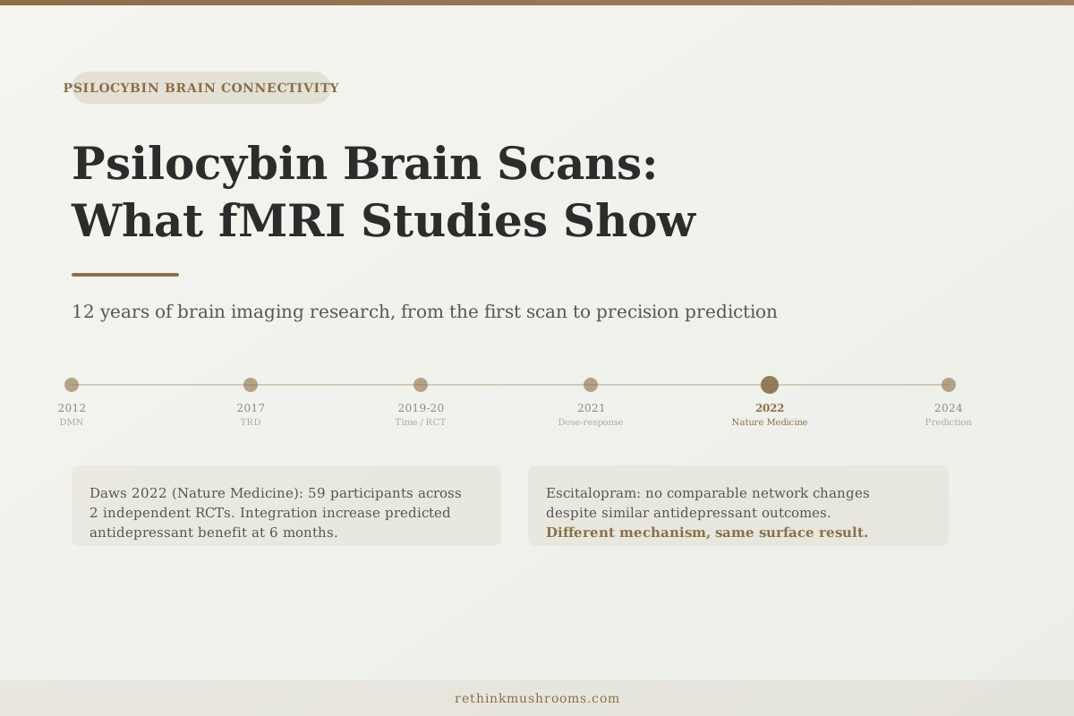
Across two independent RCTs, psilocybin therapy increased global brain integration. The degree of integration increase predicted antidepressant benefit at 6 months. An SSRI produced no comparable network changes.
Daws RE, et al. Nature Medicine. 2022
4 months
lasting DMN connectivity changes in psilocybin + mindfulness RCT
42
patients whose psilocybin response was predictable from baseline fMRI
2012
year of the first fMRI study documenting psilocybin's DMN effects
Daws RE, et al. Nature Medicine. 2022
 View as image ↗
Most articles about psilocybin skip the measurement question entirely. They report that the brain becomes "hyperconnected" or that a key circuit "quiets down" without explaining what that means, how it is measured, or why the measurement is reliable.
The answer to all three questions starts with a technique called functional magnetic resonance imaging, or fMRI. Understanding what fMRI actually captures changes how you evaluate every psilocybin brain study published since 2012. This article covers both the methodology and the major findings chronologically, from the original DMN discovery to the 2024 precision imaging work showing that a patient's baseline brain state predicts their treatment response.
For a deep dive specifically into the default mode network and what psilocybin does to it, see Psilocybin and the Default Mode Network: How It Quiets the Ego. What follows zooms out to cover the full range of functional connectivity findings across the whole brain.
Key Takeaways
- fMRI measures a proxy signal for neural activity (blood oxygenation), and "functional connectivity" means two brain regions whose activity rises and falls together over time
- Carhart-Harris et al. (PNAS, 2012) showed psilocybin reduces activity in the default mode network, with the reduction correlating with subjective experience intensity
- Preller et al. (Biological Psychiatry, 2020) found connectivity changes are time-dependent: early disruption within networks, then cross-network hyperconnectivity as the experience peaks
- Madsen et al. (2021) documented a linear dose-response: more psilocin in the blood means more network disruption, and the subjective experience tracks both
- Daws et al. (Nature Medicine, 2022) found that psilocybin increased global brain integration across two independent RCTs, and this integration predicted antidepressant benefit at 6 months; an SSRI produced no comparable network changes
- Alcohol dependence can blunt psilocybin's connectivity effects, suggesting pre-existing network alterations modulate response (Reinwald 2023)
- Copa et al. (2024) found that baseline fMRI patterns can predict who will respond to psilocybin for depression before treatment begins
Before the Findings: What fMRI Actually Measures
A functional MRI scanner does not photograph neurons firing. It cannot see electrical signals directly. What it measures is a secondary proxy: the blood oxygenation level-dependent response, abbreviated as BOLD.
When neurons in a brain region become active, local blood flow increases to supply them with oxygen. Oxygenated blood and deoxygenated blood have different magnetic properties, and the scanner detects this difference as a BOLD signal. Researchers use changes in the BOLD signal as a practical approximation of neural activity in that region.
Functional connectivity is a specific way of analyzing these signals. If two brain regions show BOLD signals that consistently rise and fall together across time, they are described as "functionally connected." Their activity is synchronized. This synchronization can persist even when a person is lying still with no task, which is called the "resting-state" fMRI paradigm. Most psilocybin research uses resting-state scans before and after drug administration, or in some cases continuously across a session.
One nuance matters for interpreting psilocybin findings. Functional connectivity is not the same as anatomical wiring. Two regions can show synchronized activity without a direct structural connection between them, because both may be receiving input from a third region. What fMRI measures is coordination patterns, not physical pathways. Psilocybin research describes how these coordination patterns change, not necessarily which neurons are being directly connected.
With that foundation established, here is what the research actually found.
The First Scans: What Carhart-Harris Found in 2012
The foundational psilocybin fMRI study was published in PNAS in 2012. Robin Carhart-Harris and colleagues scanned 15 healthy volunteers at rest before and after intravenous psilocybin administration, then compared the BOLD signals between conditions.[1]
The finding that changed the field: psilocybin significantly reduced blood flow and BOLD signal in the brain's self-referential circuit, known as the default mode network (DMN). The two most affected regions were the medial prefrontal cortex, which becomes active when a person is thinking about themselves, planning, or ruminating, and the posterior cingulate cortex, which acts as a connectivity hub integrating information from across the brain.
The reduction was not uniform across the brain. It was concentrated in these DMN hubs, and it correlated directly with the subjective intensity of the psychedelic experience. The more the DMN quieted, the more profound the experience participants reported.[1]
This established the framework that most subsequent psilocybin fMRI research builds on: psilocybin disrupts the brain's default self-referential processing, and that disruption is dose-linked and experience-linked. The study also introduced the methodology that has become standard: resting-state fMRI comparing conditions, with subjective experience ratings collected in parallel.
Beyond the DMN: Specific Changes in Depressed Brains
The 2012 study used healthy volunteers. A later team examined what the same measurements show in people whose brains are already altered by treatment-resistant depression.
Carhart-Harris and colleagues published a cohort study in Scientific Reports in 2017, administering psilocybin to 19 patients who had failed at least two antidepressant treatments.[2] The fMRI findings differed meaningfully from the healthy-volunteer data.
In depressed patients, psilocybin increased amygdala reactivity after treatment. The amygdala is the brain's primary region for processing emotionally significant signals. In depression, it often shows blunted or dysregulated responses. The post-treatment increase in amygdala responsiveness correlated with antidepressant benefit.
Simultaneously, resting-state connectivity decreased between the parahippocampus and the prefrontal cortex. The parahippocampus is associated with memory retrieval and the maintenance of fixed cognitive patterns. Its tight coupling to the prefrontal cortex is one mechanism hypothesized to maintain rigid, ruminative depressive thinking. The loosening of this specific connection after psilocybin was another predictor of who responded.
The 2017 study suggested that the mechanism of psilocybin in depression is not simply "quieting the DMN" as in healthy volunteers. It appears to involve something more targeted: loosening the specific network connections that hold depressive patterns in place, while simultaneously restoring emotional responsiveness in regions that had gone quiet.[2]
The Experience Has Phases, and So Does the Brain
Earlier studies captured connectivity as a before-and-after comparison, losing all temporal information about what happens during the experience itself. Preller and colleagues addressed this with a 2020 study in Biological Psychiatry that scanned participants continuously across a psilocybin session.[4]
The design was randomized and placebo-controlled, with 25 participants. The finding was that psilocybin's connectivity effects are not static. They evolve across the session in two distinguishable phases.
The early phase shows disruption within normal brain networks. Networks that usually maintain internal coherence begin losing it. Regions that normally fire together start decoupling. The late phase shows the emergence of cross-network communication. Regions from normally separate networks begin synchronizing with each other, producing the hyperconnectivity pattern that earlier studies captured in static snapshots.
This two-phase pattern resolved a confusion in the literature. Some studies found primarily disruption; others found primarily hyperconnectivity. Both findings were correct, and the discrepancy reflected when during the session the measurements were taken.[4]
For more on how cross-network communication produces the altered experience, including the role of specific gateway structures in the brain, see How Psilocybin Makes Brain Regions Talk to Each Other.
More Drug, More Change: Dose-Response in Network Architecture
A 2021 cohort study by Madsen and colleagues in European Neuropsychopharmacology established the dose-response relationship quantitatively, using a measurement approach that earlier studies had not employed.[5]
The key methodological addition: they collected blood samples from 18 participants during scanning to measure plasma psilocin levels. Psilocybin itself is not active in the brain. The body converts it into psilocin, the compound that actually crosses into brain tissue and binds to serotonin receptors. Measuring blood psilocin levels gives a continuous pharmacokinetic marker during the session.
The result: changes in brain network architecture correlated linearly with psilocin blood levels. Two metrics were tracked. Network integrity refers to how coherently the regions within a given network fire together. Network segregation refers to how well-separated different networks remain from each other. Both decreased as psilocin levels rose.
The relationship was not a threshold. There was no point below which nothing happened and above which effects began. The relationship was a gradient across the entire concentration range measured.
Subjective experience intensity tracked the same gradient. More psilocin in the blood, more network disruption in the scanner, more intense the experience reported. The pharmacokinetics (drug concentration), the brain measurement (network architecture), and the experience (subjective report) were all moving together.[5]
What Stays Different: Changes That Outlast the Drug
The connectivity changes during a psilocybin session are acute and dramatic. What surprised researchers was that certain changes persist measurably after the drug has cleared the system.
An RCT by Smigielski and colleagues, published in NeuroImage in 2019, tested psilocybin combined with mindfulness meditation training against mindfulness alone in 38 participants.[3] The researchers measured brain connectivity four months after the session, well past any pharmacological effect.
The psilocybin-plus-mindfulness group showed lasting decreases in DMN self-referential connectivity compared to the mindfulness-only group at four months. The drug's acute disruption, combined with contemplative practice during the integration period, produced network changes that were still measurable four months later. Mindfulness training alone did not produce changes of the same magnitude or duration.
The largest lasting-effects finding came from Daws and colleagues in Nature Medicine in 2022, combining data from two independent RCTs with 59 total participants.[6]
Their analysis focused on global brain integration, a measure of how much cross-network communication is occurring across the whole brain, as opposed to network-to-network segregation. A brain with high modularity is divided into tightly separated modules that rarely communicate. A brain with high integration has more communication crossing those module boundaries.
Psilocybin therapy increased global integration. The increase was measured after the drug had cleared, meaning it represented a persistent post-treatment shift. The degree of integration increase correlated with antidepressant benefit at six months. Patients whose brains showed greater post-treatment integration showed greater reductions in depression.
The comparison condition was escitalopram, a standard SSRI antidepressant. Escitalopram produced no comparable change in global integration. Both drugs produced antidepressant effects in their respective groups, but through measurably different brain mechanisms. Psilocybin reorganized the network architecture; escitalopram did not.[6]
For the theoretical framework explaining why global integration increases after psilocybin, see How Psilocybin Increases Brain Entropy (And Why That's Good).
An Important Asterisk: When the Expected Changes Are Blunted
Not all brains respond the same way to psilocybin.
Reinwald and colleagues published a 2023 study in Translational Psychiatry examining psilocybin's DMN-suppressing effect in an animal model of alcohol dependence.[7] Alcohol-dependent rats showed blunted DMN suppression after psilocybin compared to controls. Their brains, which had been altered by chronic alcohol exposure, did not produce the same connectivity response.
This is a preclinical study, and extrapolating from rat brain connectivity to human therapy requires caution. But the finding raises a question that human research has not yet fully answered: do pre-existing network alterations from chronic substance use reduce psilocybin's ability to produce the connectivity changes associated with therapeutic benefit?
If so, treatment protocols for alcohol and addiction-related indications may need to account for this. The standard psilocybin dosing protocols were largely developed in healthy volunteers or patients with depression. Participants whose brain networks have been reorganized by years of substance use may require different parameters to achieve comparable connectivity disruption.
The broader lesson from Reinwald 2023 is that the brain psilocybin acts on is not a blank slate. The starting conditions of the network architecture matter for the outcome.[7]
Reading Tomorrow's Response from Today's Scan
The most clinically significant recent development in psilocybin fMRI research is predictive precision.
Copa and colleagues published a 2024 study in the Journal of Affective Disorders examining whether baseline fMRI patterns, measured before any treatment, could predict which patients would respond to psilocybin for depression.[8]
In 42 patients, certain default mode network connectivity patterns at baseline predicted antidepressant benefit after psilocybin therapy. Patients with particular network configurations before treatment showed greater reductions in depression after it.
The practical implication is a potential route toward precision psychiatry in psychedelic medicine. Rather than treating all depressed patients identically and observing the distribution of responses, baseline fMRI might eventually identify in advance who is most likely to respond. Patients with network profiles not associated with response could receive different treatments or modified protocols.
Copa 2024 is early evidence. Forty-two patients is a modest sample size, and the predictive patterns require replication in larger cohorts before they could influence clinical practice. But the finding is consistent with two earlier threads: Madsen 2021 showed that individual variation in network disruption tracks drug concentration, meaning individual pharmacokinetics contribute to response variability; Reinwald 2023 showed that pre-existing network state modulates the response.[8]
Taken together, the current evidence points toward a model where psilocybin's fMRI-measurable effects are real, dose-linked, experience-linked, partially durable, and individually variable in ways that may be predictable from baseline brain state.
The companion article on Psilocybin and Brain Connectivity: The Hyperconnected Brain covers the broader hyperconnectivity and desynchronization findings, including the 2024 Nature paper documenting how psilocybin's disruption compares to other compounds. The Connectome on Psilocybin: Visual Maps of a Changed Brain covers the topological structure of what the brain's connectivity map looks like under psilocybin.
The Bottom Line
Twelve years of psilocybin fMRI research has built a consistent picture: psilocybin disrupts the default mode network acutely (Carhart-Harris 2012), loosens depression-specific rigid network connections (Carhart-Harris 2017), produces time-dependent phase changes across the session (Preller 2020), follows a linear dose-response with plasma psilocin (Madsen 2021), creates lasting global integration that predicts antidepressant benefit in two independent RCTs (Daws 2022), and can be modulated by pre-existing network alterations (Reinwald 2023). The field is now moving toward using baseline fMRI to predict individual treatment response (Copa 2024). The measurement tool itself is a second-order measure: fMRI's BOLD signal is a proxy for neural activity, not a direct view of neurons. The findings are robust across multiple labs and methodologies, but the gap between measuring coordination patterns and explaining consciousness remains.
Frequently Asked Questions
Sources & References
- 1. Source details for this reference are not yet available.
- 2. Source details for this reference are not yet available.
- 3. Source details for this reference are not yet available.
- 4. Source details for this reference are not yet available.
- 5. Source details for this reference are not yet available.
- 6. Source details for this reference are not yet available.
- 7. Source details for this reference are not yet available.
- 8. Source details for this reference are not yet available.