Meshima Anti-Angiogenesis
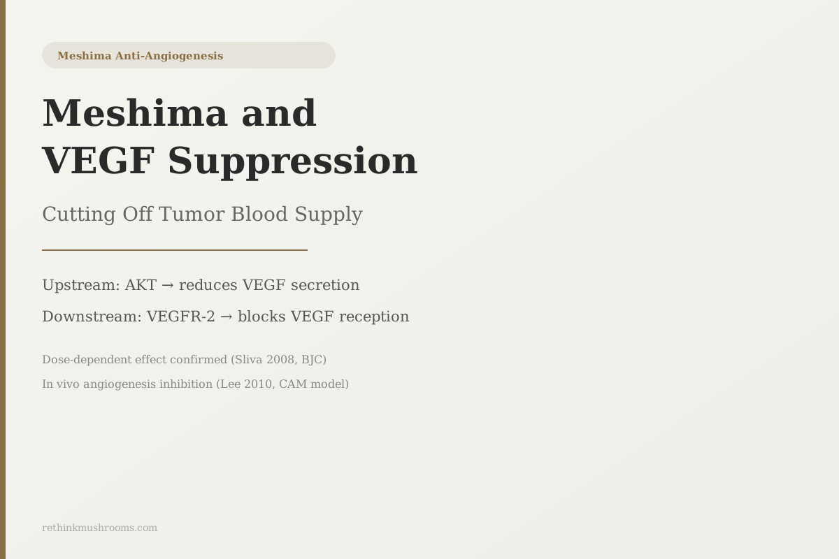
2 documented VEGF-blocking mechanisms
Phellinus linteus reduces VEGF secretion from cancer cells in a dose-dependent manner by blocking AKT kinase, and directly inhibits VEGFR-2 phosphorylation in endothelial cells, blocking VEGF reception.
Sliva et al., British Journal of Cancer, 2008
AKT Thr308
phosphorylation site suppressed in breast cancer cells, reducing VEGF production (Sliva 2008)
VEGFR-2
receptor phosphorylation directly blocked in human endothelial cells (Lee 2010)
dose-response
VEGF suppression scaled with PL extract concentration, confirming pharmacological mechanism (Sliva 2008)
Sliva et al., British Journal of Cancer, 2008
 View as image ↗
VEGF is the molecular signal tumors send to recruit blood vessels. Without it, a tumor cannot grow beyond about 2 millimeters. Without a response to it, the capillaries that would feed a growing tumor cannot form. VEGF sits at the center of a strategy called anti-angiogenesis therapy, and the drugs designed around it have become standard-of-care treatments in multiple cancers.
Research on Phellinus linteus (meshima) has found that the mushroom interferes with VEGF signaling at two documented points. The first reduces how much VEGF cancer cells produce, working through a kinase called AKT that drives VEGF production inside tumor cells. The second blocks the receptor that VEGF needs to activate on blood vessel cells, preventing the signal from triggering new capillary growth even when VEGF is present in the tissue.
This is the more VEGF-specific companion to the broader anti-angiogenesis article. For the full three-mechanism picture including the Wnt/beta-catenin pathway, see Meshima and Anti-Angiogenesis. For the foundational introduction to meshima, see What Is Meshima Mushroom.
Key Takeaways
- VEGF (vascular endothelial growth factor) is the primary molecular signal tumors use to recruit blood vessel formation; blocking VEGF is the basis for approved cancer drugs including bevacizumab (Avastin), sorafenib, and sunitinib
- Sliva et al. (2008, British Journal of Cancer) found Phellinus linteus reduced VEGF secretion from MDA-MB-231 breast cancer cells in a dose-dependent manner by blocking AKT phosphorylation at Thr308 and Ser473
- Lee et al. (2010) found PL methanol extract directly blocked VEGFR-2 phosphorylation in human umbilical vein endothelial cells, preventing VEGF from triggering the capillary-formation cascade even when VEGF was present
- The dose-response pattern in Sliva 2008 is mechanistically important: VEGF suppression scaled with extract concentration, confirming the AKT inhibition is a genuine pharmacological effect rather than non-specific toxicity
- A 2023 systematic review by Qin et al. identified beta-1,3/1,6-glucan polysaccharides as the primary anti-angiogenic compound class; potency depends on molecular weight and branching pattern, which vary across commercial extracts
- No human trial has measured VEGF levels in cancer patients before and after Phellinus linteus supplementation; the VEGF-suppression evidence is entirely from cell culture and animal models
What VEGF Does and Why Tumors Overproduce It
VEGF is a protein that healthy tissue uses to regulate blood vessel growth during wound healing and development. It is a normal signaling molecule with a legitimate biological role: when tissue is damaged, cells secrete VEGF to signal blood vessels to grow toward the injury and supply the healing process.
Tumors hijack this system. As a tumor grows, its cells experience hypoxia (oxygen starvation) because they have outgrown the diffusion range of nearby capillaries. Hypoxia activates a transcription factor called HIF-1alpha, which turns on VEGF production. The tumor begins secreting VEGF into the surrounding tissue, which attracts nearby endothelial cells (the cells lining blood vessels) to grow new capillaries toward the tumor. This is tumor angiogenesis: the process by which a tumor builds its own blood supply.
AKT kinase is one of the primary drivers of VEGF production at the molecular level. When AKT is phosphorylated (activated), it promotes HIF-1alpha and other VEGF-activating transcription factors. Blocking AKT reduces VEGF secretion at the source. This is the upstream approach.
The pharmaceutical industry has pursued two other approaches downstream. Bevacizumab is a monoclonal antibody that binds free VEGF in the bloodstream, neutralizing it before it can reach its receptor. Sorafenib and sunitinib block VEGFR-2, the receptor on endothelial cells that VEGF activates. Both approaches are used in multiple cancer types. The point of explaining them here is not comparison, but context: they establish that VEGF suppression (upstream) and VEGFR-2 blocking (downstream) are validated anti-angiogenic strategies in clinical oncology.
A comprehensive 2010 review by Sliva documented VEGF suppression as one of Phellinus linteus's central anti-tumor mechanisms, alongside anti-proliferative, pro-apoptotic, and anti-invasive effects across multiple cancer models.[3]
How PL Reduces VEGF at the Source: The AKT Pathway
Sliva et al. (2008) tested Phellinus linteus extract on MDA-MB-231 cells, a triple-negative breast cancer line that is highly aggressive and does not express estrogen or progesterone receptors, making it a stringent test for compounds that don't depend on hormone signaling to work.[1]
VEGF secretion from MDA-MB-231 cells decreased in a dose-dependent pattern when treated with increasing concentrations of PL extract. The dose-response relationship is mechanistically important. A compound that reduces VEGF at low concentrations and suppresses it more at higher concentrations is showing a genuine pharmacological effect: the molecule is hitting a specific target. Uniform toxicity (simply killing cells) would not produce this pattern; a specific AKT inhibitor would.
The study measured AKT phosphorylation directly at two key sites: Thr308 and Ser473. Both are required for full AKT activation. Suppression at both sites means AKT activity was substantially reduced, not just partially. The study also confirmed that the extract inhibited capillary morphogenesis in human aortic endothelial cells: treated endothelial cells could not form the capillary-like structures they would normally build in response to angiogenic signals.
Three findings together: less VEGF secreted from cancer cells, lower AKT activity in those cells, and impaired capillary formation capacity in the endothelial cells that would respond to VEGF. The AKT data provides the molecular explanation for the VEGF and capillary findings.
How PL Blocks VEGF at the Receiver: VEGFR-2 Inhibition
VEGFR-2 (vascular endothelial growth factor receptor 2) is the primary receptor through which VEGF triggers capillary formation. When VEGF binds to VEGFR-2 on the surface of endothelial cells, the receptor phosphorylates itself (activates), which triggers the downstream cascade: the endothelial cell starts dividing, migrating toward the tumor, and organizing into tube-shaped capillaries.
Blocking VEGFR-2 phosphorylation prevents this cascade from starting, even when VEGF is present in the tissue. This is the downstream approach: the cancer cell has still secreted VEGF, but the blood vessel cells can no longer respond to it.
Lee et al. (2010) tested Phellinus linteus methanol extract and its fractions on human umbilical vein endothelial cells (HUVECs), the standard laboratory model for studying vascular responses.[2] The results covered four distinct stages of the angiogenic process:
HUVEC proliferation was inhibited: treated cells divided more slowly. Migration was inhibited: treated cells moved less toward angiogenic signals. Tube formation was inhibited: treated cells could not organize into the hollow, capillary-like structures they form in the Matrigel tube formation assay. And VEGFR-2 phosphorylation was directly suppressed: the receptor was present but could not activate.
Lee et al. also used the chick chorioallantoic membrane (CAM) assay, a whole-organism angiogenesis model. The CAM is the membrane inside a fertilized chicken egg that contains a rapidly developing vascular network. PL extract reduced new vessel formation in the CAM, confirming that the effects seen in HUVECs translate to actual blood vessel formation in a living tissue.
Upstream vs. Downstream: Why Two Mechanisms Together Are Significant
The pharmaceutical anti-angiogenesis experience has shown that blocking only one point in the VEGF pathway often leads to resistance or limited response. Bevacizumab blocks free VEGF; cancer cells can adapt by upregulating VEGF production or activating alternative angiogenic signals. VEGFR inhibitors are often combined with VEGF antibodies for this reason.
Phellinus linteus appears to operate at two points simultaneously: reducing VEGF production (AKT pathway) and blocking VEGF reception (VEGFR-2 pathway). The upstream and downstream mechanisms do not require each other: AKT inhibition can reduce VEGF secretion regardless of VEGFR-2 status, and VEGFR-2 blocking can prevent angiogenesis regardless of how much VEGF is present.
Whether the combination of these two effects produces a synergistic result in PL-treated tissue has not been studied directly. Whether it compares to pharmaceutical anti-angiogenic drugs in any clinical sense has not been studied at all. The point is not equivalence but mechanism: PL hits the VEGF pathway from two separate angles, not one.
For a direct examination of how PL's mechanisms map against those of bevacizumab and related drugs, see Meshima vs. Pharmaceutical Angiogenesis Inhibitors.
Which Polysaccharide Fractions Drive VEGF Suppression
The Sliva 2008 and Lee 2010 studies used whole or methanol extracts that contain multiple compound classes. Qin et al. (2023) conducted a systematic review of Phellinus linteus polysaccharides and found that the anti-angiogenic activity attributed to these fractions depends critically on structural features: specifically, the branching pattern and molecular weight of the beta-glucan chains.[5]
Beta-1,3/1,6-glucan linkages (branched polysaccharides) show stronger biological activity than linear beta-1,3-glucans across multiple endpoints. High-molecular-weight fractions and low-molecular-weight fractions also perform differently depending on the biological assay. This means that "Phellinus linteus extract" is not a single compound with a fixed biological profile: different extracts with different polysaccharide structures will show different anti-angiogenic potency.
A 2020 systematic review by Wong et al. placed PL among the most studied medicinal mushrooms for breast cancer suppression, noting that the anti-angiogenic and anti-proliferative evidence in breast cancer models represents the most concentrated preclinical data for any single mushroom species in this cancer type.[4]
Does This Make PL Similar to Bevacizumab?
Mechanistically, there is a parallel. Bevacizumab neutralizes free VEGF; Phellinus linteus reduces VEGF production via AKT. Both reduce VEGF availability to endothelial cells. Sorafenib blocks VEGFR-2 kinase activity; PL also blocks VEGFR-2 phosphorylation in endothelial cells. The molecular approach points overlap.
The honest answer to whether PL is "similar to bevacizumab" is: similar in approach, incomparably different in evidence level and clinical application.
Bevacizumab has been tested in randomized controlled trials with thousands of patients across multiple cancer types. Dose, pharmacokinetics, drug interactions, combination protocols, and adverse effects are fully characterized. It is an approved drug with defined clinical indications.
Phellinus linteus's VEGF suppression data consists of cell culture studies and an in vivo chick embryo assay. Its pharmacokinetics in humans (how much of the active compound reaches the blood after oral consumption, at what concentration, and for how long) have not been measured. No clinical trial has tested anti-angiogenic effects in human cancer patients.
This is not a dismissal of the preclinical data. The dose-response VEGF suppression from Sliva 2008 is specific and mechanistically coherent. The VEGFR-2 findings from Lee 2010 replicate a validated pharmaceutical target. What the data does not do is support clinical equivalence with or substitution for pharmaceutical anti-angiogenic therapy. For the broader mechanistic picture, see Phellinus linteus and VEGF Inhibition. For clinical cancer adjunct context from a more studied mushroom, see Ganoderma lucidum as a Cancer Adjunct.
The Bottom Line
Phellinus linteus (meshima) suppresses tumor VEGF signaling through two documented mechanisms: reducing VEGF secretion via AKT kinase inhibition in cancer cells (Sliva 2008, British Journal of Cancer) and directly blocking VEGFR-2 phosphorylation in endothelial cells (Lee 2010). The dose-response pattern in Sliva 2008 confirms pharmacological specificity. Qin 2023 identifies beta-1,3/1,6-glucan polysaccharide fractions as the primary anti-angiogenic compound class, with potency depending on molecular structure. No human VEGF suppression data exists for Phellinus linteus.
Frequently Asked Questions
Sources & References
- 1RTHM-00271·Sliva, D et al. (2008). “Phellinus linteus Extract in Breast-Cancer Cell Models: What This 2008 Study Actually Found.” British journal of cancer.Study breakdown →PubMed →↩
- 2RTHM-00349·Lee, Yeon Sil et al. (2010). “Anti-angiogenic activity of methanol extract of Phellinus linteus and its fractions..” Journal of ethnopharmacology.Study breakdown →PubMed →↩
- 3RTHM-00364·Sliva, Daniel (2010). “Medicinal mushroom Phellinus linteus as an alternative cancer therapy..” Experimental and therapeutic medicine.Study breakdown →PubMed →↩
- 4RTHM-00664·Wong, Jack Ho et al. (2020). “Mushroom extracts and compounds with suppressive action on breast cancer: evidence from studies using cultured cancer cells, tumor-bearing animals, and clinical trials..” Applied microbiology and biotechnology.Study breakdown →PubMed →↩
- 5RTHM-01770·Qin, Dandan et al. (2023). “Polysaccharides from Phellinus linteus: A systematic review of their extractions, purifications, structures and functions..” International journal of biological macromolecules.Study breakdown →PubMed →↩