Meshima Anti-Angiogenesis Research
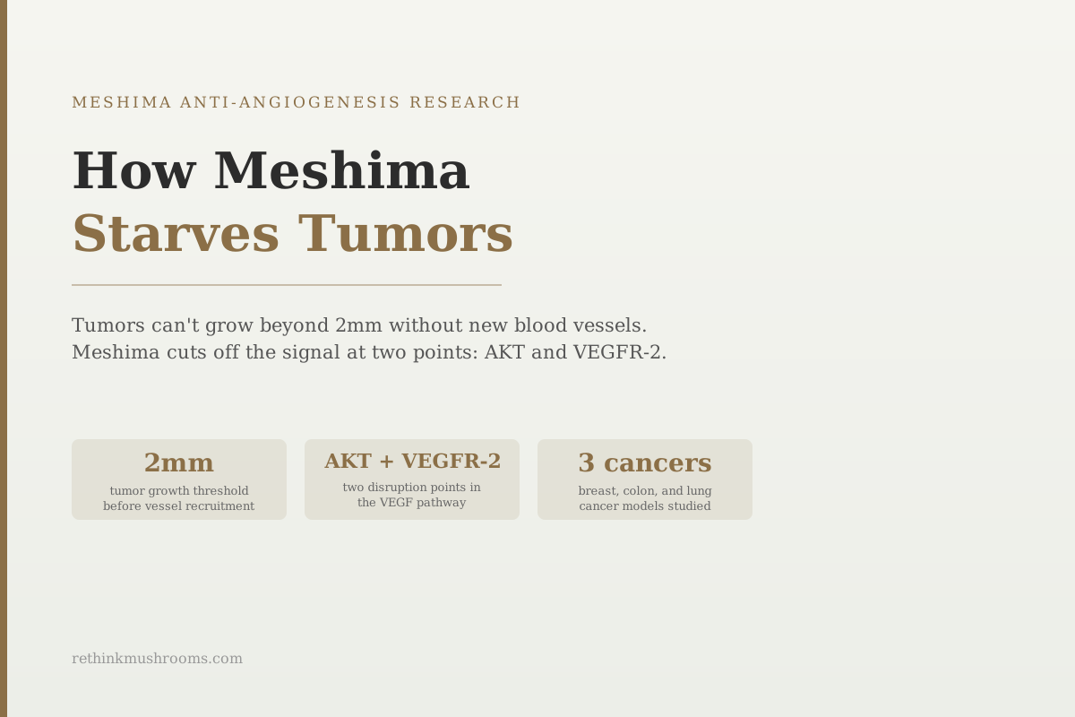
2mm growth threshold
A solid tumor cannot grow beyond 2 millimeters without recruiting new blood vessels. Meshima (Phellinus linteus) disrupts this process by suppressing the growth factor that triggers vessel formation.
Sliva D, Experimental and Therapeutic Medicine, 2010
AKT + VEGFR-2
the two signal points where Meshima disrupts tumor blood vessel recruitment (Sliva 2008, Lee 2010)
3 cancer types
breast, colon, and lung cancer models with documented Meshima anti-angiogenic activity
2023
most recent published evidence: Sanghuangporus sanghuang extract inhibited lung cancer growth and invasion in vivo
Sliva D, Experimental and Therapeutic Medicine, 2010
 View as image ↗
A solid tumor growing in the body hits a hard biological limit at around 2 millimeters in diameter. Beyond that size, the cells at the center can't get oxygen and nutrients through diffusion alone. To keep growing, the tumor has to build its own blood supply: a process called angiogenesis, the formation of new blood vessels. Every tumor that becomes dangerous has solved this problem.
Tumors solve it by secreting a signaling protein called VEGF (vascular endothelial growth factor). VEGF travels to nearby blood vessel cells and tells them to grow toward the tumor, forming new capillaries. Without this signal, tumor growth stalls. Cutting off that signal, or blocking the cells from receiving it, is the basis of a significant class of cancer drugs and an active area of research into natural compounds including Meshima (Phellinus linteus).
This article covers what the preclinical research shows about how Meshima disrupts tumor angiogenesis, which specific pathways are involved, and where the evidence currently stands. For the broader Meshima overview, see what is Meshima mushroom: Korea's anti-cancer mushroom, and for the compound sources behind these effects, see Phellinus linteus bioactive compounds: polysaccharides and phenols.
Key Takeaways
- A solid tumor cannot grow beyond approximately 2 millimeters without recruiting new blood vessels through VEGF signaling; disrupting this process is the basis of anti-angiogenic cancer therapy
- A 2008 British Journal of Cancer study found Phellinus linteus reduced VEGF secretion from breast cancer cells in a dose-dependent manner by inhibiting AKT phosphorylation at two specific sites (Thr308 and Ser473)
- A 2010 study found Phellinus linteus methanol extract directly blocked VEGFR-2 phosphorylation in endothelial cells and inhibited their proliferation, migration, and tube formation, cutting off the signal at the receiver rather than just the source
- These are two distinct inhibition points: AKT suppresses VEGF production inside cancer cells; VEGFR-2 blockade prevents endothelial cells from responding to VEGF already released
- A 2011 study confirmed anti-angiogenic activity extends beyond breast cancer: protein-bound polysaccharides from Phellinus linteus reduced microvessel density in colon cancer xenograft tumors and disrupted Wnt/β-catenin signaling
- All mechanistic evidence is preclinical; no human clinical trial has tested Meshima specifically for anti-angiogenic activity in cancer patients
The 2mm Problem: Why Every Solid Tumor Must Recruit New Blood Vessels
A cancer cell that breaks free and starts dividing can grow into a cluster of cells without any special requirements, for a while. Simple diffusion of oxygen and nutrients from surrounding tissue keeps cells alive up to roughly 2 millimeters across.
Beyond that threshold, cells at the center of the growing mass start to suffocate. Without a blood supply delivering oxygen continuously, the central cells die from oxygen starvation (called hypoxia). Most early tumors die here. The ones that survive do so by sending a chemical distress signal that asks the body's blood vessel machinery to come help.
That distress signal is VEGF. When tumor cells are hypoxic, they activate a molecular cascade that ends with VEGF being secreted in large quantities into the surrounding tissue. VEGF binds to receptors on endothelial cells (the cells that line blood vessels), and those cells start multiplying and migrating toward the VEGF source. New capillaries form. The tumor now has a blood supply and the metabolic fuel to keep expanding.
This is why VEGF inhibition became one of the central strategies in cancer drug development. Bevacizumab (brand name Avastin), approved in 2004, is a monoclonal antibody that directly neutralizes free VEGF in the bloodstream. VEGFR-targeted kinase inhibitors like sorafenib block the receptor signaling. Both approaches aim to cut off the tumor's recruitment of new vessels.
A comprehensive review by Sliva in Experimental and Therapeutic Medicine in 2010 identified AKT inhibition and VEGF suppression as central mechanisms in Phellinus linteus's documented anti-tumor activity across breast, prostate, lung, and colon cancer models.[3]
The Dual Mechanism: Two Interruption Points in the VEGF Pathway
What separates Meshima's anti-angiogenic profile from many natural compound claims is that the research documents two distinct points of interference in the VEGF signaling chain, not just one.
The first point is upstream: inside the cancer cell, where VEGF is produced. The second point is downstream: at the endothelial cells that receive the VEGF signal and respond by growing new vessels.
This dual-point mechanism means that even if some VEGF molecules escape the first suppression step and reach blood vessel cells, there is a second barrier blocking the response. These are not redundant effects: they act on different cell types (cancer cells vs. endothelial cells) through different molecular targets (AKT vs. VEGFR-2).
For readers of this cluster, the companion articles on Meshima and anti-angiogenesis: cutting off tumor blood supply and Meshima and VEGF suppression: cutting off tumor blood supply go deeper on each mechanism separately.
AKT and VEGF: Dose-Dependent Suppression in Breast Cancer Cells
The upstream mechanism was documented by Sliva and colleagues in a 2008 study published in the British Journal of Cancer.[1]
The researchers tested Phellinus linteus extract on MDA-MB-231 cells, an aggressive triple-negative breast cancer cell line commonly used in oncology research. VEGF secretion from these cells decreased in a dose-response pattern as Phellinus linteus concentration increased; more extract produced less VEGF.
The researchers then traced why VEGF was declining. They found that Phellinus linteus inhibited phosphorylation of AKT at two specific sites: Thr308 and Ser473.
AKT is a signaling protein that acts as a central node in cancer cell survival. When AKT is active (phosphorylated), it drives a chain of downstream effects including increased VEGF production, resistance to apoptosis, and enhanced cell growth. When AKT is inhibited, VEGF production falls along with other survival signals. The Phellinus linteus extract appeared to suppress AKT activity, and VEGF levels dropped as a consequence.[1]
The study also tested the functional consequence: it put the Phellinus linteus-treated breast cancer conditioned medium (liquid that the cancer cells had been sitting in, containing their secretions) onto human aortic endothelial cells and measured capillary formation. Endothelial cells exposed to conditioned medium from Phellinus linteus-treated cancer cells formed fewer capillary-like structures than those exposed to untreated conditioned medium. Less VEGF in the medium meant less endothelial cell activation.
VEGFR-2: Lee 2010 Blocks the Receiver
The downstream mechanism, blocking the VEGF receptor itself on endothelial cells, was documented by Lee and colleagues in a 2010 study.[2]
The researchers used HUVEC cells, which stand for human umbilical vein endothelial cells, a standard model for studying blood vessel biology. They put endothelial cells through three functional tests: proliferation (do they multiply?), migration (do they move toward a VEGF signal?), and tube formation (do they organize into capillary-like structures in a scaffold gel?). Phellinus linteus methanol extract reduced all three in a dose-dependent manner.
The molecular target was VEGFR-2: the main receptor on endothelial cell surfaces that VEGF binds to. When VEGF binds to VEGFR-2, it activates the receptor by adding phosphate groups to it (phosphorylation). Phosphorylated VEGFR-2 then drives the endothelial cell to proliferate and form new vessels. Phellinus linteus extract blocked VEGFR-2 from being phosphorylated even in the presence of VEGF.[2]
The study also tested this in vivo using the chick chorioallantoic membrane model, a standard method for measuring real blood vessel growth rather than cell behavior in a dish. In this model, Phellinus linteus extract reduced visible new blood vessel formation. This is a step beyond cell culture: it shows the compound disrupting actual vessel growth in a living biological system.
Together with the Sliva 2008 breast cancer data, the Lee 2010 findings establish a coherent two-point mechanism: Phellinus linteus compounds suppress VEGF production in cancer cells and block VEGF reception in endothelial cells.
Beyond Breast Cancer: Colon Tumors, Reduced Microvessel Density, and a Third Pathway
Song and colleagues published a study in BMC Cancer in 2011 testing a protein-bound polysaccharide fraction from Phellinus linteus on SW480 colon cancer cells and in a mouse xenograft model.[4]
The key angiogenesis finding from the animal model: tumors from mice treated with the Phellinus linteus polysaccharide showed reduced microvessel density compared to controls. Microvessel density is measured by staining tumor tissue sections for endothelial cell markers and counting the blood vessels present. Fewer vessels in the treated tumors confirms that angiogenesis was reduced in living tumor tissue, not just in cell culture.
The study also found that the Phellinus linteus treatment disrupted Wnt/β-catenin signaling in the colon cancer cells. Wnt/β-catenin is a developmental pathway that, when abnormally active in cancer, promotes cell growth, invasion, and blood vessel recruitment. Its disruption adds a third mechanistic route through which Phellinus linteus may suppress tumor angiogenesis, distinct from the AKT and VEGFR-2 mechanisms.[4]
The Song 2011 study is particularly significant because it moves beyond breast cancer cell lines into a different cancer type (colorectal) and provides in vivo evidence of reduced vessel formation in actual tumor tissue.
2023 Lung Cancer Data and the Taxonomic Name Change
Recent research has continued to document anti-tumor activity in what was previously classified as Phellinus linteus. Wang and colleagues published a 2023 study in Nutrition Research and Practice using what modern taxonomy now calls Sanghuangporus sanghuang, the reclassified name for a related species within the broader Phellinus linteus lineage.[5]
The researchers tested the extract against A549 and H1650 lung cancer cell lines, both common models for non-small cell lung cancer. At concentrations of 6-9 mg/mL, the extract inhibited cancer cell migration and invasion dose-dependently. The downstream targets included STAT3 (a protein that, when activated, drives cancer survival and growth), Bcl-2 (an anti-apoptotic protein), CDK4, and Cyclin D1 (both involved in cell cycle control). In vivo tumor growth inhibition was confirmed in mouse models.[5]
The taxonomic note matters for anyone searching the scientific literature. Phellinus linteus research published before approximately 2015 uses the original name; research from 2016 onward may use Sanghuangporus sanghuang. The biological material is closely related, and the mechanistic continuity across older and newer studies supports the validity of both bodies of research. The broader VEGF-focused research (Sliva 2008, Lee 2010) was conducted on species classified under the Phellinus linteus lineage; the Wang 2023 lung cancer data was conducted under the updated taxonomy.
The earliest published evidence of Phellinus linteus polysaccharides inhibiting tumor growth dates to a 1999 animal study, establishing the anti-tumor activity of these compounds more than 25 years ago.[7] The progression from that foundational finding to the specific AKT and VEGFR-2 mechanisms reflects the typical arc of preclinical research: observation, then mechanistic characterization.
The Compounds Behind the Effect: Polysaccharide Structure and Function
Understanding which specific compounds in Phellinus linteus produce these anti-angiogenic effects is relevant to supplement quality. Qin and colleagues published a 2023 systematic review in the International Journal of Biological Macromolecules characterizing the polysaccharides of Phellinus linteus, covering their extraction methods, structural features, and documented biological functions.[6]
The review identified β-(1,3)-linked glucans as the primary structural class responsible for anti-tumor and immunomodulatory activity. These are polysaccharides built on a backbone of glucose units connected at the 1 and 3 positions. The 1,3-linkage pattern is the same structural feature associated with anti-tumor activity in other medicinal mushrooms including Ganoderma, Lentinula, and Trametes species.
The protein-bound polysaccharide fraction used in the Song 2011 colon cancer xenograft study (the one that reduced microvessel density) is structurally related to these β-glucan fractions. The polysaccharide complexity of Phellinus linteus is considerable, and different fractions show different biological profiles, which is why extraction method and fraction specificity matter when evaluating supplement quality and research applicability.[6]
For a broader comparison of Meshima's anti-angiogenic approach with pharmaceutical VEGF inhibitors including bevacizumab, see Meshima vs. pharmaceutical angiogenesis inhibitors. For the other anti-tumor mechanisms operating alongside angiogenesis inhibition, the Phellinus linteus and VEGF inhibition article provides the VEGF-specific depth. For mushroom cancer research with a stronger human evidence base, Ganoderma lucidum as a cancer adjunct: clinical evidence offers a useful comparison point, and ganoderic acids and cancer: specific anti-tumor research covers Reishi's own angiogenesis inhibition mechanisms.
The Bottom Line
Meshima (Phellinus linteus) disrupts tumor angiogenesis at two molecular points. Sliva 2008 demonstrated that Phellinus linteus suppresses VEGF secretion from breast cancer cells in a dose-dependent manner by blocking AKT phosphorylation. Lee 2010 showed that Phellinus linteus also inhibits VEGFR-2 phosphorylation in endothelial cells, blocking the vessel-forming response even when VEGF is present. Song 2011 extended the evidence to colon cancer xenografts, confirming reduced microvessel density in tumor tissue and identifying Wnt/β-catenin as a third disrupted pathway. A 2023 study under the updated taxonomic name Sanghuangporus sanghuang found anti-invasion and anti-tumor activity in lung cancer models. All evidence is preclinical; no human clinical trial has tested these mechanisms in cancer patients.
Frequently Asked Questions
Sources & References
- 1RTHM-00271·Sliva, D et al. (2008). “Phellinus linteus Extract in Breast-Cancer Cell Models: What This 2008 Study Actually Found.” British journal of cancer.Study breakdown →PubMed →↩
- 2RTHM-00349·Lee, Yeon Sil et al. (2010). “Anti-angiogenic activity of methanol extract of Phellinus linteus and its fractions..” Journal of ethnopharmacology.Study breakdown →PubMed →↩
- 3RTHM-00364·Sliva, Daniel (2010). “Medicinal mushroom Phellinus linteus as an alternative cancer therapy..” Experimental and therapeutic medicine.Study breakdown →PubMed →↩
- 4RTHM-00437·Song, Kyoung-Sub et al. (2011). “Protein-bound polysaccharide from Phellinus linteus inhibits tumor growth, invasion, and angiogenesis and alters Wnt/β-catenin in SW480 human colon cancer cells..” BMC cancer.Study breakdown →PubMed →↩
- 5RTHM-01817·Wang, Weike et al. (2023). “Sanghuangporus sanghuang extract inhibits the proliferation and invasion of lung cancer cells in vitro and in vivo..” Nutrition research and practice.Study breakdown →PubMed →↩
- 6RTHM-01770·Qin, Dandan et al. (2023). “Polysaccharides from Phellinus linteus: A systematic review of their extractions, purifications, structures and functions..” International journal of biological macromolecules.Study breakdown →PubMed →↩
- 7. Source details for this reference are not yet available.