Ergothioneine & Eye Health
1 dedicated transporter
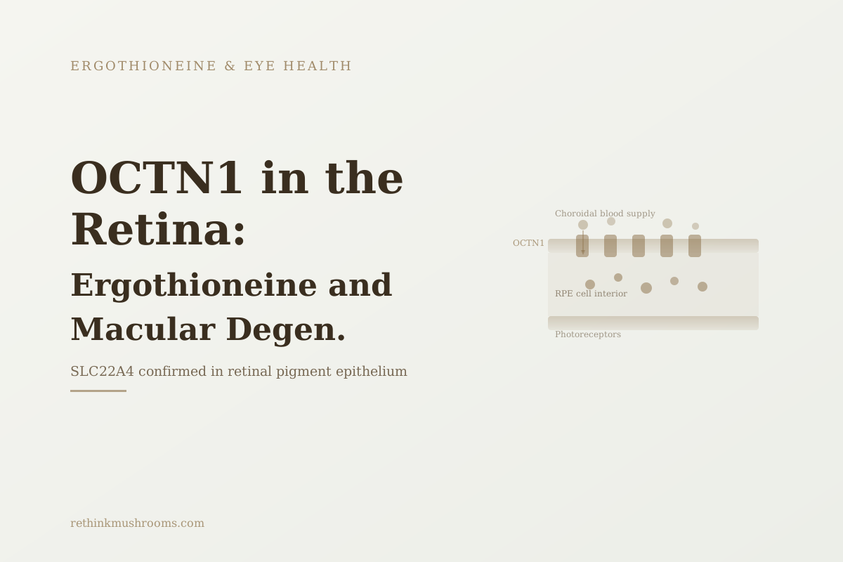
OCTN1 (SLC22A4) is the only protein the human body evolved specifically to transport ergothioneine; Gründemann's 2022 inventory confirms it is expressed in retinal pigment epithelium.
Gründemann, FEBS Lett, 2022
Extreme selectivity
OCTN1 transports ergothioneine with far higher affinity than any other tested compound (Tucker 2019)
RPE confirmed
OCTN1 expression confirmed in retinal pigment epithelium, the cells that fail in AMD (Gründemann 2022)
Neuroretina too
ergothioneine clears amyloid beta from neuroretinal tissue in mouse AMD/Alzheimer's model (Wijesinghe 2023)
Gründemann, FEBS Lett, 2022
 View as image ↗
Key Takeaways
- OCTN1 (SLC22A4) was discovered in 2005 as the specific, dedicated transporter for ergothioneine; it was the first demonstration that the body evolved an entire transport system for one dietary compound (Gründemann et al., PNAS, 2005)
- Tucker et al. 2019 showed OCTN1 has extreme substrate selectivity, transporting ergothioneine with far higher affinity and efficiency than any other compound tested, evidence the transporter evolved specifically for this molecule (Biochem Biophys Res Commun, 2019)
- Gründemann's 2022 comprehensive inventory of OCTN1-expressing tissues confirmed that retinal pigment epithelium cells are on the list, establishing why ergothioneine specifically accumulates in the cells that fail in AMD (FEBS Lett, 2022)
- Cheah and Halliwell's authoritative 2021 review confirms ergothioneine concentrates in oxidative-stress-sensitive tissues including the retina via the OCTN1 system (Redox Biol, 2021)
- Gu et al. 2024 showed that when RPE cells face oxidative stress (the condition OCTN1 exists to prevent), L-ergothioneine activating NRF2 is the top protective response among tested antioxidants (Exp Eye Res, 2024)
- Wijesinghe et al. 2023 found ergothioneine clears amyloid beta from neuroretinal tissue in mouse models, extending OCTN1's functional importance beyond the RPE into the photoreceptor layers (Front Neurosci, 2023)
Most content about OCTN1 retina ergothioneine exists only in academic journal PDFs and one 2025 bioRxiv preprint. No health or science editorial site has covered the specific question of why OCTN1 is expressed in retinal tissue and what that expression means for macular degeneration. This article covers that mechanistic ground.
The short version: OCTN1 is the only transporter protein the human body evolved specifically for ergothioneine. It is expressed in retinal pigment epithelium, the cell layer whose degeneration defines age-related macular degeneration. Ergothioneine reaches the retina because those cells have the machinery to import it. When that supply falls, the cells are exposed to the oxidative stress that the transporter exists to prevent.
For the broader eye health evidence, see Ergothioneine for Eye Health: Research and Implications. For the full AMD-specific research synthesis, see Ergothioneine and Macular Degeneration: Retinal Protection Research. For a grounding in what ergothioneine is, see what ergothioneine is and how the body hoards it.
What is OCTN1 in the retina? OCTN1 (SLC22A4) is a membrane transporter protein expressed in retinal pigment epithelium cells. It uses electrochemical gradients to actively import ergothioneine from extracellular fluid into the cell interior, concentrating it well above what passive diffusion achieves. RPE cells face continuous oxidative stress from light exposure; ergothioneine, imported via OCTN1, is part of their antioxidant defense. Its expression in the exact cells that fail in AMD creates a direct mechanistic connection between ergothioneine supply and macular degeneration risk.
What OCTN1 Is and How It Works
OCTN1 is encoded by the SLC22A4 gene. It is a member of the solute carrier family 22, a broad class of membrane transporters that move ions, organic acids, and other small molecules across cell membranes. Within that family, OCTN1 is distinguished by its substrate: ergothioneine.
In 2005, Gründemann and colleagues at the University of Cologne identified OCTN1 as the specific transporter for ergothioneine, the first study to demonstrate that the body had evolved dedicated molecular infrastructure for this single compound.[1] Before this work, ergothioneine's unusual tissue distribution (high concentrations in specific tissues exposed to oxidative stress, low concentrations in tissues that aren't) had been observed but unexplained.
The mechanism works as follows: OCTN1 sits in the plasma membrane of cells that express it. Using the electrochemical gradient across the membrane (maintained by sodium/potassium pumps), it binds ergothioneine in the extracellular space and transports it into the cell interior. This active pumping concentrates ergothioneine intracellularly at levels far above its extracellular concentration. The cell expends energy to maintain this concentration gradient because the intracellular ergothioneine serves functions the cell needs.
The protein structure of OCTN1 contains a binding site specifically shaped for ergothioneine's molecular geometry. This structural specificity is what makes it a dedicated transporter rather than a general-purpose carrier.
OCTN1 Evolved for One Molecule and One Molecule Only
The specificity of OCTN1 for ergothioneine goes beyond what a general transporter would show. In 2019, Tucker and colleagues tested OCTN1's substrate selectivity systematically, using a range of ergothioneine structural analogues and other potentially competing compounds.[2]
The results were clear: OCTN1 transports ergothioneine with far higher affinity and efficiency than any other compound tested. Structurally similar molecules showed dramatically lower transport rates. The transporter's selectivity is not just high; it is extreme.
This selectivity has an evolutionary implication. Proteins with narrow substrate specificity generally evolved under specific biological pressures. A transporter precisely tuned to one compound, maintained across hundreds of millions of years of mammalian evolution, indicates that this specific compound has been biologically important for a very long time. If ergothioneine were a dietary coincidence with no essential function, evolution wouldn't maintain the expensive infrastructure of a dedicated transporter for it.
The Tucker 2019 data reinforces the Gründemann 2005 discovery: OCTN1 isn't incidentally transporting ergothioneine. It evolved to do so. The tissues that express OCTN1 are the tissues the body has determined need reliable ergothioneine access.
For the general OCTN1 biology at a systemic level, see OCTN1: The Dedicated Transporter That Proves Ergothioneine Matters. This article focuses specifically on what OCTN1 expression means in retinal tissue.
OCTN1 in the Retina: What Gründemann's 2022 Tissue Inventory Shows
Knowing that OCTN1 is a dedicated, evolutionarily maintained ergothioneine transporter still leaves an anatomical question: which tissues actually express it?
Gründemann's 2022 comprehensive review in FEBS Letters assembled the most complete inventory of OCTN1-expressing tissues available in the literature.[3] The list includes the expected high-oxidative-stress tissues: blood cells, kidney, liver, intestinal epithelium. And it includes two eye tissues: lens epithelial cells and retinal pigment epithelium.
Retinal pigment epithelium (RPE) is a single-cell-thick layer immediately posterior to the photoreceptors. Its functions are central to retinal health: absorbing light not captured by photoreceptors (preventing scattered light), providing metabolic support to the photoreceptors above, clearing their visual cycle waste products, and maintaining the integrity of the blood-retinal barrier. When RPE cells degenerate, photoreceptors lose their support and die. The result is AMD.
The fact that RPE cells express OCTN1 is not a minor anatomical detail. It means the cells that are the primary target of AMD pathology have a dedicated import system for ergothioneine. The body invested in maintaining that import system in exactly those cells. The connection between ergothioneine supply and RPE cell health is built into the anatomy.
Cheah and Halliwell's authoritative 2021 review of ergothioneine biology confirms this within the broader context of the compound's tissue distribution: ergothioneine concentrates in tissues with the highest oxidative stress exposure, and the retina belongs in that category.[4]
What OCTN1 Expression in RPE Means for AMD
OCTN1 expression in RPE cells is the anatomical foundation. The functional significance becomes clearer when looking at what happens to RPE cells under the conditions OCTN1 exists to counteract.
In 2024, Gu and colleagues published a systematic screen of antioxidants for RPE cell protection under oxidative stress in the journal Experimental Eye Research.[5] L-ergothioneine was the top-performing compound. It activated the NRF2 pathway, a master switch for antioxidant gene expression, more effectively than all other tested candidates including vitamins C and E. In a separate mouse oxidative stress model, ergothioneine maintained better retinal structure than controls.
The Gu 2024 finding connects the anatomical OCTN1 story to the functional one. OCTN1 expresses in RPE cells. OCTN1 imports ergothioneine into those cells. When RPE cells are exposed to the oxidative stress that causes AMD-like pathology, ergothioneine is the best available defense among the antioxidants tested. The transporter exists in these cells precisely because the cells need what the transporter imports.
This is the mechanistic chain: OCTN1 in RPE cells (Gründemann 2022) imports ergothioneine (Gründemann 2005, Tucker 2019), which protects those cells from the oxidative stress that drives AMD pathology (Gu 2024). Each link in the chain is supported by independent evidence from different research groups.
The Neuroretina: OCTN1's Importance Beyond the RPE
The RPE is the primary AMD target, but the retina is more than the RPE. The neuroretina, the neural layers containing the photoreceptors, ganglion cells, and their supporting cells, is the part of the retina that actually processes visual information. Damage to the neuroretina produces the visual loss that patients experience.
A 2023 study by Wijesinghe and colleagues extended the ergothioneine story to neuroretinal tissue in a way that wasn't previously documented.[6] Working with Alzheimer's disease model mice, they found that ergothioneine supplementation reduced amyloid beta accumulation in the neuroretina.
The connection to AMD is direct. Amyloid beta deposits are a recognized feature of AMD pathology in the macula, not just Alzheimer's dementia. The same protein that accumulates in the Alzheimer's brain accumulates in the aging macula and contributes to AMD progression. Wijesinghe 2023 establishes that ergothioneine can clear amyloid beta from neuroretinal tissue in an animal model.
This finding extends the functional significance of OCTN1 expression beyond the RPE. If OCTN1 is expressed in neuroretinal cell types as well, ergothioneine would be available to address amyloid beta accumulation in photoreceptor-containing tissue. Gründemann 2022 focused on RPE expression; whether OCTN1 is also expressed in specific neuroretinal cell types remains an area for further investigation. The Wijesinghe 2023 data establishes that ergothioneine gets to neuroretinal tissue and acts there, whatever the specific import route.
How Ergothioneine Reaches the Retina: The Transport Pathway
Understanding OCTN1 in the retina requires understanding the anatomical route ergothioneine takes from a mushroom meal to a retinal cell.
Ergothioneine is absorbed from the intestine via OCTN1 transporters in intestinal epithelial cells. It enters the portal circulation and distributes in the blood. Unlike most compounds, it is not rapidly metabolized or excreted; the body retains ergothioneine. From the systemic circulation, ergothioneine reaches the choroidal blood supply behind the retina.
The RPE cells sit at the interface between the choroidal vasculature and the photoreceptors. They are bathed on their basal side by the choriocapillaris, the dense network of capillaries in the choroid. OCTN1 on the basal surface of RPE cells imports ergothioneine from the choroidal blood supply into the RPE cell interior.
This route gives dietary ergothioneine access to the exact cells where it is needed. The pathway from dietary intake to retinal accumulation depends on: adequate dietary intake from mushrooms or other sources, OCTN1 expression and function in intestinal cells (for absorption), circulating plasma levels, and OCTN1 expression and function in RPE cells (for retinal delivery). Any failure in that chain reduces ergothioneine in the very cells the body built the infrastructure to protect.
For the lens version of this transport pathway, see Ergothioneine and Eye Health: Why Your Lens Contains It, which covers OCTN1 in lens epithelial cells. Both tissues use the same transporter for the same purpose but face different primary threats.
What Happens When OCTN1 Supply Falls: The Depletion Problem
OCTN1 in RPE cells can only import what is available in the circulation. Circulating ergothioneine reflects dietary intake. In populations with low mushroom consumption and as people age, plasma ergothioneine falls.
The implication for retinal health: RPE cells with less circulating ergothioneine to import have less intracellular ergothioneine available for oxidative stress defense. The OCTN1 infrastructure exists; the substrate for it is depleted.
This is the mechanistic link between dietary ergothioneine and AMD risk that the current evidence suggests but hasn't yet confirmed in a clinical trial. The anatomy (OCTN1 in RPE), the mechanism (ergothioneine protecting RPE via NRF2), and the supply pathway (dietary ergothioneine via OCTN1 absorption) form a coherent picture. The missing piece is a human trial showing that maintaining or raising plasma ergothioneine through diet or supplementation affects AMD outcomes.
For the cataract parallel, cataractous human lenses show marked ergothioneine depletion compared to normal lenses (Shukla 1981), demonstrating that ergothioneine depletion and clinical eye disease are connected in the lens. A parallel retinal study hasn't been done. For the cataract depletion data, see Ergothioneine and Cataract Prevention.
The Bottom Line
OCTN1 (SLC22A4) is the sole dedicated transporter for ergothioneine, confirmed in retinal pigment epithelium by Gründemann's 2022 comprehensive tissue inventory. Its extreme substrate selectivity (Tucker 2019) indicates it evolved specifically for ergothioneine. When RPE cells face oxidative stress, ergothioneine activating NRF2 is the top-ranked protective response tested (Gu 2024); and ergothioneine clears amyloid beta from neuroretinal tissue in mouse models (Wijesinghe 2023). The mechanistic chain from transporter to AMD protection is well-supported preclinically; human trial evidence is absent.
Frequently Asked Questions
Sources & References
- 1RTHM-00154·Gründemann, Dirk et al. (2005). “Discovery of the ergothioneine transporter..” Proceedings of the National Academy of Sciences of the United States of America.Study breakdown →PubMed →↩
- 2RTHM-01129·Tucker, Robert A J et al. (2019). “Specificity of the ergothioneine transporter natively expressed in HeLa cells..” Biochemical and biophysical research communications.Study breakdown →PubMed →↩
- 3RTHM-01527·Gründemann, Dirk et al. (2022). “The ergothioneine transporter (ETT): substrates and locations, an inventory..” FEBS letters.Study breakdown →PubMed →↩
- 4RTHM-00678·Cheah, Irwin K et al. (2021). “Ergothioneine, recent developments..” Redox biology.Study breakdown →PubMed →↩
- 5RTHM-01896·Gu, Sijie et al. (2024). “Screening and evaluation of antioxidants for retinal pigment epithelial cell protection: L-ergothioneine as a novel therapeutic candidate through NRF2 activation..” Experimental eye research.Study breakdown →PubMed →↩
- 6RTHM-01819·Wijesinghe, Printha et al. (2023). “Ergothioneine, a dietary antioxidant improves amyloid beta clearance in the neuroretina of a mouse model of Alzheimer's disease..” Frontiers in neuroscience.Study breakdown →PubMed →↩