Ergothioneine & Eye Health
Top antioxidant for RPE cells
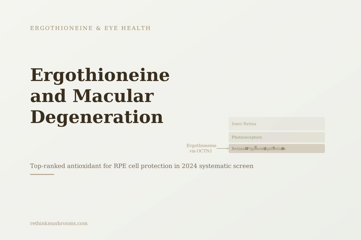
L-ergothioneine was identified as the lead cytoprotective antioxidant for retinal pigment epithelium cells in a 2024 systematic screen, protecting them from oxidative stress through NRF2 pathway activation.
Gu et al., Exp Eye Res, 2024
3 mechanisms
NRF2 activation, amyloid clearance, and senescent cell elimination identified in 2023-2024 research
Lower
blood ergothioneine in older adults with mild cognitive impairment vs age-matched controls, declining beyond age 60 (Cheah 2016)
Zero human trials
all current evidence for AMD is preclinical; no RCT has tested ergothioneine supplementation
Gu et al., Exp Eye Res, 2024
 View as image ↗
Key Takeaways
- Gu et al. 2024 screened a library of antioxidants for retinal pigment epithelium cell protection and identified L-ergothioneine as the lead cytoprotective candidate, acting through NRF2 pathway activation (Exp Eye Res, 2024)
- Wijesinghe et al. 2023 showed ergothioneine supplementation reduced amyloid beta accumulation in the neuroretina of Alzheimer's model mice; the same amyloid deposits appear in AMD retinas (Front Neurosci, 2023)
- Apparoo et al. 2024 found ergothioneine-rich mushroom extracts eliminated neuronal senescent cells; RPE cellular senescence is a recognized driver of AMD progression (Brain Res, 2024)
- OCTN1 transporters are expressed in retinal pigment epithelium specifically, meaning the retina actively imports ergothioneine from the bloodstream (Gründemann, FEBS Lett, 2022)
- Cheah et al. 2016 found whole blood ergothioneine declines significantly beyond age 60, and older adults with mild cognitive impairment had significantly lower plasma ergothioneine than age-matched controls (Biochem Biophys Res Commun, 2016)
- All current evidence for ergothioneine and AMD is preclinical; no human randomized controlled trial has been conducted for any retinal disease indication
In 2024, researchers at a Chinese ophthalmology institute published a study in Experimental Eye Research specifically designed to find the best antioxidant for protecting retinal pigment epithelium cells. The retinal pigment epithelium sits behind the photoreceptors and is the primary site of damage in age-related macular degeneration. The researchers screened multiple candidates under oxidative stress conditions. L-ergothioneine came out on top, and the research on ergothioneine macular degeneration has been building steadily since.
This article covers the three mechanistic threads that 2023-2024 research has identified, what they mean for understanding AMD, and where the evidence honestly ends. For a broad survey of ergothioneine across all eye tissues, see Ergothioneine for Eye Health: Research and Implications. For the foundational biology of the OCTN1 transport system, see what ergothioneine is and how the body concentrates it.
What ergothioneine does for macular degeneration risk: L-ergothioneine is a dietary antioxidant from mushrooms that concentrates in retinal pigment epithelium via the OCTN1 transporter. It activates the NRF2 antioxidant pathway, clears amyloid beta from neuroretinal tissue, and reduces RPE cellular senescence in preclinical models. No human clinical trial has tested it for AMD. The evidence is mechanistically compelling and preclinical only.
What AMD Does to the Retina: Why RPE Cells Are the Target
Age-related macular degeneration is the leading cause of irreversible vision loss in people over 50 in high-income countries. It affects the macula, the central portion of the retina responsible for sharp, detailed vision.
The primary pathological event is the degeneration of retinal pigment epithelium cells. RPE cells perform several functions critical to retinal health: they absorb stray light, supply nutrients to the photoreceptors above them, clear the metabolic waste photoreceptors generate, and recycle visual pigments. When RPE cells degenerate, photoreceptors lose their support structure and die. The result is progressive loss of central vision.
What drives RPE degeneration? The research points to sustained oxidative stress, accumulation of abnormal deposits, chronic inflammation, and cellular aging. The RPE is among the most metabolically active cell types in the body, exposed to high light levels and constant oxidative burden. Antioxidant defense systems are therefore central to its long-term survival.
Understanding AMD means understanding what makes RPE cells die, and what antioxidants can protect them under the conditions they actually face.
The 2024 Ophthalmology Finding: Ergothioneine Tops the RPE Antioxidant Screen
The most directly relevant study in the current ergothioneine and macular degeneration literature was published in Experimental Eye Research in 2024 by Gu and colleagues.[1]
The study's design was systematic: apply oxidative stress to RPE cells in culture, test multiple antioxidant candidates for their ability to protect those cells, and rank the results. L-ergothioneine was the highest-performing compound.
The mechanism Gu's team identified was NRF2 pathway activation. NRF2 (nuclear factor erythroid 2-related factor 2) is a transcription factor that, when activated, switches on a battery of antioxidant and cytoprotective genes. In RPE cells facing oxidative stress, robust NRF2 activation is associated with better survival. Ergothioneine emerged as the standout candidate in the screen, and NRF2 activation was central to its protective effect: when the researchers blocked NRF2, ergothioneine's protection was abolished.
Gu's team also tested ergothioneine in a mouse model of oxidative stress, where it maintained retinal structure better than controls. The mouse model data provides a step toward in vivo relevance, though it remains preclinical.
This is the kind of finding that redirects future research. A compound that tops a systematic antioxidant screen in a dedicated ophthalmology journal, for the specific cell type that fails in AMD, is a serious candidate for clinical investigation.
Three Mechanistic Threads: What 2023-2024 Research Identified
The Gu 2024 finding sits within a cluster of related studies from the same period. Each addresses a distinct mechanism relevant to AMD, and together they form a more complete mechanistic picture than any single study could provide.
Thread 1: NRF2 activation in RPE cells. Gu 2024 establishes ergothioneine as the lead cytoprotective candidate identified for RPE protection among the antioxidants screened.[1] NRF2 activation is well-established in the AMD literature as a protective mechanism, and this study shows ergothioneine engages it in the relevant cell type: blocking NRF2 abolished ergothioneine's protective effect, confirming the pathway is central to how it protects RPE cells.
Thread 2: Amyloid beta in the neuroretina. In 2023, Wijesinghe and colleagues published a study on ergothioneine in Alzheimer's model mice and found something directly relevant to AMD: ergothioneine supplementation reduced amyloid beta accumulation in the neuroretina.[2] The neuroretina is the neural layer of the retina containing the photoreceptors and their support cells. Amyloid beta deposits in retinal tissue are a recognized feature of AMD as well as Alzheimer's disease. The same pathological protein that accumulates in the Alzheimer's brain accumulates in the aging macula. Wijesinghe 2023 establishes that ergothioneine can clear amyloid beta from neuroretinal tissue in an animal model, providing a mechanistic bridge between ergothioneine, Alzheimer's pathology, and AMD.
Thread 3: Cellular senescence in RPE. Apparoo and colleagues' 2024 study used ergothioneine-rich mushroom extracts in a cellular model and found they eliminated neuronal senescent cells.[3] Cellular senescence is a state in which cells stop dividing but remain metabolically active, secreting inflammatory signals that damage surrounding tissue. RPE senescence is recognized as a driver of AMD pathology; senescent RPE cells create the inflammatory microenvironment that accelerates photoreceptor death. Clearing senescent cells from the RPE region addresses AMD upstream of visible photoreceptor damage.
These three mechanisms (NRF2 activation, amyloid clearance, and senescent cell elimination) are independent. They address different stages and aspects of AMD pathology. No other antioxidant in the current literature has preclinical data in all three categories for retinal tissue.
How Ergothioneine Reaches the Retina: The OCTN1 System
A mechanistic case for ergothioneine in AMD requires understanding how it gets into retinal tissue at all. The answer involves the same dedicated transport system that explains its presence in the lens.
Cheah and Halliwell's 2021 authoritative review confirms that ergothioneine concentrates in oxidative-stress-sensitive tissues including the retina, driven by OCTN1 transport.[4] Gründemann's 2022 comprehensive tissue inventory confirmed OCTN1 expression in retinal pigment epithelium specifically.[5] The RPE cells that face the highest oxidative burden in AMD are also the cells with a dedicated ergothioneine importer.
This anatomical fact is important for the mechanistic argument. Ergothioneine doesn't reach the RPE by accident or passive diffusion. The RPE cells that OCTN1 serves are the same cells that degenerate in AMD. The body built delivery infrastructure specifically to those cells.
For the deep mechanistic treatment of OCTN1 in retinal tissue, including what happens when the transporter is silenced, see OCTN1 in the Retina: Ergothioneine and Macular Degeneration.
Why Macular Degeneration Risk Rises as Ergothioneine Falls With Age
Age-related macular degeneration is, by definition, age-related. The strongest risk factor is age. The ergothioneine literature provides a plausible biological connection between aging and the retinal vulnerability that characterizes AMD.
In 2016, Cheah and colleagues measured ergothioneine in older adults and found that whole blood levels decline significantly beyond age 60, with a subset who had mild cognitive impairment showing significantly lower plasma ergothioneine than age-matched controls.[6] Ergothioneine also declined further with advancing age across the entire sample. The cognitive finding is relevant to retinal health because the retina is an extension of the central nervous system. Conditions that deplete ergothioneine systemically in the aging brain also affect the retina, which is brain tissue.
The animal-level confirmation came in 2024. Katsube and colleagues showed ergothioneine supplementation extended lifespan and reduced oxidative damage markers across multiple tissues in male mice.[7] This is the first direct longevity study for ergothioneine and it supports the broader framework: ergothioneine depletion with age leads to increased oxidative damage in tissues that depend on it, including the retina.
The temporal picture that emerges: ergothioneine levels fall as people age; AMD risk rises as people age; the retina expresses dedicated ergothioneine importers; and preclinical evidence shows ergothioneine addresses three mechanisms behind AMD pathology. These correlations don't establish causation, but they are coherent and point in the same direction.
For more on the age depletion trajectory, see Ergothioneine Declines with Age: What This Means for Health. For the neuroretina-brain connection in more depth, see Ergothioneine and Brain Health: Neuroprotective Research.
The Evidence Ceiling: Preclinical Data, No Human Trials
The research on ergothioneine and macular degeneration is the strongest it has ever been. The 2023-2024 wave of studies addressed three distinct AMD mechanisms, all in retinal or neural tissue, across dedicated journals including Experimental Eye Research and Frontiers in Neuroscience.
The ceiling of what that evidence can claim is still preclinical. Every mechanism study is from cell culture or animal models. The population data on ergothioneine and age-related health outcomes is observational. No randomized controlled trial has tested ergothioneine supplementation for AMD, AMD prevention, or RPE cell function in humans.
- Cell & Lab Studiescompleted
RPE cell antioxidant screen identifying ergothioneine as the lead cytoprotective agent via NRF2 (Gu 2024), and elimination of neuronal senescent cells by ergothioneine-rich extracts (Apparoo 2024)
- Animal Studiescurrent
Ergothioneine maintained retinal structure in mice under oxidative stress (Gu 2024), cleared amyloid beta from the neuroretina of Alzheimer's-model mice (Wijesinghe 2023), and extended lifespan while lowering oxidative-damage markers in male mice (Katsube 2024)
- Human Observational Datacompleted
Blood ergothioneine declines with age and is lower in people with mild cognitive impairment (Cheah 2016) — systemic and neurological, not a retinal or AMD outcome
- Small Human Trials (AMD)missing
No human trial has tested ergothioneine for AMD, AMD prevention, or RPE function
- Large Randomized Trials (AMD)missing
None
- Meta-Analyses & Systematic Reviewsmissing
No AMD-specific ergothioneine trial evidence exists to synthesize
For AMD specifically, the evidence has climbed through cell and animal models, with supporting human observational data on ergothioneine and aging. No human trial rung has been reached — the AMD-specific clinical evidence base is empty.
Gu 2024; Apparoo 2024; Wijesinghe 2023; Katsube 2024; Cheah 2016
This is an important distinction. The preclinical evidence is the kind that typically motivates clinical trials. It shows mechanism, identifies the tissue, explains why the compound gets there, and demonstrates protective effects in model systems. None of that is the same as showing that taking ergothioneine supplements reduces AMD incidence or progression in humans.
The honest position is that the mechanistic case is well-developed, the preclinical evidence is consistent across independent research groups, and the human clinical data is entirely missing. That's where the field is right now.
The lens evidence follows the same pattern. Cataractous human lenses contain markedly lower ergothioneine than normal lenses, yet no human supplementation trial exists there either. For that side of the eye health evidence, see Ergothioneine and Cataract Prevention.
Medical context
ModerateErgothioneine is not an AMD treatment
Concern
Because the preclinical findings are striking, people worried about macular degeneration may be tempted to take ergothioneine supplements in place of evidence-based eye care. No human trial has tested ergothioneine for AMD prevention or treatment, and it is not a substitute for established management.
What the research says
For people with or at risk of AMD, the interventions with human randomized-trial support are the AREDS2 formulation (lutein, zeaxanthin, vitamins C and E, zinc) for intermediate disease, alongside not smoking and regular ophthalmology follow-up. Treat mushrooms as a healthy food, not a therapy. This article is educational and not medical advice.
Particularly relevant for: Adults with or concerned about age-related macular degeneration
What to do
Do not replace AREDS2 supplementation, smoking cessation, or ophthalmology monitoring with ergothioneine; discuss any supplement with your eye doctor.
AREDS2 Research Group, JAMA, 2013; Gu et al., Exp Eye Res, 2024
The Bottom Line
Ergothioneine was identified as the lead antioxidant for retinal pigment epithelium protection in the most rigorous published screen to date, acting via NRF2 activation. Two additional 2023-2024 studies identified ergothioneine's capacity to clear amyloid beta from neuroretinal tissue and eliminate senescent RPE cells, two independent AMD-relevant mechanisms. All current evidence is preclinical; no human clinical trial has tested ergothioneine for AMD.
Frequently Asked Questions
Sources & References
- 1RTHM-01896·Gu, Sijie et al. (2024). “Screening and evaluation of antioxidants for retinal pigment epithelial cell protection: L-ergothioneine as a novel therapeutic candidate through NRF2 activation..” Experimental eye research.Study breakdown →PubMed →↩
- 2RTHM-01819·Wijesinghe, Printha et al. (2023). “Ergothioneine, a dietary antioxidant improves amyloid beta clearance in the neuroretina of a mouse model of Alzheimer's disease..” Frontiers in neuroscience.Study breakdown →PubMed →↩
- 3RTHM-01850·Apparoo, Yasaaswini et al. (2024). “Potential role of ergothioneine rich mushroom as anti-aging candidate through elimination of neuronal senescent cells..” Brain research.Study breakdown →PubMed →↩
- 4RTHM-00678·Cheah, Irwin K et al. (2021). “Ergothioneine, recent developments..” Redox biology.Study breakdown →PubMed →↩
- 5RTHM-01527·Gründemann, Dirk et al. (2022). “The ergothioneine transporter (ETT): substrates and locations, an inventory..” FEBS letters.Study breakdown →PubMed →↩
- 6RTHM-00420·Cheah, Irwin K et al. (2016). “Ergothioneine levels in an elderly population decrease with age and incidence of cognitive decline; a risk factor for neurodegeneration?.” Biochemical and biophysical research communications.Study breakdown →PubMed →↩
- 7RTHM-00884·Katsube, Makoto et al. (2024). “Ergothioneine promotes longevity and healthy aging in male mice..” GeroScience.Study breakdown →PubMed →↩