King Trumpet / King Oyster (Pleurotus eryngii)
3 apoptosis pathways
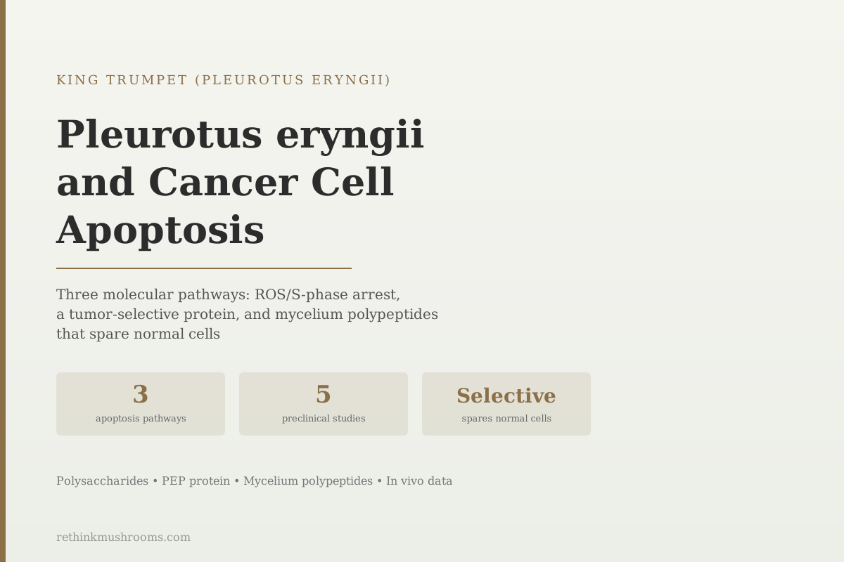
A novel protein from Pleurotus eryngii induced extensive apoptosis in colon cancer cells and suppressed tumor growth in mice, while leaving normal cells unharmed. Selectivity between cancer and normal cells is one of the central challenges in cancer pharmacology, and P. eryngii's PEP protein meets it in preclinical testing.
Yuan et al., 2017
S-phase arrest
PEP-2 polysaccharide from P. eryngii caused S-phase cell cycle arrest in HepG-2 hepatoblastoma cells via reactive oxygen species (Ren et al., 2016)
Tumor-selective
PEP protein from P. eryngii induced apoptosis in colon cancer cells in vitro and in vivo without damaging normal cells, a key selectivity finding (Yuan et al., 2017)
Mycelium active
PEMP polypeptide from P. eryngii mycelium showed concentration-dependent antitumor, antioxidant, and immunostimulatory activity (Sun et al., 2017)
Yuan et al., 2017
 View as image ↗
One of the central challenges in cancer pharmacology is selectivity: killing cancer cells without damaging the normal cells surrounding them. Most chemotherapy agents have poor selectivity, causing the off-target damage that produces side effects. In a 2017 laboratory study, a protein extracted from Pleurotus eryngii (king trumpet mushroom) induced extensive apoptosis in colon cancer cells while leaving normal cells unharmed, in both cell culture and in tumor-bearing mice.[2]
This article covers the molecular mechanisms by which P. eryngii compounds trigger cancer cell apoptosis, the built-in cell self-destruction program that cancer cells typically disable. All of the evidence reviewed here is preclinical (cell culture or animal models). No clinical trials on P. eryngii for human cancer have been conducted.
For the broader picture of P. eryngii cancer research including all compound classes studied, see King Trumpet and Cancer: Antitumor Research. To understand how mushroom polysaccharides trigger apoptosis across species, see How Mushroom Polysaccharides Kill Cancer Cells: Four Distinct Mechanisms.
For the complete overview of king trumpet's nutritional and health profile, see the king trumpet mushroom overview.
Key Takeaways
- Pleurotus eryngii compounds trigger cancer cell apoptosis through at least three distinct molecular pathways, documented across five preclinical studies
- PEP-2, a high molecular mass polysaccharide from P. eryngii, induced apoptosis and S-phase cell cycle arrest in HepG-2 hepatoblastoma cells via elevated intracellular reactive oxygen species (Ren et al., 2016)
- A novel protein called PEP from P. eryngii induced extensive apoptosis in colon cancer cells in vitro and inhibited tumor growth in mice in vivo, while sparing normal cells, demonstrating tumor selectivity not seen in most chemotherapy agents (Yuan et al., 2017)
- Polypeptides from P. eryngii mycelium (PEMP) showed concentration-dependent antitumor, antioxidant, and immunostimulatory activities simultaneously, suggesting multi-mechanism anti-cancer action from the mycelium fraction (Sun et al., 2017)
- Polysaccharides extracted from P. eryngii processing residue (waste material after extraction) also suppressed HepG-2 hepatoma cell proliferation in a dose-dependent manner, showing that bioactive anti-cancer compounds are retained even in processing by-products (Ma et al., 2014)
- In vivo: water-soluble P. eryngii polysaccharide (PEPw) significantly suppressed tumor growth in mice bearing renal cancer at 50, 100, and 200 mg/kg in a dose-dependent manner (Yang et al., 2013)
What Apoptosis Is and Why Cancer Cells Disable It
Every cell in the human body contains an internal program for self-destruction called apoptosis. When a cell becomes damaged beyond repair, infected, or potentially malignant, apoptosis normally activates and the cell dismantles itself in an orderly way, without triggering inflammation or damaging nearby tissue.
Cancer cells typically disable this program. Mutations accumulate that allow cells to survive signals that would normally trigger apoptosis. The result is uncontrolled proliferation: cells that should die, continue dividing. This is why restoring apoptosis sensitivity is a major goal in cancer drug development.
The most common apoptosis pathways involve mitochondria (the intrinsic pathway) and death receptor signaling on the cell surface (the extrinsic pathway). Both eventually activate enzymes called caspases that carry out the cell's self-destruction. Reactive oxygen species (ROS) accumulating inside a cell is a trigger for the intrinsic mitochondrial pathway. Cell cycle arrest, stopping a cell from moving through the stages of division, is often a prelude to apoptosis induction.
P. eryngii compounds documented to trigger apoptosis in cancer cells include specific polysaccharide fractions, a novel protein, and polypeptides from the mycelium. Each works through a somewhat different molecular entry point.
Three Doors, One Endpoint
How P. eryngii Compounds Converge on Cancer Cell Death
Five preclinical studies describe three different molecular entry points — a direct ROS route, a tumor-selective protein, and an immune-mediated route. They start from different compounds and different parts of the mushroom, but all arrive at the same measured outcome.
Mechanism 1
[1] [3]Compound
Polysaccharides (PEP-2; PEPE-1/2/3)
Fruiting body + processing residue
What it does
PEP-2 raises intracellular reactive oxygen species and forces S-phase arrest in HepG-2 hepatoblastoma cells; residue fractions suppress the same line dose- and time-dependently (PEPE-1 < PEPE-2 < PEPE-3).
Mechanism 2
[2]Compound
PEP protein
Fruiting body
What it does
PEP suppresses HCT116 / MC38 colon cancer cells and drives extensive apoptosis (p53, cleaved-PARP up; cyclin B/E, cdc-2 down) with no effect on normal CCD-18Co colon cells at the same doses. Confirmed in tumor-bearing mice.
Mechanism 3
[4] [5]Compound
Polypeptide PEMP; polysaccharide PEPw
Mycelium (PEMP) + fruiting body (PEPw)
What it does
PEMP activates macrophages (NO, TNF-α, IL-6, TLR2/4) alongside direct antitumor and antioxidant action; PEPw suppresses Renca renal tumors in mice at 50–200 mg/kg via NK-cell and CTL activation.
Cancer cell apoptosis + tumor growth suppression
Endpoint reached in cell culture and in tumor-bearing mice only. No human clinical trial of any P. eryngii compound for cancer exists — every route stops at preclinical.
Source: Ren 2016; Yuan 2017; Ma 2014; Sun 2017; Yang 2013 (see references)
Mechanism 1: ROS-Mediated Apoptosis and S-Phase Cell Cycle Arrest
Ren and colleagues in 2016 extracted and characterized polysaccharide fractions from P. eryngii and tested their effects against HepG-2 human hepatoblastoma cells.[1] A high molecular mass fraction called PEP-2 showed the strongest anti-cancer activity.
What PEP-2 did to the cancer cells: it raised intracellular ROS levels, increased S-phase cell cycle arrest, and induced apoptosis. These three effects are connected. Elevated ROS destabilizes mitochondrial membrane integrity, signaling the intrinsic apoptosis pathway. S-phase arrest prevents the cell from completing DNA replication, blocking division and creating more time for apoptosis signals to trigger self-destruction.
Ma and colleagues in 2014 tested three polysaccharide fractions (PEPE-1, PEPE-2, PEPE-3) extracted from P. eryngii processing residue, the waste material left after conventional polysaccharide extraction.[3] All three fractions suppressed HepG-2 proliferation and enhanced LDH release (a marker of cell membrane damage) in a dose-dependent and time-dependent manner. The potency order was PEPE-1 less than PEPE-2 less than PEPE-3, suggesting the more complex or larger molecular fractions are more active.
The Ma 2014 finding that processing residue retains bioactive anti-cancer compounds is notable from both a research and practical standpoint: the compounds are present throughout the mushroom material, not limited to the primary extract fraction. For the mechanism of how polysaccharides activate immune cells against cancer, see How Mushroom Sugars Activate Cancer-Fighting Immune Cells.
Mechanism 2: The PEP Protein and Selective Cancer Cell Apoptosis
Yuan and colleagues in 2017 isolated a novel protein from P. eryngii called PEP and tested it against colon cancer cells in both cell culture and in tumor-bearing mice.[2] The results are among the most compelling in the P. eryngii cancer literature for two reasons: tumor selectivity and in vivo confirmation.
In cell culture: PEP suppressed colon cancer cell proliferation and induced cell cycle arrest and extensive cellular apoptosis.
In tumor-bearing mice: PEP inhibited tumor growth in vivo, confirming the effect is not limited to the artificial environment of a cell culture dish.
The selectivity finding: PEP induced apoptosis in cancer cells without damaging normal cells. In the study, normal human colon cells treated with PEP showed no significant toxicity, while cancer cells showed substantial apoptosis induction.
This selectivity is significant because it addresses the primary limitation of most cytotoxic cancer treatments: off-target damage to normal tissue. Most chemotherapy agents kill rapidly dividing cells generally, which is why they also damage intestinal lining, hair follicles, and bone marrow. A compound that preferentially targets cancer cells while sparing normal cells is pharmacologically much more attractive. The mechanism of PEP's selectivity has not been fully characterized in published literature, but one likely factor is differential surface receptor expression: cancer cells often overexpress certain surface proteins that PEP may preferentially bind.
Yuan et al. (2017) · Food & Function
- Population
- Human & murine colon cancer cell lines (HCT116, MC38) plus tumor-bearing mice; normal human colon cells (CCD-18Co) as the selectivity control
- Intervention
- PEP, a novel protein isolated from P. eryngii
- Comparator
- Same PEP doses applied to normal CCD-18Co colon cells
- Finding
- PEP suppressed cancer cell proliferation dose- and time-dependently and induced extensive apoptosis (p53 and cleaved-PARP up; cyclin B, cyclin E, cdc-2 down), while showing no inhibitory effect on normal colon cells at the same concentrations. In mice, PEP significantly suppressed allograft tumor growth.
preliminary A standout selectivity result with in vivo confirmation — but confined to cell lines and mouse allografts. No human data exists, and the mechanism behind the cancer-vs-normal selectivity is not yet characterized.
Yuan et al., International Journal of Biological Macromolecules, 2017 (RTHM-00932, PMID 28880032)
Mechanism 3: Mycelium Polypeptides with Multi-Mode Anti-Cancer Activity
Sun and colleagues in 2017 extracted polypeptides from P. eryngii mycelium (the underground network, not the fruiting body) and tested the fraction called PEMP.[4]
PEMP showed three concurrent activities: antioxidant (scavenging of reactive oxygen species), antitumor (concentration-dependent inhibition of cancer cell proliferation), and immunostimulatory (activation of immune cells involved in anti-cancer surveillance).
The immunostimulatory component distinguishes this mechanism from the direct apoptosis induction of PEP protein. Rather than attacking cancer cells directly, the immune activation pathway enhances the immune system's natural cancer surveillance function. In Sun 2017, PEMP stimulated macrophages (increasing nitric oxide, TNF-α, and IL-6 output and raising TLR2/TLR4 expression), the frontline innate immune cells that engulf abnormal cells and signal the wider immune response. A related in vivo finding, that P. eryngii polysaccharide PEPw elevated NK cell and cytotoxic T-lymphocyte activity in tumor-bearing mice (Yang et al., 2013), extends this immune-activation theme to a second compound and a whole-animal model.[5]
The fact that PEMP comes from the mycelium rather than the fruiting body is relevant for anyone interested in P. eryngii-derived supplements: mycelium-derived products are not simply inferior substitutes for fruiting body extracts. They may contain distinct bioactive compounds with different pharmacological profiles. For how these immunostimulatory mechanisms work in detail, see King Trumpet and Cancer: Antitumor Research.
In Vivo Confirmation: From Cell Culture to Tumor-Bearing Mice
The movement from cell culture results to in vivo animal data is a significant step in preclinical evidence. Cell culture experiments measure what happens to isolated cells in artificial conditions; animal studies test whether the compound can reach the target tissue, survive metabolism, and produce measurable effects in a living organism.
Yang and colleagues in 2013 extracted a water-soluble polysaccharide (PEPw) from P. eryngii fruiting bodies and tested it in mice bearing Renca renal cancer, a validated model of kidney cancer.[5] PEPw significantly suppressed tumor growth at all three doses tested (50, 100, and 200 mg/kg), with a dose-dependent relationship: higher doses produced greater suppression.
This in vivo data, combined with Yuan 2017's PEP protein animal model results, establishes that P. eryngii anti-cancer activity is not purely an in vitro artifact. The compounds produce measurable tumor suppression in animals.
The caveat remains: the distance from mouse tumor models to human cancer clinical outcomes is large. Mouse models of cancer use tumor cells grafted into immunocompromised mice under controlled conditions. Human cancer is biologically more complex, develops differently, and exists in immune contexts that differ substantially from these experimental conditions.
How to Read This Evidence
The five studies reviewed here establish a mechanistic picture:
P. eryngii contains multiple compound classes (polysaccharides, proteins, polypeptides from both fruiting body and mycelium) that trigger cancer cell apoptosis and suppress tumor growth in laboratory settings. The mechanisms are distinct for each compound class: ROS-mediated apoptosis and cell cycle arrest (polysaccharides), selective tumor cell protein-mediated apoptosis (PEP protein), and immunostimulatory antitumor activity (mycelium polypeptides). In vivo animal data confirms the anti-cancer activity transfers beyond cell culture for at least two compound types.
What this evidence does not establish: clinical efficacy in human cancer. There are no clinical trials testing P. eryngii compounds in cancer patients. The concentrations used in cell culture experiments are often higher than what could be achieved in human tissue through oral consumption. The cancer cell lines used (HepG-2, colon cancer cells) are laboratory models, not primary patient tumors.
The PEP protein's normal-cell selectivity is the most pharmacologically interesting finding in this cluster because it suggests a safety profile that conventional cytotoxics lack. Whether this selectivity holds in human clinical testing is unknown.
- Cell & Lab Studiescompleted
ROS-mediated apoptosis and S-phase arrest in HepG-2 (Ren 2016); dose-dependent suppression of HepG-2 by residue polysaccharides (Ma 2014); tumor-selective apoptosis of colon cancer cells with no harm to normal colon cells (Yuan 2017); antitumor and immunostimulatory action of mycelium polypeptide (Sun 2017)
- Animal Studiescurrent
PEP protein suppressed allograft colon tumors in mice (Yuan 2017); PEPw polysaccharide suppressed Renca renal tumors in mice at 50–200 mg/kg via immune activation (Yang 2013)
- Human Case Reportsmissing
No case reports of P. eryngii compounds used in human cancer
- Small Human Trialsmissing
No clinical trials of any P. eryngii compound for cancer in humans
- Large Randomized Trialsmissing
None
- Meta-Analyses & Systematic Reviewsmissing
None
The mechanisms are documented in cell culture and confirmed in mouse tumor models for two compound classes, which is genuine preclinical progress. But the ladder stops there: no human case reports, no clinical trials, no meta-analyses. 'P. eryngii triggers cancer cell apoptosis' is well-supported in the laboratory and unproven in people.
Aggregate of the five cited preclinical studies (Ren 2016; Ma 2014; Yuan 2017; Sun 2017; Yang 2013)
Reading this as health information
ModeratePreclinical anti-cancer activity is not a reason to self-treat cancer with king trumpet mushrooms or extracts
Concern
Every result on this page comes from cancer cells in a dish or tumors in mice. The concentrations that killed cancer cells in the lab are often far higher than anything reachable by eating mushrooms or taking a supplement, and no P. eryngii compound has been tested against cancer in a single human being. 'Kills cancer cells in the lab' is where most compounds that never become drugs also start.
What the research says
Treat these findings as early mechanistic research, not treatment guidance. King trumpet is a nutritious culinary mushroom and can be enjoyed as food; that is different from using it, or an extract, as a cancer therapy.
Particularly relevant for: Anyone with cancer, or supporting someone who does
What to do
Do not delay, replace, or modify any cancer treatment based on mushroom research. Discuss any supplement with your oncologist first — some interact with chemotherapy or radiation. This article is educational and is not medical advice.
Consistent limitation across all five cited studies (Ren 2016; Ma 2014; Yuan 2017; Sun 2017; Yang 2013)
The Bottom Line
Pleurotus eryngii compounds trigger cancer cell apoptosis through three distinct mechanisms: ROS-mediated S-phase arrest from polysaccharides, selective apoptosis induction from a novel protein (PEP) without harming normal cells, and immunostimulatory antitumor activity from mycelium polypeptides. In vivo mouse data confirms anti-cancer activity for polysaccharide and protein fractions. All evidence is preclinical; no human clinical trials exist for P. eryngii cancer treatment. The tumor selectivity of PEP protein is the most pharmacologically significant finding, addressing a key challenge in cancer pharmacology that existing treatments have not solved.
Frequently Asked Questions
Sources & References
- 1. Source details for this reference are not yet available.
- 2. Source details for this reference are not yet available.
- 3. Source details for this reference are not yet available.
- 4. Source details for this reference are not yet available.
- 5. Source details for this reference are not yet available.