Psilocybin Brain Scans Depression
3x Greater Changes
Psilocybin produced more than 3-fold greater acute changes in functional brain networks than methylphenidate, driven by widespread desynchronization across spatial scales.
Siegel et al., Nature, 2024
15+ fMRI Studies
visualizing psilocybin brain effects
DMN Dissolution
default mode network loses coherence
Weeks-Long Changes
connectivity alterations persist post-dose
Siegel et al., Nature, 2024
 View as image ↗
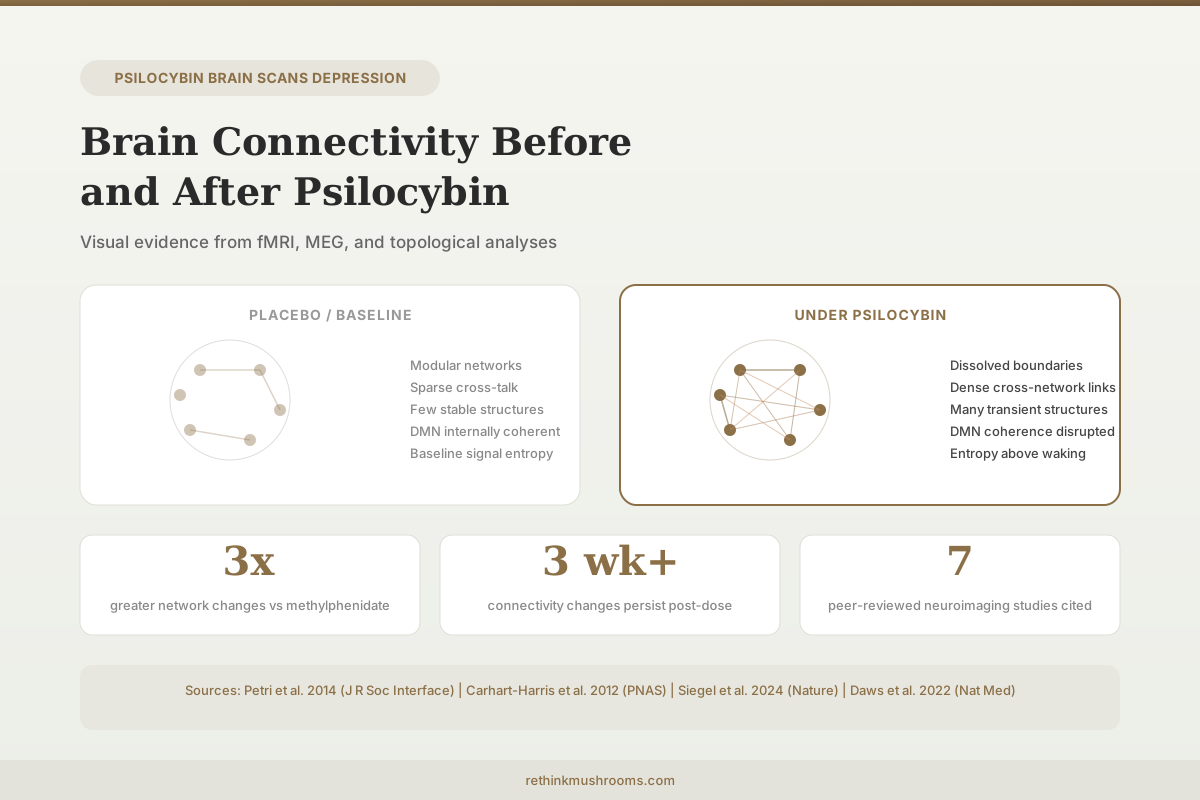
When Petri et al. published their 2014 paper on brain connectivity under psilocybin, one image went viral: a circular graph of brain regions showing sparse, orderly connections under placebo and a dense web of cross-network links under psilocybin.[1] That visualization captured something neuroimaging data had been showing since 2012: psilocybin does not simply activate or suppress brain regions. It reorganizes how they communicate. This article maps the visual evidence from fMRI, MEG, and topological analyses that show what happens to brain connectivity before, during, and after psilocybin exposure.
For related coverage of how these connectivity changes relate to depression treatment, see how brain scans are proving psilocybin rewires depression and psilocybin and default mode network changes in depression.
Key Takeaways
- Psilocybin produced 3-fold greater acute changes in functional brain networks than methylphenidate, with effects driven by desynchronization across cortical and subcortical structures (Siegel et al., Nature, 2024)
- Topological analysis revealed dramatically increased cross-network connectivity under psilocybin, with many transient low-stability structures appearing that are absent under placebo (Petri et al., 2014)
- The default mode network loses internal coherence while gaining connections to networks it normally does not communicate with (Carhart-Harris et al., PNAS, 2012)
- MEG signal diversity exceeded normal waking consciousness levels under psilocybin, representing the first measured increase above baseline waking entropy (Schartner et al., 2017)
- Post-treatment fMRI in depression patients showed increased global brain integration and decreased network modularity that correlated with symptom improvement (Daws et al., Nature Medicine, 2022)
- Psilocybin decreased claustrum connectivity with the default mode network while increasing connectivity with the fronto-parietal task control network (Barrett et al., 2020)
The 2012 Foundation: First fMRI Maps Under Psilocybin
Carhart-Harris et al. (2012) published the first controlled fMRI study of psilocybin's effects on human brain function, scanning 30 healthy volunteers after intravenous administration of psilocybin or placebo.[2]
The results defied the initial hypothesis. The researchers expected to see increased brain activity (more blood flow, higher BOLD signal) given the intensity of the psychedelic experience. Instead, psilocybin produced widespread decreases in cerebral blood flow and BOLD signal, with the largest reductions in hub regions: the medial prefrontal cortex (mPFC) and the posterior cingulate cortex (PCC). These two regions are core nodes of the default mode network (DMN), the brain system most active during self-referential thinking, mind-wandering, and autobiographical memory.
The magnitude of DMN deactivation correlated with the intensity of subjective effects. Volunteers who reported the strongest ego dissolution experienced the greatest decreases in mPFC and PCC activity. This suggested that the DMN functions as a kind of neurological constraint on consciousness, and psilocybin's subjective effects arise not from adding activity but from removing it.
Connectivity analyses revealed that psilocybin reduced the coupling between the mPFC and PCC, the two strongest hubs in the DMN. Under normal conditions, these regions maintain a tight functional link. Under psilocybin, that link weakened, and the DMN lost its internal coherence as a network.
The Homological Scaffold: Making Cross-Talk Visible
Petri et al. (2014) applied algebraic topology to the same dataset, using a mathematical framework called persistent homology to map the structure of brain connectivity at a level conventional analyses miss.[1]
Standard functional connectivity analyses measure correlations between pairs of brain regions. Persistent homology goes further: it identifies higher-dimensional structures, loops and cavities in the network that reveal how groups of regions communicate in coordinated patterns rather than just pairwise links.
Under placebo, the brain's homological scaffold was organized into a small number of stable, persistent structures. These represent the brain's normal network architecture: well-defined communities (visual, auditory, motor, default mode) with relatively little cross-communication.
Under psilocybin, the scaffold transformed. Many new transient structures appeared, connecting brain regions that normally operate in separate networks. These cross-network links were individually unstable (they formed and dissolved rapidly) but collectively represented a massive increase in the diversity of communication patterns. Regions in the visual cortex became transiently connected with regions in the default mode network, auditory cortex, and frontal areas in combinations that never appeared under placebo.
The resulting circular visualization became iconic: a dense, colorful web of connections replacing the sparse, modular pattern of normal brain function. The image did not show random noise. The underlying edges were more stable than those in the placebo condition; it was the higher-level structures built from those edges that were transient. The brain was not falling apart. It was exploring connectivity patterns outside its normal repertoire.
The mathematical framework matters because it captures phenomena invisible to standard correlation-based connectivity analyses. Two brain regions might show the same pairwise correlation under placebo and psilocybin, but participate in completely different higher-order communication structures. Persistent homology identifies these topological features: closed loops of coordinated activity involving three, four, or more regions that form geometric structures in network space. Under psilocybin, the landscape of these structures shifted from a few deep, stable cycles to many shallow, transient ones spanning brain regions that typically operate independently.
Signal Diversity: More Than Placebo, More Than Waking
Schartner et al. (2017) approached the same question from an information-theoretic perspective, analyzing MEG (magnetoencephalography) recordings from participants given psilocybin, LSD, or ketamine.[3]
Using measures of signal diversity (Lempel-Ziv complexity, amplitude coalition entropy, and synchrony coalition entropy), the researchers found that all three psychedelics increased spontaneous signal diversity above the level of normal waking consciousness. This was the first empirical demonstration that any manipulation could push neural signal complexity above baseline waking levels. Previous research had only shown that anesthesia, sleep, and disorders of consciousness reduce signal diversity below waking baseline.
For psilocybin specifically, the increase was most pronounced in single-channel Lempel-Ziv complexity, and changes in signal diversity correlated with subjective reports of psychedelic experience intensity. The finding provided a quantitative metric for what the connectivity studies showed qualitatively: psilocybin puts the brain into a state of expanded informational repertoire.
The theoretical framework for interpreting this, the entropic brain hypothesis, proposes that the psychedelic state represents an increase in the entropy of spontaneous brain activity, loosening the constraints that normally keep brain function organized into a narrow range of states. Whether higher entropy represents "more consciousness" or simply "different consciousness" remains debated, but the measurement itself is reproducible across studies and imaging modalities.
Desynchronization at Scale: The 2024 Nature Study
Siegel et al. (2024) published the largest and most comprehensive neuroimaging study of psilocybin to date in Nature, combining fMRI data from multiple doses and comparing effects against the stimulant methylphenidate as an active control.[4]
Psilocybin produced more than 3-fold greater acute changes in functional networks than methylphenidate. The effect was driven by desynchronization: brain activity across cortical networks and subcortical structures became temporally uncoupled. Regions that normally fire in synchrony within a network lost their coordinated timing.
This desynchronization was not uniform. It disrupted connectivity across spatial scales, from local cortical circuits to long-range corticocortical connections. The pattern was dose-dependent, with higher doses producing greater desynchronization. The study combined data from multiple scanning sessions and dosing protocols, providing the most statistically powered analysis of psilocybin's neural effects to date.
The methylphenidate comparison was important because it controls for nonspecific arousal effects. Stimulants alter brain activity and subjective experience, but they do not produce the cross-network reorganization seen with psilocybin. The 3-fold difference indicated that psilocybin's connectivity effects are qualitatively different from general pharmacological activation, not just quantitatively larger. This distinction helps rule out the possibility that psilocybin's connectivity changes simply reflect increased arousal, attention, or cardiovascular effects of drug administration.
The Claustrum: A Hidden Hub Reshaped
Barrett et al. (2020) focused on a specific structure that most psilocybin imaging studies had overlooked: the claustrum, a thin sheet of neurons between the insula and the putamen that sends and receives connections to nearly every cortical region.[5]
Using task-free fMRI in 15 participants who received psilocybin or placebo, the researchers found that psilocybin decreased both the amplitude of low-frequency fluctuations and the variance of BOLD signal in the left and right claustrum. The claustrum essentially became quieter.
Connectivity changes were directional. The right claustrum decreased its connectivity with the default mode network and auditory network but increased connectivity with the fronto-parietal task control network (FPTC). The left claustrum decreased connectivity with the FPTC.
This was the first empirical evidence in any species for a role of 5-HT2A receptor signaling in claustrum function. The claustrum is one of the most 5-HT2A-dense structures in the brain, and psilocybin's primary mechanism involves 5-HT2A agonism. The finding suggested that the claustrum may act as a synchronizing hub under normal conditions, and psilocybin disrupts this synchronization, contributing to the cross-network connectivity changes observed in other studies.
For the receptor pharmacology, see 5-HT2A receptor density and psilocybin response. For more on brain imaging comparisons, see before and after psilocybin: brain imaging comparisons.
After the Trip: Lasting Connectivity Changes
The acute effects of psilocybin last 4-6 hours. The connectivity changes do not necessarily resolve on the same timeline.
Carhart-Harris et al. (2017) scanned patients with treatment-resistant depression before and one day after psilocybin therapy (two doses: 10 mg and 25 mg, seven days apart).[6] Post-treatment scans showed decreased cerebral blood flow in the temporal cortex, including the amygdala, with decreased amygdala blood flow correlating with reduced depressive symptoms. Within the default mode network, resting-state functional connectivity increased post-treatment, a pattern opposite to the acute DMN disruption.
This apparent reversal is important: during psilocybin, the DMN loses coherence; after psilocybin, DMN connectivity increases. One interpretation is that psilocybin temporarily disrupts rigid, pathological DMN connectivity patterns (associated with rumination in depression) and allows them to reform in a healthier configuration.
Daws et al. (2022) extended this to a randomized controlled design, comparing psilocybin therapy against escitalopram (an SSRI) in patients with depression.[7] fMRI was recorded at baseline and three weeks after treatment. Psilocybin produced decreased brain network modularity and increased global integration, with higher-order cortical networks (rich in 5-HT2A receptors) becoming more functionally interconnected and flexible. The antidepressant response correlated with the degree of modularity decrease.
Escitalopram produced no measurable changes in brain network organization despite similar clinical improvement on some measures. This suggested that psilocybin and SSRIs achieve antidepressant effects through fundamentally different neural mechanisms: psilocybin by reorganizing network architecture, escitalopram by modulating neurotransmitter levels without visible structural network change.
The network cartography analysis in the Daws study revealed that the changes were not random. Higher-order networks with the greatest density of 5-HT2A receptors showed the most pronounced increases in between-network connectivity. The selectivity of this pattern aligns with psilocybin's known pharmacology as a 5-HT2A agonist and suggests the receptor binding directly mediates the network reorganization rather than triggering a nonspecific cascade.
For deeper analysis of amygdala-specific findings, see psilocybin and amygdala reactivity: what brain scans reveal about depression relief. For the full fMRI evidence in depression, see what psilocybin does to the depressed brain: fMRI studies.
Limitations of the Visual Evidence
The brain connectivity images from psilocybin research are visually striking but require careful interpretation.
Sample sizes are small. The Petri and Carhart-Harris 2012 studies used 15 participants. Barrett used 15. Even Siegel (2024), the largest study, used a relatively modest sample by pharmaceutical standards. Small samples increase the risk that observed patterns reflect individual variation rather than reliable drug effects.
Acute vs. post-treatment effects differ. The iconic cross-network connectivity images represent the acute psychedelic state, not the lasting therapeutic effect. The post-treatment changes (increased DMN coherence, decreased modularity) look nothing like the acute state. Using acute-state images to illustrate therapeutic mechanisms is misleading.
Visualization choices matter. The Petri circular graph uses a threshold to determine which connections are displayed. Different thresholds produce different-looking images. The visual impression of "chaos vs. order" depends partly on display parameters, not just underlying neurobiology.
fMRI measures blood flow, not neural activity. The BOLD signal is an indirect proxy for neural activity. Whether psilocybin changes neural firing patterns in the way fMRI images suggest, or whether the hemodynamic response itself is altered, remains an active question. Psilocybin's vasodilatory effects could influence cerebral blood flow independent of neural activity.
Correlation, not mechanism. Connectivity changes correlate with subjective effects and clinical outcomes, but correlation does not establish that the connectivity change causes the experience or the therapeutic benefit. The connectivity change could be an epiphenomenon of some other, unmeasured mechanism.
The Bottom Line
Neuroimaging studies spanning 2012 to 2024 have produced converging visual evidence that psilocybin reorganizes brain connectivity rather than simply increasing or decreasing activity. The acute state features dissolved network boundaries, increased cross-network communication, and elevated signal entropy above normal waking levels. Post-treatment, the brain shows increased global integration and decreased modularity that correlate with antidepressant response. The evidence is reproducible across fMRI, MEG, and topological analysis methods, though sample sizes remain small and the relationship between connectivity changes and therapeutic mechanisms is correlational.
Frequently Asked Questions
Sources & References
- 1RTHM-00345·Petri, G et al. (2014). “Homological scaffolds of brain functional networks..” Journal of the Royal Society.Study breakdown →PubMed →↩
- 2RTHM-00245·Carhart-Harris, Robin L et al. (2012). “Neural correlates of the psychedelic state as determined by fMRI studies with psilocybin..” Proceedings of the National Academy of Sciences of the United States of America.Study breakdown →PubMed →↩
- 3RTHM-00489·Schartner, Michael M et al. (2017). “Increased spontaneous MEG signal diversity for psychoactive doses of ketamine, LSD and psilocybin..” Scientific reports.Study breakdown →PubMed →↩
- 4RTHM-00911·Siegel, Joshua S et al. (2024). “Psilocybin desynchronizes the human brain..” Nature.Study breakdown →PubMed →↩
- 5RTHM-00608·Barrett, Frederick S et al. (2020). “Psilocybin acutely alters the functional connectivity of the claustrum with brain networks that support perception, memory, and attention..” NeuroImage.Study breakdown →PubMed →↩
- 6RTHM-00463·Carhart-Harris, Robin L et al. (2017). “Psilocybin for treatment-resistant depression: fMRI-measured brain mechanisms..” Scientific reports.Study breakdown →PubMed →↩
- 7RTHM-00752·Daws, Richard E et al. (2022). “Increased global integration in the brain after psilocybin therapy for depression..” Nature medicine.Study breakdown →PubMed →↩