Psilocybin & Depression
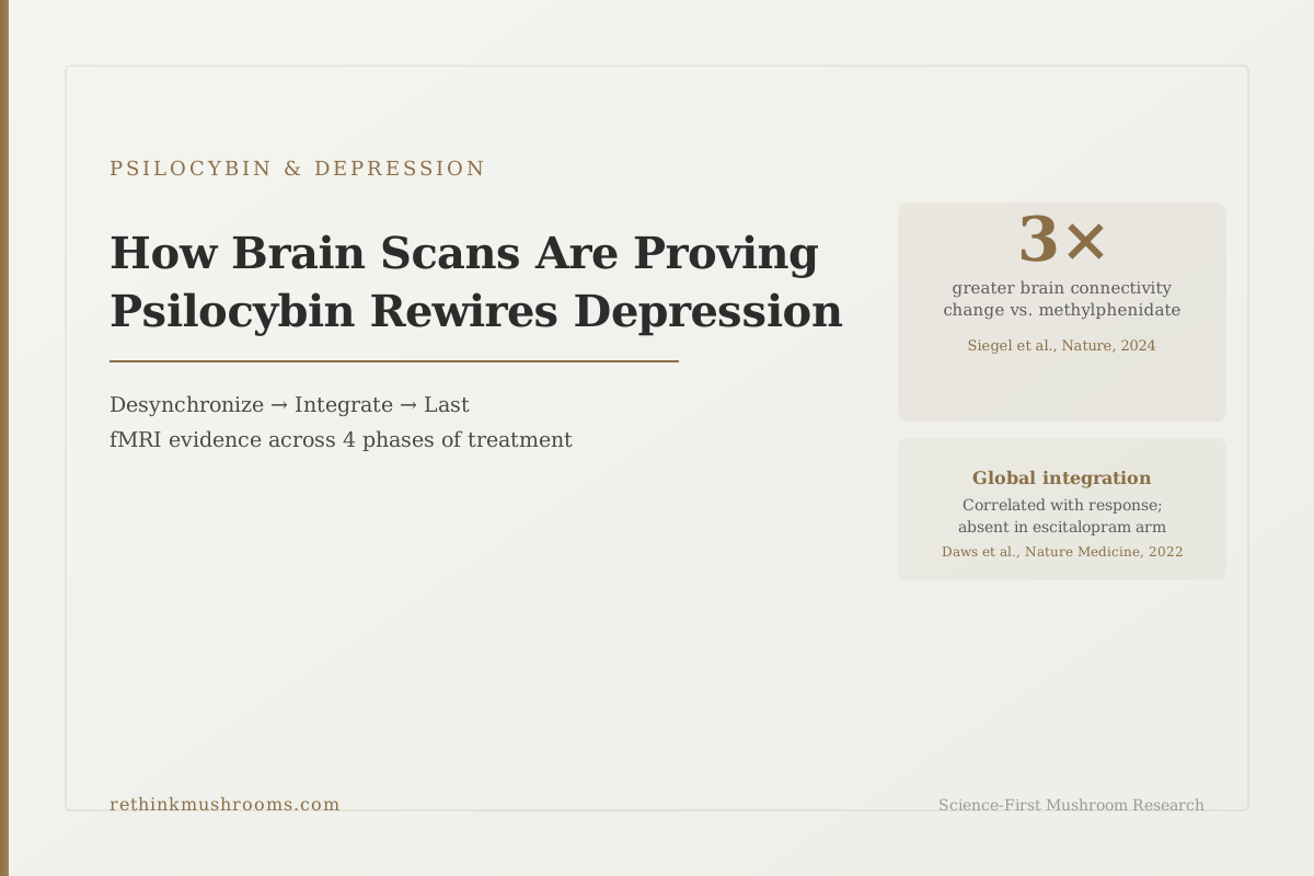
3× greater brain change
In a 2024 Nature study, psilocybin caused more than three times greater change in brain connectivity compared to methylphenidate (Ritalin) as an active control. The scale of disruption did not require a mystical experience, suggesting the therapeutic mechanism is pharmacological, not experiential.
Siegel et al., Nature, 2024
n=43 trial
Daws 2022 (Nature Medicine): psilocybin therapy increased global brain network integration; previously siloed depression networks reconnected, an effect absent in the escitalopram comparison arm
1 month duration
Barrett 2020: significant changes in brain function and emotional processing persisted for up to one month following a single high-dose psilocybin session
Predictive
Copa 2024: baseline fMRI connectivity patterns taken before any treatment now predict who will respond to psilocybin, opening a path to patient selection biomarkers
Siegel et al., Nature, 2024
 View as image ↗
In 2022, a headline spread from UCSF: "Psilocybin rewires the brain for people with depression." It was based on a Nature Medicine study measuring brain connectivity before and after psilocybin therapy in 43 depressed patients. The data showed real, measurable rewiring, not a metaphor.
Then in 2024, a Nature paper put a harder number on it. Psilocybin caused more than three times greater change in brain network connectivity than methylphenidate, the stimulant used as an active control in the same experiment.[1]
Most coverage of psilocybin and depression is still catching up to the 2022 data. Almost none has integrated the 2024 finding.
The brain scan evidence for psilocybin and depression now covers three distinct phases. First, the acute session produces massive network disruption: brain regions that normally fire in coordinated patterns become temporarily desynchronized to a degree no other psychiatric intervention produces. Second, in the days after treatment, brain networks that were previously isolated from each other begin exchanging signals; the rigid, self-reinforcing depression circuits lose their tight boundaries. Third, both of these changes last well past the acute experience; in one study, measurably so for up to a month following a single session.
A fourth finding may be the most consequential for clinical practice: a 2024 study found that the brain's connectivity pattern before any treatment can now predict whether a patient will respond.
For the clinical outcome data that this imaging evidence is explaining, see psilocybin for major depression: the clinical evidence.
Key Takeaways
- A 2024 Nature study found psilocybin caused more than 3× greater change in brain network connectivity than methylphenidate as an active control; the scale of disruption did not require a mystical experience (Siegel et al., Nature, 2024)
- Psilocybin therapy increased global brain network integration in 43 depressed patients; tightly siloed networks became more interconnected, and this integration correlated with antidepressant response; the escitalopram comparison arm showed no such change (Daws et al., Nature Medicine, 2022)
- In treatment-resistant depression patients, psilocybin increased amygdala reactivity to emotional faces one day after treatment, and this increase predicted better outcomes, the opposite of what SSRIs do, suggesting emotional re-engagement rather than blunting (Roseman et al., Neuropharmacology, 2018)
- Brain function and emotional processing changes persisted for up to one month following a single high-dose psilocybin session, confirming that imaging-measured changes are not acute artifacts but lasting neural reorganization (Barrett et al., Scientific Reports, 2020)
- Baseline (pre-treatment) fMRI connectivity patterns now predict psilocybin treatment outcomes for depression, turning brain scans from retrospective observation tools into prospective patient selection tools (Copa et al., Journal of Affective Disorders, 2024)
- The 2021 Davis JAMA Psychiatry RCT (71% response rate, 54% remission at 1 month) provides the clinical anchor that all of this neuroimaging evidence is mechanistically explaining
The Mechanism, Phase by Phase
How the Depressed Brain Changes on Psilocybin — Across Four Measurement Windows
Acute connectivity change vs active stimulant control
Before treatment
Overmodular networks — and a scan that predicts response
- Depression networks are overmodular: the default-mode "self-talk" circuit is overactive and walled off from other networks.
- Pre-treatment fMRI connectivity can now predict who will respond, before any dose is given.
Copa 2024
During the session
More than 3x the connectivity change of a stimulant control
- Psilocybin caused >3x greater change in brain network connectivity than methylphenidate (Ritalin) as an active control.
- The scale of desynchronization did not require a mystical or intense subjective experience.
Siegel 2024
Days after treatment
Networks reintegrate; the amygdala re-engages
- Global network integration rises and correlates with antidepressant response — a change absent in the escitalopram (SSRI) arm.
- Amygdala reactivity to emotional faces increases one day post-dose and predicts better outcomes — the opposite of SSRIs.
- Resting amygdala blood flow falls, correlating with reduced symptom scores.
Daws 2022 · Roseman 2018 · Carhart-Harris 2017
Weeks later
The changes hold, and so does the clinical benefit
- Brain function and emotional processing remain measurably altered up to one month after a single high dose.
- Clinical anchor: 71% response and 54% remission at the one-month follow-up.
Barrett 2020 · Davis 2021
Sources: Copa (2024); Siegel (2024); Daws (2022); Roseman (2018); Carhart-Harris (2017); Barrett (2020); Davis (2021)
What "Rewires the Brain" Actually Means When Measured
"Rewires the brain" has become a common phrase in psilocybin coverage. Before it becomes meaningless, it's worth being specific about what brain scanning actually measures and what "rewiring" looks like in data.
fMRI (functional magnetic resonance imaging) tracks blood flow to brain regions. When a region is more active, it draws more blood. By measuring which regions show synchronized blood flow changes, researchers identify which brain regions are communicating; their activity rises and falls in the same rhythm, like instruments playing the same beat.
Researchers then map these communication patterns into networks. The default mode network, for example, is a group of brain regions involved in self-referential thinking and mind-wandering; its regions fire together. The salience network monitors the environment for important signals. In a healthy brain, these networks maintain some independence but also exchange signals with each other.
In depression, this exchange gets disrupted in a specific way. The default mode network (the self-talk circuit) becomes overactive and increasingly isolated from other networks. It gets stuck running its own loops, the biological substrate of rumination. Other networks can't interrupt it effectively.
When neuroimaging researchers describe psilocybin as "rewiring" the brain, they are describing two measurable events: first, the acute disruption of these overactive network patterns, and second, the post-treatment reorganization in which previously isolated networks start connecting more broadly.
For how this plays out specifically in the default mode network, see brain connectivity before and after psilocybin: visual evidence.
Step 1: The Acute Session Produces Massive Network Disruption
The most dramatic quantification of what psilocybin does to the brain during a session comes from Siegel and colleagues, published in Nature in 2024.[1]
The study used both fMRI and EEG to measure brain connectivity in participants who received either psilocybin or methylphenidate (Ritalin, an active control used to rule out placebo effects from simply taking a pill and expecting an experience). The result: psilocybin caused more than three times greater change in brain network connectivity than methylphenidate.
The specific pattern was desynchronization: brain regions that normally fire in coordinated patterns became temporally independent. Networks that usually maintain their boundaries became porous. The brain's usual hierarchy of signal processing, in which higher-level regions constrain lower-level ones, flattened.
An important finding from Siegel 2024 bears on the ongoing debate about whether the therapeutic effect of psilocybin requires a mystical or emotionally intense experience. The degree of network disruption did not correlate with the subjective intensity of the experience. People who reported more ordinary sessions showed the same magnitude of brain connectivity change as those who reported profound experiences. This suggests the therapeutic mechanism is pharmacological at the network level, not dependent on what the patient consciously experiences during the session.
This is contentious. Other researchers argue the experience does matter for outcomes. But the imaging data puts pressure on the "mystical experience is essential" position.
Step 2: In the Days After Treatment, Isolated Networks Start Connecting
The 2022 Daws study in Nature Medicine measured something different from the acute effect: what happens to brain networks in the week after psilocybin treatment.[2]
The study ran fMRI on 43 depressed patients before treatment and one week after, comparing psilocybin therapy to escitalopram (a standard SSRI antidepressant). The key measure was network modularity (how tightly clustered brain networks are into separate groups that mostly communicate internally).
In depression, modularity is increased: the brain's networks talk more to themselves and less to each other. Regions are overspecialized and underconnected globally.
After psilocybin therapy, modularity decreased. Brain regions that were previously confined mostly to local network conversations began connecting across longer distances. This is what "global integration" means in the data: not a metaphorical opening up, but a measurable shift in which brain regions exchange signals with which other brain regions.
Critically, this change correlated with antidepressant response. Patients who showed greater increases in global integration reported greater reductions in depression symptoms.
The escitalopram arm showed no such change in global integration. The two treatments produced equivalent clinical outcomes on standard rating scales in the parent trial, but their brain scan signatures were completely different. If two treatments produce similar symptom improvement through different brain mechanisms, that's not just academic; it informs which patients might benefit from which treatment, how long effects last, and what happens when one approach stops working.
The Comparison That Proves the Mechanism Is Distinct From SSRIs
The escitalopram comparison in Daws 2022 is one of the most important findings in the neuroimaging literature, but it receives less attention than it deserves.
SSRIs work by keeping more serotonin available in the spaces between neurons, gradually changing how serotonin receptor sensitivity is tuned over weeks. They do produce brain changes visible on imaging, but the pattern is different. SSRIs are associated with reduced amygdala reactivity, a kind of emotional smoothing that can help with anxiety but sometimes produces the emotional blunting patients describe as feeling flat.
Psilocybin therapy produced global integration of brain networks that SSRIs don't. Psilocybin produced the amygdala reactivity pattern that is the opposite of SSRIs. On brain scans, they look like different interventions even when behavioral rating scales show similar score improvements.
This matters because patients who don't respond to SSRIs are not simply "treatment-resistant" in a way that should preclude trying anything else. They may be treatment-resistant to one mechanism while remaining fully responsive to a mechanistically distinct one. The imaging evidence supports the clinical observation that psilocybin helps people whom SSRIs did not.
For a detailed look at how psilocybin and SSRIs compare on emotional processing specifically, see psilocybin and 5-HT2A receptors: the gateway to psychedelic effects.
The Amygdala Paradox: The Counterintuitive Finding That Predicts Better Outcomes
The amygdala is the brain's emotional alarm system. It activates in response to emotional stimuli, particularly threats. In depression, the amygdala's processing of emotional signals is often dysregulated: in some states overactive (driving anxiety), in others underresponsive (contributing to emotional numbness).
SSRIs tend to reduce amygdala reactivity. Patients on SSRIs show decreased amygdala responses to threatening or emotional faces. This is part of the mechanism for reducing anxiety, but it's also part of the blunting effect.
The Roseman 2018 study measured amygdala reactivity the day after psilocybin treatment in 19 treatment-resistant depression patients.[4] The finding was the opposite of SSRIs: amygdala responses to fearful and neutral faces increased after psilocybin. And this increase correlated with treatment efficacy; patients whose amygdala reactivity increased most showed the greatest depression symptom reduction.
The Carhart-Harris 2017 study added a complementary imaging measure: blood flow to the amygdala (as distinct from its reactivity to emotional stimuli) decreased post-treatment, and this decrease correlated with reduced symptom scores.[3]
Together, these findings suggest a nuanced picture: psilocybin reduces the amygdala's chronic, trait-level overactivation in depression (lower baseline blood flow), while simultaneously restoring its healthy ability to respond to emotional signals (increased phasic reactivity to faces). The net effect is an amygdala that is less perpetually agitated but more emotionally responsive, the opposite of SSRI-induced blunting.
Carhart-Harris 2017 also proposed the "brain reset" framing: that psilocybin breaks self-reinforcing negative cognitive and neural patterns, allowing the brain to reconfigure toward more flexible states. The amygdala data fits this framing; depression's rigid emotional suppression is interrupted, and emotional processing capacity is restored.
For the full amygdala-focused analysis, see psilocybin and amygdala reactivity: what brain scans reveal about depression relief.
Step 3: How Long the Brain Changes Last
One of the most practically important questions in psilocybin research is duration. If the brain changes produced by psilocybin therapy dissipate within days, the clinical significance is limited. If they persist for weeks or months, they represent a genuine therapeutic reorganization, not a temporary perturbation.
Barrett and colleagues addressed this in a 2020 Scientific Reports study following 12 participants through one month after a single high-dose psilocybin session.[5] Significant changes in brain function and emotional processing were detected at the one-month mark. The imaging-measured changes were not acute artifacts of the drug's direct pharmacology during the session; they reflected lasting neural reorganization.
The sample was small (n=12), which limits how much confidence to place in specific effect sizes. But the directional finding (that brain function is measurably different one month later) is consistent with clinical outcome data showing that depression remission rates remain elevated at one-month follow-up in multiple RCTs.
The Davis 2021 JAMA Psychiatry RCT reported 71% of psilocybin participants showed a clinically significant response and 54% met criteria for remission at the one-month follow-up.[7] Those outcomes align with what the imaging data would predict: lasting neural reorganization producing lasting symptom relief, not a transient drug effect that wears off when the molecule clears.
For what happens at six and twelve months, see how long does psilocybin's antidepressant effect last?.
Brain Scans Before Treatment Now Predict Who Will Respond
The most forward-looking development in psilocybin neuroimaging is that baseline scans, taken before any treatment, are beginning to predict who will respond.
Copa and colleagues published a 2024 study in the Journal of Affective Disorders demonstrating that pre-treatment fMRI functional connectivity patterns predicted psilocybin treatment outcomes for depression.[6]
The implication is significant. If a patient's brain scan before treatment can indicate whether that specific patient will respond to psilocybin, brain scans stop being retrospective tools for understanding mechanism and become prospective tools for clinical decision-making. A clinician could theoretically use a baseline fMRI to determine whether psilocybin or a different treatment is more likely to work for a given patient.
This remains early-stage work. The Copa 2024 study is one investigation, and the predictive model would need validation in larger samples before informing clinical practice. But it represents the trajectory of the field: from "psilocybin changes brains" to "we can predict whose brain, and in what direction, before giving anything."
This pairs directly with research on who responds best to psilocybin overall. See who responds best to psilocybin for depression? Predictors of outcome.
What the Imaging Evidence Adds Up To
Taken together, the neuroimaging research provides something more coherent than a collection of individual study findings: a mechanistic story that spans four phases of treatment.
Before treatment: brain networks in depression are overmodular; the self-talk circuit is overactive and isolated, and other networks can't interrupt it. Baseline connectivity patterns predict who will respond.
During treatment: psilocybin produces a massive, pharmacologically driven desynchronization of these rigid patterns, more than three times greater than an active stimulant comparison. This disruption doesn't require intense subjective experience to occur at scale.
After treatment: the previously siloed networks begin integrating more broadly. The amygdala's chronic overactivation decreases, but its responsiveness to emotional signals increases. Both changes correlate with symptom improvement.
Weeks later: these brain-measured changes persist, consistent with clinical remission rates that hold at one-month follow-up.
For a detailed look at the default mode network's specific role in this sequence, see psilocybin and default mode network changes in depression. For what psilocybin does to the depressed brain across all imaging modalities, see what psilocybin does to the depressed brain: fMRI studies.
Context
ModerateThis is neuroimaging research on supervised clinical dosing, not a treatment protocol
Concern
Every imaging finding here comes from psilocybin administered under medical supervision in a research setting, at defined doses, with psychological support. Psilocybin remains a Schedule I substance in the United States and is illegal in most jurisdictions. The brain changes described do not transfer to unsupervised, self-directed, or recreational use, and psilocybin can be harmful for people with personal or family histories of psychosis or bipolar disorder, or those taking serotonergic medications such as SSRIs, MAOIs, or lithium.
What the research says
The evidence supports psilocybin as a mechanistically distinct antidepressant when delivered inside a supervised clinical framework. It does not establish safety or efficacy for any at-home or unsupervised approach, and the predictive imaging work remains early-stage and not yet validated for clinical use.
Particularly relevant for: Anyone reading this as a basis for personal treatment decisions, especially with a psychiatric history or on serotonergic medication
What to do
This is educational information, not medical advice. Do not stop, start, or change any depression treatment based on it. Consult a qualified clinician about evidence-based options, and only consider psilocybin through a legal, supervised clinical trial or approved program.
Siegel et al., Nature, 2024; Daws et al., Nature Medicine, 2022; Davis et al., JAMA Psychiatry, 2021
The Bottom Line
Neuroimaging now provides a four-phase mechanistic account of how psilocybin treats depression. Before treatment, overmodular brain networks predict response. During treatment, psilocybin produces more than 3× greater network disruption than stimulant controls. After treatment, global brain integration increases while amygdala baseline overactivation decreases, both correlating with symptom relief. These changes persist for at least one month. A 2024 study now shows baseline fMRI connectivity can predict who will respond before dosing, moving brain scans from observation tools toward clinical selection tools. No SSRI produces this pattern of brain changes.
Frequently Asked Questions
Sources & References
- 1RTHM-00911·Siegel, Joshua S et al. (2024). “Psilocybin desynchronizes the human brain..” Nature.Study breakdown →PubMed →↩
- 2RTHM-00752·Daws, Richard E et al. (2022). “Increased global integration in the brain after psilocybin therapy for depression..” Nature medicine.Study breakdown →PubMed →↩
- 3RTHM-00463·Carhart-Harris, Robin L et al. (2017). “Psilocybin for treatment-resistant depression: fMRI-measured brain mechanisms..” Scientific reports.Study breakdown →PubMed →↩
- 4RTHM-01001·Roseman, Leor et al. (2018). “Increased amygdala responses to emotional faces after psilocybin for treatment-resistant depression..” Neuropharmacology.Study breakdown →PubMed →↩
- 5RTHM-01169·Barrett, Frederick S et al. (2020). “Emotions and brain function are altered up to one month after a single high dose of psilocybin..” Scientific reports.Study breakdown →PubMed →↩
- 6RTHM-00872·Copa, Débora et al. (2024). “Predicting the outcome of psilocybin treatment for depression from baseline fMRI functional connectivity..” Journal of affective disorders.Study breakdown →PubMed →↩
- 7RTHM-00682·Davis, Alan K et al. (2021). “Effects of Psilocybin-Assisted Therapy on Major Depressive Disorder: A Randomized Clinical Trial..” JAMA psychiatry.Study breakdown →PubMed →↩