Psilocybin Brain Scans Depression
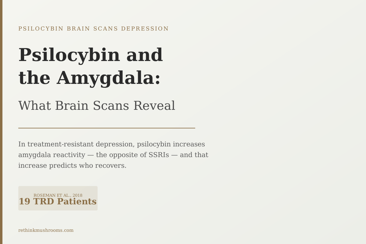
19 TRD patients
One day after psilocybin treatment, amygdala reactivity to fearful faces increased. That increase predicted better outcomes. SSRIs move the amygdala in the opposite direction.
Roseman et al., Neuropharmacology, 2018
< 24 hours
when amygdala changes appear after treatment
Opposite to SSRIs
direction of amygdala effect in TRD patients
Up to 1 month
emotional processing changes persist after one session
Roseman et al., Neuropharmacology, 2018
 View as image ↗
When psilocybin researchers scanned the brains of people with treatment-resistant depression, they found something that contradicted decades of thinking about how antidepressants should work. One day after treatment, psilocybin amygdala reactivity had increased, not decreased.[1] And the patients whose amygdala responded most strongly showed the greatest recovery from depression.
This is the central puzzle in the imaging literature on psilocybin and the amygdala. Understanding it requires knowing what the amygdala does, how depression disrupts it, and why psilocybin's opposite effect in healthy versus depressed people is not a contradiction but a clue. The broader clinical evidence for psilocybin in depression is covered in our overview of psilocybin for major depression. This article focuses on what brain scans, specifically, reveal about this one region.
Key Takeaways
- In healthy volunteers, psilocybin decreased amygdala reactivity to emotional faces, and that decrease correlated with improved mood (Kraehenmann et al., Biological Psychiatry, 2015, n=13)
- In treatment-resistant depression patients, amygdala reactivity to fearful faces increased one day after psilocybin treatment, and the increase predicted better clinical outcomes (Roseman et al., Neuropharmacology, 2018, n=19)
- Decreased connectivity between the brain's prefrontal emotional regulator and the amygdala correlated with reduced rumination at one week post-dosing (Mertens et al., Journal of Psychopharmacology, 2020, n=19)
- A second imaging method, resting cerebral blood flow, showed amygdala blood flow dropped after psilocybin treatment in TRD patients and correlated with symptom reduction (Carhart-Harris et al., Scientific Reports, 2017, n=19)
- Significant changes in brain function and emotional processing persisted for up to one month after a single high-dose psilocybin session (Barrett et al., Scientific Reports, 2020, n=12)
- SSRIs suppress amygdala reactivity in depressed patients; psilocybin increases it, pointing to a fundamentally different therapeutic mechanism
What the Amygdala Does — and How Depression Disrupts Emotional Processing
The amygdala is the brain's threat-detection hub and a core part of its emotional processing system. When you recognize a fearful face, process an emotionally charged memory, or sense danger, the amygdala fires. It initiates the body's stress response and encodes the emotional significance of events.
In a healthy brain, the amygdala works in dynamic balance with the prefrontal cortex. The prefrontal cortex acts as a regulator. It evaluates whether a perceived threat warrants a full stress response, calibrates emotional reactions, and can suppress amygdala activation when the situation doesn't call for it.
Depression disrupts this balance in ways that show up on brain scans. In many people with major depression, the amygdala becomes hyperreactive to negative stimuli. The prefrontal regulation system becomes less effective at applying the brake. And the brain's default mode network, which generates self-referential thought, becomes so dominant that emotional experience is filtered through a persistent loop of negative prediction.
But depression's effect on the amygdala is not uniformly one of excess. Many patients, particularly those who have been depressed for years or who have taken SSRIs for extended periods, experience emotional blunting. Their whole emotional range flattens, including positive emotions, curiosity, and the capacity for grief. For these patients, the amygdala has not gone hyperactive. It has gone quiet.
This distinction, between a hyperactive amygdala and a blunted one, is essential to understanding why psilocybin's imaging data looks the way it does. The drug does not push the amygdala in a single fixed direction. The direction it moves depends on where the amygdala is starting from.
Psilocybin and Amygdala Reactivity: Opposite Directions in Healthy vs. Depressed People
The psilocybin amygdala paradox, plainly stated: in healthy volunteers, psilocybin decreases amygdala reactivity to emotional faces, correlating with improved mood (Kraehenmann et al., 2015, n=13). In patients with treatment-resistant depression, psilocybin increases amygdala reactivity one day after treatment, predicting better clinical outcomes (Roseman et al., 2018, n=19). The same drug moves the same brain region in opposite directions, depending on the patient's starting state.
In 2015, Kraehenmann and colleagues gave psilocybin to 13 healthy adults in a randomized, placebo-controlled design and used fMRI to measure amygdala response to neutral faces.[3] Psilocybin decreased amygdala reactivity to those faces, and this decrease correlated with enhanced positive mood. The finding matches what people typically report during a psilocybin experience: reduced threat-based emotional responding, increased openness, a shift toward calm or positive affect.
In treatment-resistant depression patients, the picture after treatment runs in the opposite direction.
Roseman and colleagues, in a 2018 study of 19 TRD patients, scanned participants one day after a psilocybin session.[1] The scans showed increased amygdala response to fearful faces. And the magnitude of this increase predicted treatment outcomes. The patients whose amygdala responded most strongly to fearful faces one day after dosing showed the greatest antidepressant benefit.
The most coherent interpretation is not that the drug does two contradictory things. It is that the drug restores appropriate reactivity from whatever starting point the brain occupies. Healthy volunteers with normal emotional processing experience a temporary calming of the amygdala. Depressed patients whose emotional reactivity has been suppressed, by the illness itself or by prior medication, experience something more like a restoration. The amygdala comes back online.
The Roseman Finding: Why More Amygdala Reactivity Predicted Better Outcomes, Not Worse
The Roseman 2018 result is counterintuitive enough to deserve close examination.
The conventional model of antidepressant action says: reduce arousal, reduce reactivity, reduce distress. SSRIs, the most commonly prescribed antidepressants, decrease amygdala reactivity in depressed patients. This is generally treated as a core therapeutic mechanism, reducing the overactivation of the brain's threat-response circuit.
In that framework, finding that increased amygdala reactivity predicts better outcomes looks like a contradiction. If lowering reactivity is good, why is raising it also good?
The answer depends on which clinical problem you are solving.
For a patient whose amygdala is hyperactive and who experiences intense negative emotional responses to ordinary situations, lowering reactivity may help. This is the patient population SSRIs are often described as treating.
But treatment-resistant depression is a different clinical situation. These patients have, by definition, not responded to conventional approaches. Many have experienced significant emotional blunting, particularly those with long histories of antidepressant use. For them, the relevant problem may not be excess reactivity but a suppressed system that has lost its range.
What Roseman's brain scans may be capturing is not "more fear" but "more emotional range."[1] An amygdala that responds more robustly to fearful faces one day after psilocybin may be one that has recovered its ability to process emotional signals, including negative ones. This is not the same as being more distressed. It is being more emotionally present.
Patients often describe this in qualitative terms after psilocybin treatment. They report accessing feelings that depression had made inaccessible: grief, connection, warmth. The Roseman finding offers a neuroimaging correlate for that subjective shift. The amygdala is participating in emotional life again.
The Prefrontal Brake: How Psilocybin Loosens the Circuit That Drives Rumination
A 2020 study by Mertens and colleagues examined the same cohort of 19 TRD patients using a different analytic lens: functional connectivity between specific brain regions, rather than overall amygdala reactivity.[2]
The region they focused on was the ventromedial prefrontal cortex, which functions as the brain's main top-down regulator of the amygdala. Think of it as the prefrontal brake. When this circuit is active, the prefrontal regulator suppresses how strongly the amygdala responds to emotional signals. When it operates with excessive force, it can block emotional processing entirely, which may manifest as the emotional blunting common in long-term depression.
Mertens found that after psilocybin treatment, the connection between the prefrontal brake and the right amygdala became weaker during emotional face processing.[2] The tether loosened. And this loosening correlated with reduced rumination at one week post-dosing.
The finding connects a specific circuit change to a specific symptom: when the prefrontal regulator's grip on the amygdala weakens, the repetitive self-referential negative thinking that characterizes depression decreases. Rumination, in this framing, requires active prefrontal suppression of emotional input to sustain itself. When that suppression loosens, the feedback loop breaks.
The Mertens result complements the Roseman finding from opposite ends of the same circuit. Roseman showed the amygdala's response to external emotional stimuli increased. Mertens showed the prefrontal region that was over-suppressing the amygdala lost some of its grip. Both findings point to the same underlying change: emotional signals are getting through more freely.
For a visual account of how these connectivity shifts appear in fMRI data, the brain connectivity before and after psilocybin article maps the broader network-level evidence. The default mode network's contribution to rumination and how psilocybin disrupts it is explored in psilocybin and the default mode network.
A Second Imaging Method: Blood Flow Scans Tell the Same Story
Measuring brain region communication during emotional tasks is one window into amygdala function. Cerebral blood flow, measured at rest, is another.
In 2017, Carhart-Harris and colleagues measured cerebral blood flow in 19 TRD patients using arterial spin labeling, a technique that maps how much blood each brain region receives at rest as a proxy for baseline neural activity.[4] After psilocybin treatment, resting amygdala blood flow decreased, and this decrease correlated with symptom reduction.
At first, this appears to contradict Roseman 2018. Roseman found increased amygdala reactivity to emotional stimuli. Carhart-Harris found decreased amygdala blood flow at rest. How can both be true?
The two methods are measuring different states of the same region. Resting blood flow captures the amygdala's baseline activity, a kind of idle background level. Task-based reactivity measures the amygdala's range of response when called upon to process an emotional stimulus.
A brain region can have lower baseline activity while simultaneously showing greater reactivity to specific stimuli. This combination describes healthy emotional processing more accurately than either finding alone. The amygdala is not constantly aroused at a high level, which characterizes hypervigilance. But it responds robustly when genuine emotional signals arrive, which is what being emotionally engaged looks like.
Psilocybin appears to shift the amygdala toward this pattern from whichever pathological state a depressed patient is in. The blood flow findings add a third imaging modality, alongside Roseman's reactivity data and Mertens' connectivity data, all pointing at the same directional change.
The 2017 paper also found decreased connectivity between the parahippocampal region and the prefrontal cortex, and this decrease correlated with improved mood.[4] This additional finding is consistent with disruption of the rigid, memory-linked negative thinking patterns that define much of depressive experience.
One Session, Month-Long Changes: How Long the Brain Keeps Reorganizing
The Roseman and Mertens findings were measured one day after treatment. The Carhart-Harris blood flow data came from shortly after dosing. A reasonable question is whether these changes are extended pharmacological aftereffects that dissipate as psilocybin clears the bloodstream.
Barrett and colleagues addressed this directly. In a 2020 study of 12 participants, they measured brain function and emotional processing at multiple time points following a single high-dose psilocybin session.[5] Significant changes in both brain function and emotional processing persisted for up to one month.
This is critical for interpreting the amygdala data. The changes are not acute drug effects that resolve when psilocybin metabolizes. The brain continues reorganizing for weeks.
The mechanism behind lasting change from a single session remains an active research question. The leading theoretical framework, the REBUS model developed by Carhart-Harris and Friston, proposes that psilocybin temporarily loosens the brain's rigid top-down predictive patterns, creating a window in which the brain can reorganize around different, less pathological states. Under this model, the amygdala changes reflect the brain settling into a new way of weighting emotional signals rather than reverting to its pre-treatment default.
The Barrett study was conducted in healthy volunteers, so extending its duration estimate to depressed populations requires caution. But it establishes that the one-day and one-week measurements from the TRD studies are not capturing peak effects that are already fading. They may be capturing early stages of a reorganization that is still underway.
How This Compares to SSRIs: Why Psilocybin Takes the Opposite Approach
SSRIs consistently reduce amygdala reactivity in both healthy people and depressed patients. This is documented across multiple studies and is broadly understood as central to how the medications reduce negative emotional bias and distress.
The clinical downside is that SSRIs suppress the amygdala's response indiscriminately. Negative emotional responses become muted, but so do positive ones. Emotional blunting, described by patients as feeling behind glass or unable to access their own feelings, is one of the most common complaints about long-term SSRI use.
Psilocybin's effect in treatment-resistant depression patients runs in the opposite direction.[1] Rather than suppressing amygdala reactivity, it appears to restore it, particularly in patients whose emotional system has been dampened by the illness or by prior medication. The brain scan evidence points to a mechanism of emotional re-engagement rather than emotional dampening.
This distinction may have implications for patient selection. A person who has been on SSRIs for years and experiences significant emotional blunting might be starting from a different amygdala baseline than a person with acute, hyperactive negative affect and no prior medication history. The imaging literature suggests these are patients with different starting states, and psilocybin's direction of effect may differ accordingly.
The clinical experience side of this contrast is explored in greater depth in psilocybin and emotional processing: the opposite of blunting and in psilocybin and emotional reconnection vs. SSRI emotional blunting. For the full fMRI evidence base covering what psilocybin does to the depressed brain beyond the amygdala alone, see what psilocybin does to the depressed brain: fMRI studies.
What the Evidence Doesn't Answer Yet
The amygdala imaging findings are consistent across multiple methods and research groups, but several key questions remain open.
Sample sizes are small. Roseman 2018 included 19 patients. Mertens 2020 used the same cohort. Carhart-Harris 2017 also included 19 participants. Kraehenmann 2015 had 13 healthy volunteers. Barrett 2020 had 12. These samples are sufficient to detect effects and generate hypotheses, but not to draw firm conclusions about generalizability or the relative magnitude of effects across patient subgroups.
The Roseman 2018 finding, the central result in this article, has not been replicated in a larger controlled trial. The correlation between increased amygdala reactivity and treatment outcomes is striking, but it remains a correlation. Patients who showed greater amygdala reactivity post-dosing may have differed in other unmeasured ways, including baseline depression severity, prior treatment history, or the intensity of their acute session experience, any of which could independently explain better outcomes.
The Barrett 2020 duration data comes from healthy volunteers, not depressed patients. Clinical studies have not yet systematically tracked amygdala-specific changes across the one-month timeframe in TRD populations.
What the evidence does support is a directional hypothesis: psilocybin's effect on the amygdala is mechanistically distinct from SSRI effects, involves restoring rather than suppressing emotional reactivity, and produces imaging-detectable changes that correlate with both clinical outcomes and reductions in rumination. Larger trials with neuroimaging endpoints will extend or refine these findings.
The Bottom Line
Five brain imaging studies, using three different methods across healthy and depressed populations, point to a coherent picture: psilocybin restores amygdala reactivity rather than suppresses it, loosens the prefrontal circuit that drives rumination, and produces changes that persist for weeks after a single session. The direction of effect runs opposite to SSRIs, suggesting a mechanistically different therapeutic pathway. All five studies involve small samples, and replication with larger groups and active control conditions is needed before these findings translate into clinical applications.
Frequently Asked Questions
Sources & References
- 1. Source details for this reference are not yet available.
- 2. Source details for this reference are not yet available.
- 3. Source details for this reference are not yet available.
- 4. Source details for this reference are not yet available.
- 5. Source details for this reference are not yet available.