Chaga Bioactive Compounds
Allomelanin vs Eumelanin
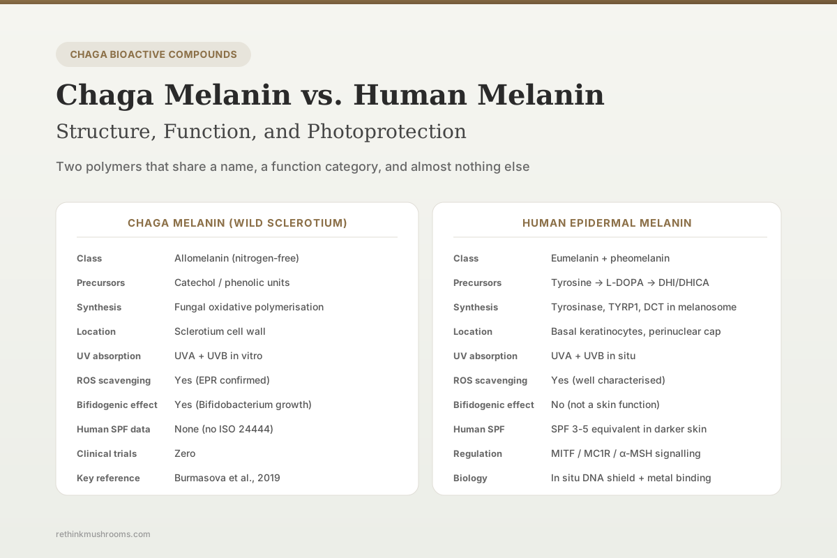
Wild chaga melanin is classified as an allomelanin (nitrogen-free catechol polymer), while human epidermal melanin is predominantly eumelanin (nitrogen-containing DHI/DHICA polymer from tyrosine). Both absorb UV and scavenge free radicals, but they arise from different biosynthetic pathways, differ in elemental composition, and interact with biological targets in different ways.
Burmasova et al., Biomolecules, 2019
Different precursors
chaga melanin derives from catechol and phenolic precursors; human eumelanin derives from tyrosine via L-DOPA
Both UV-absorbing
both polymer classes absorb across UVB and UVA and scavenge reactive oxygen species in vitro
0 human trials
no randomised clinical trial has tested topical or oral chaga melanin for any skin outcome
Burmasova et al., Biomolecules, 2019
 View as image ↗
Melanin is a family of biological pigments, not a single molecule. Across kingdoms, different organisms synthesise structurally related but not identical polymers from different phenolic precursors through related but not identical enzymatic and non-enzymatic pathways. Chaga (Inonotus obliquus) is among the most melanin-rich fungi, producing the dark pigment that defines its characteristic black sclerotium. Human skin produces melanin through melanocytes in the basal layer of the epidermis, with pigment transferred to keratinocytes in protective capsules over nuclei. Chaga melanin and human melanin share broad functional properties (UV absorption, radical scavenging, metal binding) but differ in biosynthetic origin, elemental composition, and the specific biological interactions documented in the laboratory.[1] For the UV-exposure-focused sibling, see chaga and UV damage: photoprotective properties.
Key Takeaways
- Burmasova et al. (2019) applied spectrophotometry, EPR spectroscopy, and dynamic light scattering to chaga melanins and classified wild sclerotium melanin as allomelanin (nitrogen-free, derived from catechol-type phenolic precursors) while cultivated mycelium melanin was classified closer to eumelanin
- Human epidermal melanin is predominantly eumelanin, a nitrogen-containing polymer biosynthesised from tyrosine via L-DOPA and dopaquinone intermediates under tyrosinase catalysis in melanosomes, with pheomelanin (sulphur-containing, cysteine-modified) as a minority component
- Both polymer classes absorb UVA and UVB strongly and scavenge reactive oxygen species in vitro, but the molecular mechanisms differ: human eumelanin operates through extended π-conjugation of DHI/DHICA units; chaga allomelanin operates through extended π-conjugation of oxidised catechol units
- Lin et al. (2023) documented that chaga polysaccharide fractions reduced UVB-induced photodamage in HaCaT keratinocytes via Nrf2/HO-1 activation; the role of co-extracted melanin in this effect was not isolated
- Yun et al. (2011) reported that I. obliquus extract suppressed UV-induced skin thickening and wrinkle formation in hairless mice and reduced MMP-1 and MMP-9 activity in fibroblasts; again the melanin contribution was not isolated from other bioactive fractions
- Chaga melanin has documented bifidogenic effects (stimulation of Bifidobacterium growth), which is absent from human melanin biology; this is one area where chaga melanin and human melanin do not share functional space
What Melanin Actually Is: A Polymer Family, Not a Molecule
The term "melanin" covers several structurally related classes of polymeric pigments. In humans, eumelanin and pheomelanin are the two canonical types. Eumelanin (brown-black) polymerises from 5,6-dihydroxyindole (DHI) and 5,6-dihydroxyindole-2-carboxylic acid (DHICA) units derived from tyrosine oxidation. Pheomelanin (red-yellow) incorporates sulphur through cysteine conjugation with dopaquinone and produces benzothiazine and benzothiazole monomer units. Both are synthesised in melanocyte-specific organelles (melanosomes) under the catalytic action of tyrosinase (TYR), tyrosinase-related protein 1 (TYRP1), and dopachrome tautomerase (DCT). Human hair and skin colour reflect the ratio and total content of these two pigment types.
Fungal melanins are a separate subfamily. DHN-melanin (dihydroxynaphthalene melanin) is common in many fungi, synthesised from acetyl-CoA via polyketide intermediates. Allomelanin is a less structurally defined class including nitrogen-free melanins built from catechol and related phenolic precursors. Tyrosine-derived eumelanin-like fungal melanin also exists, particularly in yeasts and some filamentous species. All share the polymeric, insoluble, redox-active character that defines melanin functionally, but they differ in monomer identity, polymerisation chemistry, and elemental composition.
This background matters because "chaga melanin" and "human melanin" share a functional name but not a biosynthetic or structural identity. Any claim that chaga melanin directly substitutes for human melanin, or supplies melanin precursors, needs to be evaluated against the biochemistry rather than assumed from the shared word.
Chaga Melanin Characterisation: Burmasova 2019
Burmasova et al. (2019) applied spectrophotometry, electron paramagnetic resonance (EPR) spectroscopy, and dynamic light scattering to compare melanins isolated from wild chaga sclerotium and cultivated mycelium.[1] The analyses showed measurable structural differences between the two sources: particle size, surface charge, antioxidant activity, and redox potential all differed. Elemental and functional group analyses placed wild sclerotium melanin closer to the allomelanin class (nitrogen-poor, built predominantly from catechol-type phenolic units), while cultivated mycelium melanin showed features closer to eumelanin-like polymers with higher nitrogen content.
The bifidogenic effect (stimulation of Bifidobacterium growth in the intestine) was documented for chaga melanin fractions. This is a functional property absent from human skin eumelanin; it reflects chaga melanin's chemistry interacting with gut commensal microorganisms rather than any human melanin-like role. The antioxidant properties observed (EPR evidence of free radical scavenging, reduced ROS in in vitro assays) are the clearest point of functional overlap with human melanin biology, where eumelanin scavenges UV-generated radicals in keratinocytes.
Glamočlija et al. (2015) performed broader chemical characterisation of chaga samples from multiple geographic origins and documented melanin content alongside phenolic acids, flavonoids, and triterpenoids.[2] The melanin fraction contributes substantially to the total antioxidant capacity observed in crude chaga extracts.
Human Epidermal Melanin: What It Does and How
Human epidermal melanin performs a defined set of protective functions. In the basal epidermis, melanosomes loaded with eumelanin are transferred from melanocytes to surrounding keratinocytes. The melanosomes redistribute to a perinuclear location, forming a supranuclear cap that absorbs incident UV before it reaches nuclear DNA. This physical UV shielding is the primary human melanin function.
Beyond shielding, eumelanin scavenges ROS generated by UV exposure (superoxide, hydrogen peroxide, hydroxyl radicals), binds metal ions (reducing Fenton-reaction ROS generation), and modulates local inflammation through interactions with immune mediators. Darker skin types, which have higher eumelanin content per melanocyte, show reduced sunburn risk and reduced skin cancer incidence compared with lighter skin types at equivalent UV exposure, though the risk reduction is not absolute and does not obviate the need for additional photoprotection in high-exposure contexts.
Human melanin biosynthesis is tightly regulated. Tyrosinase activity is controlled by MITF transcription factor, UV-induced p53 signalling to α-MSH release from keratinocytes, and receptor binding at the melanocortin-1 receptor (MC1R). Genetic variation at MC1R and related genes produces the phenotypic range of human skin and hair colour.
Functional Comparison: Where They Overlap and Where They Do Not
UV absorption: Both chaga allomelanin and human eumelanin absorb across UVB (280–315 nm) and UVA (315–400 nm) through extended π-conjugation in their aromatic polymer backbones. In vitro SPF-style absorbance testing of chaga extracts has reported SPF >30 at 10 mg/mL in aqueous or ethanolic extract, but this is not equivalent to validated in vivo SPF by ISO 24444 standards. Human eumelanin's SPF contribution has been estimated at 3–5 equivalents for darker skin types across typical exposure patterns.
Free radical scavenging: Both operate through single-electron transfer mechanisms characteristic of conjugated phenolic polymers. EPR studies of chaga melanin (Burmasova 2019) document stable free radicals consistent with redox-active polymer behaviour similar to human melanin EPR signatures.
Metal binding: Both bind transition metals (iron, copper) through chelation by catechol or indole hydroxyl groups. This binding reduces metal-catalysed ROS generation and contributes to antioxidant activity.
DNA protection: Indirect evidence for chaga melanin-containing extracts. Park et al. (2004) showed that chaga mushroom extract inhibited oxidative DNA damage in human lymphocytes by comet assay, consistent with melanin and polyphenol fractions contributing to DNA protection.[3] Eid et al. (2021) documented DNA repair gene upregulation in UVB-exposed zebrafish embryos treated with chaga polysaccharide; the melanin contribution to this effect was not isolated.[4]
Non-overlapping functions: Chaga melanin's bifidogenic effect is unique to the fungal context. Human eumelanin's role in visual system pigmentation (retinal pigment epithelium, iris) and immune modulation within melanocytes has no chaga parallel.
Preclinical UV Skin Studies: What the Chaga Evidence Shows
Three direct UV studies with chaga are available, all using extracts or polysaccharide fractions rather than isolated melanin:
Lin et al. (2023) used a UVB-induced HaCaT keratinocyte photoaging model with chaga polysaccharides (IOP).[5] IOP reduced cell senescence markers (p16, p21, p53), activated the Nrf2/HO-1 antioxidant pathway (HO-1, SOD, CAT upregulation), and restored autophagy (p-AMPK, LC3B, Beclin-1). The study attributes effects to polysaccharide fractions, though co-extracted melanin-rich phenolic material may contribute.
Yun et al. (2011) documented in vivo photoprotection.[6] I. obliquus suppressed UV-induced skin thickening and wrinkle formation in hairless mice, and inhibited MMP-1 and MMP-9 in hydrogen peroxide-treated human fibroblasts. The extract character is not a purified melanin fraction.
The isolated chaga-melanin UV study remains outstanding. Definitive attribution of chaga's photoprotective effects to its melanin, polyphenol, polysaccharide, or triterpenoid fractions has not been established. Polyphenol antioxidants characterised by Lee et al. (2007), specifically inonoblins A–C and phelligridins D, E, G, likely contribute alongside melanin rather than independently of it.[7]
Bioavailability: The Topical vs Oral Question
Human melanin is produced in situ in skin; its photoprotective action is local to the melanocyte-keratinocyte system where it was synthesised. Chaga melanin from an external source faces delivery challenges.
For topical application, the relevant questions are skin penetration depth, stability in cosmetic matrices, photostability under UV exposure, and interaction with the stratum corneum. Insoluble polymeric melanin particles do not penetrate the stratum corneum to any substantial degree; topical chaga melanin would act primarily at the skin surface as a UV-absorbing film rather than integrating into epidermal architecture. Whether this provides meaningful photoprotection depends on concentration, formulation, and uniformity of application, none of which has been validated to regulatory standards for a chaga-containing SPF product.
For oral intake, chaga melanin is not significantly absorbed systemically as an intact polymer. Its documented bifidogenic effect reflects interaction with gut microbiota rather than systemic distribution. The idea that oral chaga provides melanin to skin via the bloodstream is not supported by absorption pharmacology and is inconsistent with how dietary melanin behaves across published studies. Oral chaga may deliver phenolic monomer metabolites to skin through circulation, but this is different from delivering intact melanin.
The Commercial Claim: Evaluating "Chaga Is Like Sunscreen"
A substantial commercial market positions chaga as a natural sunscreen alternative or a source of melanin for skin support. The evidence base for this positioning is limited. Chaga extracts absorb UV in vitro and reduce UV-induced damage in cell and animal models, but:
- No validated in vivo SPF data exist for any chaga product (ISO 24444 or equivalent)
- No randomised clinical trial has tested chaga for sunburn prevention, photoaging reduction, or skin cancer incidence
- Chaga melanin does not restructure human skin melanin content or function
- Substituting chaga for regulated sunscreen increases UV exposure risk
For the specific topical skincare angle, see chaga in skincare products: topical application research.
What the Evidence Supports and What It Does Not
| Question | Evidence | Confidence |
|---|---|---|
| Is chaga melanin structurally identical to human melanin? | No: wild chaga melanin is allomelanin; human melanin is predominantly eumelanin | High |
| Does chaga melanin absorb UV light? | Yes in vitro | Moderate |
| Does chaga melanin scavenge free radicals? | Yes by EPR and cell-based assays (Burmasova 2019) | Moderate |
| Does chaga melanin protect skin from UV in humans? | Not tested; no clinical trial | None |
| Can oral chaga supplement human skin melanin? | No pharmacokinetic basis; melanin is not absorbed intact | Very low |
| Is chaga melanin a validated sunscreen ingredient? | No: no ISO 24444 in vivo SPF data; not approved as a UV filter by FDA, EMA, or similar regulators | None |
| Does chaga melanin reduce UV-induced DNA damage? | Indirect preclinical evidence from mixed extracts (Park 2004; Eid 2021) | Low to moderate |
The Bottom Line
Chaga melanin and human melanin share the general functional category of UV-absorbing, radical-scavenging phenolic polymers but differ in biosynthetic origin, elemental composition, and specific biological interactions. Wild chaga melanin is an allomelanin built predominantly from catechol-type phenolic precursors (Burmasova et al., 2019), while human epidermal melanin is predominantly eumelanin from tyrosine via L-DOPA. Both absorb UVA and UVB and scavenge ROS, but human melanin operates in situ through perinuclear melanosome shielding while chaga melanin, when consumed, interacts with gut microbiota (bifidogenic effect) or applied topically acts as a surface UV-absorbing film. Preclinical UV studies of chaga (Lin 2023; Yun 2011; Eid 2021) use mixed extracts rather than isolated melanin, so direct attribution of photoprotective effects to chaga melanin specifically is incomplete. No clinical trial has tested chaga melanin for any human skin outcome, and no validated in vivo SPF data exist.
Frequently Asked Questions
Sources & References
- 1RTHM-00545·Burmasova, Marina A et al. (2019). “Melanins of Inonotus Obliquus: Bifidogenic and Antioxidant Properties..” Biomolecules.Study breakdown →PubMed →↩
- 2RTHM-00373·Glamočlija, Jasmina et al. (2015). “Chemical characterization and biological activity of Chaga (Inonotus obliquus), a medicinal "mushroom"..” Journal of ethnopharmacology.Study breakdown →PubMed →↩
- 3RTHM-00076·Park, Yoo Kyoung et al. (2004). “Chaga mushroom extract inhibits oxidative DNA damage in human lymphocytes as assessed by comet assay..” BioFactors (Oxford.Study breakdown →PubMed →↩
- 4RTHM-00683·Eid, Jehane Ibrahim et al. (2021). “Chaga mushroom (Inonotus obliquus) polysaccharides exhibit genoprotective effects in UVB-exposed embryonic zebrafish (Danio rerio) through coordinated expression of DNA repair genes..” Heliyon.Study breakdown →PubMed →↩
- 5RTHM-00840·Lin, Jun et al. (2023). “Chaga Medicinal Mushroom, Inonotus obliquus (Agaricomycetes), Polysaccharides Alleviate Photoaging by Regulating Nrf2 Pathway and Autophagy..” International journal of medicinal mushrooms.Study breakdown →PubMed →↩
- 6RTHM-00241·Yun, Jong Seok et al. (2011). “Inonotus obliquus protects against oxidative stress-induced apoptosis and premature senescence..” Molecules and cells.Study breakdown →PubMed →↩
- 7RTHM-00118·Lee, In-Kyoung et al. (2007). “New antioxidant polyphenols from the medicinal mushroom Inonotus obliquus..” Bioorganic & medicinal chemistry letters.Study breakdown →PubMed →↩