Cordyceps and Cancer
3'-deoxyadenosine the active molecule
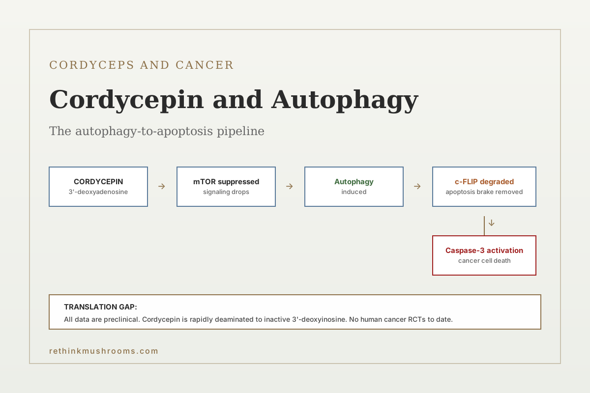
Cordycepin, the 3'-deoxyadenosine nucleoside from Cordyceps militaris, induces both autophagy and apoptosis in cancer cells, with the two processes interacting in a way that often makes the autophagy a driver of cell death rather than a survival response.
Chen et al., Molecules, 2021
mTOR
principal pathway cordycepin suppresses
c-FLIP
anti-apoptotic protein degraded via autophagy
Preclinical
no human cancer RCTs to date
Chen et al., Molecules, 2021
 View as image ↗
Autophagy is the cellular process of degrading and recycling damaged proteins and organelles. In cancer biology, autophagy has a dual character: it can help cancer cells survive metabolic stress (a pro-survival function), or it can drive cancer cells into a non-apoptotic form of cell death when sustained past a threshold (autophagy-mediated cell death). Cordycepin, the 3'-deoxyadenosine nucleoside produced by Cordyceps militaris and related species, is one of the better-studied natural products that pushes cancer cells toward the second outcome. The preclinical literature is consistent across cell types: cordycepin induces autophagy through suppression of the mTOR pathway, and in many cancer cell lines the autophagy then amplifies rather than blocks apoptotic cell death.[1]
This article walks through the specific molecular events documented in the literature, the cancer cell types where the effect has been characterized, the bioavailability and stability problems that limit translation, and the honest answer to whether this preclinical story has moved into human cancer treatment. For the broader Cordyceps-cancer context, see Cordyceps and cancer: what the research shows. For the chemotherapy-support angle, see Cordyceps as a cancer adjunct: evidence for chemotherapy support. For leukemia-specific data, see cordycepin and leukemia research: preclinical findings.
Key Takeaways
- Cordycepin (3'-deoxyadenosine) induces autophagy in cancer cells primarily through suppression of the mTOR signaling pathway, a master regulator of cellular growth and metabolism (Chen et al., 2021)
- In human non-small cell lung cancer cells, cordycepin-induced autophagy degrades c-FLIPL (a key anti-apoptotic protein), which then removes the brake on caspase-mediated apoptosis
- In LNCaP prostate carcinoma cells, cordycepin induces autophagy-associated apoptosis and the two processes interact such that blocking autophagy (Atg5 siRNA or LY294002) attenuates the apoptotic response (Lee et al., 2014)
- In human glioblastoma and U251 brain cancer cells, cordycepin-induced apoptosis proceeds through mitochondrial membrane potential collapse, caspase-9 and -3 activation, and modulation of autophagy (Chaicharoenaudomrung et al., 2018)
- In breast cancer cell lines, cordycepin-induced apoptosis and autophagy are independent of estrogen receptor status, indicating that the mechanism does not depend on hormone-responsive signaling (Choi et al., 2011)
- Cordycepin is rapidly degraded in vivo by adenosine deaminase, giving it a very short half-life and limiting direct oral bioavailability (Chen et al., 2023)
- All human-relevant evidence is preclinical (cell culture and animal); no randomized controlled human cancer trials of cordycepin or cordycepin-standardized Cordyceps preparations have been published
The Autophagy-Apoptosis Relationship in Cancer
Autophagy and apoptosis are distinct forms of programmed cell fate. Apoptosis is caspase-dependent, characterized by cytochrome c release, caspase-3 activation, and ordered cellular dismantling. Autophagy is caspase-independent, characterized by formation of double-membrane autophagosomes that engulf cellular contents and deliver them to lysosomes for degradation. In healthy cells, basal autophagy is a housekeeping function. In cancer cells under stress, autophagy can either:
- Support survival by recycling nutrients when supply is limited or clearing damaged components; this is the pro-tumor role exploited by some cancers.
- Drive death when autophagy is induced beyond a threshold or when it degrades specific pro-survival proteins; this is the cell-death-promoting role that natural products like cordycepin can activate.
Whether autophagy helps or kills a cancer cell depends on the cellular context, the level and duration of autophagy induction, and what proteins are degraded. In the cordycepin case, autophagy targets anti-apoptotic brakes (c-FLIP) and tumor-supporting proteins, which tilts the outcome toward cell death rather than survival.
Chen et al. (2021) reviewed the anti-cancer role of autophagy in cordycepin's mechanism of action. The review identifies suppression of the mTOR signaling pathway as the initial trigger for autophagy induction, followed by downstream effects on specific protein substrates that depend on cancer cell type.[1]
The mTOR Connection
The mammalian target of rapamycin (mTOR) complex is a master regulator of cellular metabolism and growth. When nutrients and growth signals are abundant, mTOR signaling is high, suppressing autophagy and promoting protein synthesis, cell growth, and proliferation. When nutrients are scarce or stress signals accumulate, mTOR signaling drops, releasing autophagy and slowing growth. Many cancer cells have constitutively elevated mTOR signaling, which drives their proliferation and also makes them dependent on that signaling.
Cordycepin suppresses mTOR signaling in cancer cells through mechanisms that include its structural similarity to adenosine (it is 3'-deoxyadenosine) and its ability to interfere with purine metabolism. The practical consequence is that cancer cells treated with cordycepin experience a sudden drop in mTOR activity, which releases autophagy and disrupts the growth program the cells were running. For cells that had become mTOR-dependent for survival, this withdrawal is lethal.
The mTOR suppression by cordycepin is not as specific as that of pharmaceutical mTOR inhibitors like rapamycin (sirolimus) or everolimus, and cordycepin has multiple additional effects (adenosine receptor modulation, polyadenylation inhibition, DNA damage response activation). But the mTOR pathway is the one most consistently identified across cell types as the entry point for autophagy induction.
Cancer Cell Type-Specific Evidence
Non-small cell lung cancer (NSCLC)
In human NSCLC cell lines, cordycepin stimulates autophagy through suppression of mTOR signaling. The autophagic process then targets c-FLIPL (cellular FLICE-inhibitory protein long form), an anti-apoptotic protein that normally blocks death receptor-mediated caspase-8 activation. Autophagy-mediated degradation of c-FLIPL removes this brake, allowing apoptosis to proceed. When autophagy is blocked (Atg5 siRNA or PI3K inhibitor LY294002), the apoptotic response to cordycepin is attenuated. This establishes autophagy as upstream of apoptosis in this cell line, not a parallel process.
Brain cancer (glioblastoma)
Chaicharoenaudomrung et al. (2018) treated U251 human glioblastoma cells with cordycepin and documented a dose-dependent increase in pro-apoptotic Bax, a decrease in anti-apoptotic Bcl-2, mitochondrial membrane potential collapse, cytochrome c release, and caspase-9 and caspase-3 activation. Autophagy markers (LC3-II increase, p62 degradation) were also elevated. The apoptotic pathway in brain cancer cells is mitochondrial-mediated (intrinsic) rather than death receptor-mediated, but the autophagy-apoptosis coupling is consistent with the NSCLC findings.[2]
Prostate cancer
Lee et al. (2014) studied cordycepin in LNCaP human prostate carcinoma cells, documenting autophagy induction (LC3 conversion and autophagosome formation) alongside apoptosis. Pharmacological or genetic blockade of autophagy reduced the apoptotic response, confirming autophagy's upstream role. The LNCaP model is androgen-sensitive, but the cordycepin effect did not require androgen manipulation, suggesting the mechanism is androgen-independent.[3]
Breast cancer
Choi et al. (2011) tested cordycepin in both estrogen receptor-positive (MCF-7) and estrogen receptor-negative (MDA-MB-231) human breast cancer cell lines. Cordycepin induced apoptosis and autophagy in both lines with comparable potency, demonstrating that the mechanism does not depend on estrogen receptor status. This is clinically relevant because estrogen receptor status is one of the principal axes of breast cancer classification and dictates response to many existing therapies.[4]
The Bioavailability Problem
If cordycepin were easily translatable to human cancer therapy, it would already be a drug. The binding constraint is not the mechanism but the pharmacokinetics.
Chen et al. (2023) reviewed strategies to improve cordycepin bioavailability and efficacy. The fundamental problem is that cordycepin is rapidly deaminated by adenosine deaminase (ADA) to 3'-deoxyinosine, which has minimal biological activity. This gives orally administered cordycepin a very short half-life and low systemic exposure, severely limiting the dose that reaches tumor tissue.[5]
Strategies to address this include:
- Co-administration with adenosine deaminase inhibitors like pentostatin, which block cordycepin degradation and increase effective half-life
- Chemical modification to create cordycepin analogs resistant to ADA (e.g., protected phosphate forms)
- Encapsulation in nanoparticles or liposomes to shield cordycepin from ADA during circulation and deliver it preferentially to tumor tissue
- Intravenous rather than oral administration to bypass first-pass metabolism, though ADA is active systemically
An older cordycepin-pentostatin combination entered clinical trial for leukemia (NSC-63878 / deoxycoformycin + cordycepin) and demonstrated that the pharmacology is tractable with appropriate delivery strategy, though no approved cancer drug has emerged.
For the same molecule's non-cancer applications, see cordycepin and leukemia research: preclinical findings and cordycepin and tumor growth inhibition: mechanisms of action.
What This Does Not Mean for Cordyceps Supplements
The preclinical cordycepin-cancer literature is substantial and mechanistically coherent. It is also, as of 2026, not translated to human cancer therapy. The implications for Cordyceps supplement users:
-
Dose gap. Cordyceps supplement products contain cordycepin at roughly 0.1-3 mg per gram of material, with wide variation by product. The doses used in cell-line and rodent studies are orders of magnitude higher than what a supplement delivers to serum in a human.
-
Bioavailability gap. Oral cordycepin bioavailability without ADA inhibition is very low. A supplement taken with a meal delivers even less cordycepin to systemic circulation than a fasted oral dose of pure cordycepin.
-
Evidence level. No randomized controlled trial of Cordyceps or cordycepin in any human cancer indication has been published. Claims that Cordyceps supplements treat cancer are not supported by the clinical literature, even where the preclinical mechanism is well-characterized.
-
Chemotherapy adjunct framing. Some in vitro work suggests cordycepin may sensitize cancer cells to chemotherapy agents. This is a potentially meaningful research direction but has not been established in human trials and should not be framed as a current treatment option.
A fair summary for consumers is that cordycepin is a genuine object of cancer pharmacology research with a clear mechanism and a difficult pharmacokinetic profile, that Cordyceps supplements are not a delivery vehicle for meaningful cordycepin doses in humans, and that anyone with cancer should work with their oncologist on evidence-based therapy rather than rely on cordycepin for treatment effects.
The Bottom Line
Cordycepin induces autophagy in cancer cells primarily through suppression of the mTOR signaling pathway, and in several cell types (NSCLC, glioblastoma, prostate LNCaP, breast MCF-7/MDA-MB-231), the autophagy is upstream of and required for cordycepin-induced apoptosis. The mechanism is mechanistically coherent and cell-type-robust but translation to human cancer therapy is blocked by rapid adenosine deaminase-mediated degradation and consequent poor oral bioavailability. All human-relevant cordycepin-cancer data are preclinical; no randomized controlled trials of cordycepin or cordycepin-standardized Cordyceps preparations in any cancer indication have been published.
Frequently Asked Questions
Sources & References
- 1RTHM-00680·Chen, Yu-Ying et al. (2021). “The Role of Autophagy in Anti-Cancer and Health Promoting Effects of Cordycepin..” Molecules (Basel.Study breakdown →PubMed →↩
- 2RTHM-00501·Chaicharoenaudomrung, Nipha et al. (2018). “Cordycepin induces apoptotic cell death of human brain cancer through the modulation of autophagy..” Toxicology in vitro : an international journal published in association with BIBRA.Study breakdown →PubMed →↩
- 3RTHM-00334·Lee, Hye Hyeon et al. (2014). “Involvement of autophagy in cordycepin-induced apoptosis in human prostate carcinoma LNCaP cells..” Environmental toxicology and pharmacology.Study breakdown →PubMed →↩
- 4RTHM-00212·Choi, Sunga et al. (2011). “Cordycepin-induced apoptosis and autophagy in breast cancer cells are independent of the estrogen receptor..” Toxicology and applied pharmacology.Study breakdown →PubMed →↩
- 5RTHM-00817·Chen, Min et al. (2023). “Cordycepin: A review of strategies to improve the bioavailability and efficacy..” Phytotherapy research : PTR.Study breakdown →PubMed →↩