Cordyceps and Anti-Aging
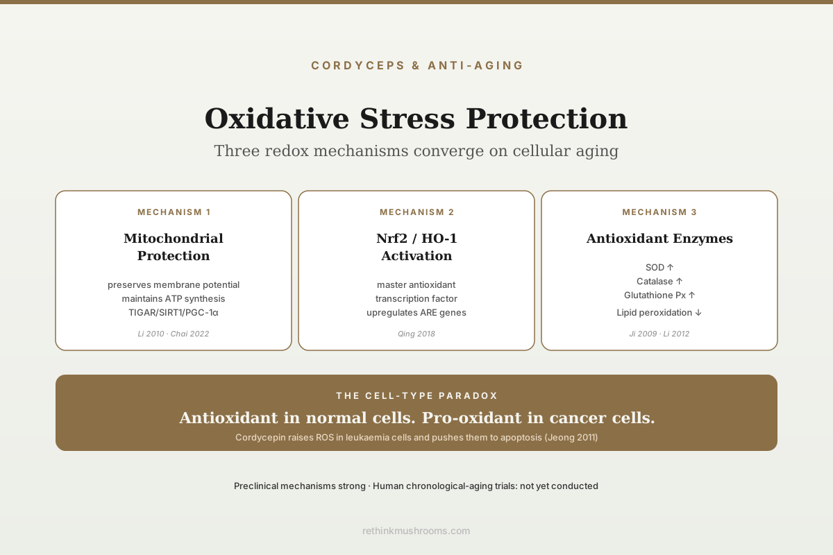
3 mechanisms converge on redox
Cordyceps polysaccharides and whole extracts protect mitochondria and raise endogenous antioxidant enzymes (SOD, catalase, GSH-Px) in rodents, while the isolated compound cordycepin activates Nrf2/HO-1. In cancer cells cordycepin does the opposite and acts as a pro-oxidant — a cell-type-specific distinction that matters.
Ji et al., Phytother. Res., 2009; Li et al., Am. J. Chin. Med., 2010; Qing et al., Int. Immunopharmacol., 2018; Bai et al., Biomed. Pharmacother., 2020
Mitochondrial Protection
blocks swelling (Li 2010); restores membrane potential and ATP (Bai 2020)
Nrf2 Activation
cordycepin drives Nrf2 nuclear translocation and HO-1 (Qing 2018)
Cell-Type-Specific
antioxidant in normal cells, pro-oxidant in cancer cells
Ji et al., Phytother. Res., 2009; Li et al., Am. J. Chin. Med., 2010; Qing et al., Int. Immunopharmacol., 2018; Bai et al., Biomed. Pharmacother., 2020
 View as image ↗
The oxidative stress theory of aging argues that cumulative reactive oxygen species damage to mitochondria, proteins, lipids and DNA drives cellular aging and age-related disease. The theory is not a complete account of aging, but it captures a real biological process, and compounds that protect against oxidative damage have a plausible role in cellular-aging research. Cordyceps polysaccharides and the small molecule cordycepin both have documented antioxidant and cytoprotective activity in preclinical models, and the mechanism is multi-pathway. Ji and colleagues (2009) demonstrated an antiaging effect of Cordyceps sinensis extract in rodent aging models, establishing the integrated biological endpoint.[1] Li and colleagues (2010) characterised the mitochondrial protection and anti-aging activity of polysaccharides from cultivated Cordyceps militaris fruiting bodies, providing the mechanistic backbone.[2]
For related context see how cordyceps may slow biological aging, cordyceps and anti-aging: research on longevity, cordyceps and nad+ metabolism: the anti-aging energy connection and cordyceps and skin aging: antioxidant research.
Key Takeaways
- Ji et al. (2009) reported anti-aging effects of Cordyceps sinensis extract in D-galactose-aged mice, improving superoxide dismutase, glutathione peroxidase and catalase activity while lowering lipid peroxidation and monoamine oxidase
- Li et al. (2010) showed Cordyceps militaris fruiting-body polysaccharides inhibit chemically induced mitochondrial swelling concentration-dependently and raise liver antioxidant enzyme activity in D-galactose-aged mice
- Li et al. (2012) demonstrated Cordyceps militaris extract attenuation of D-galactose-induced memory impairment in mice, a standard accelerated-aging model
- Liao et al. (2025) independently replicated the D-galactose result with a third species, Cordyceps cicadae, implicating the AMPK/SIRT1 pathway
- Chueaphromsri et al. (2023) found cordycepin raised SIRT1 in human mesenchymal stem cells and delayed cellular senescence, lowering senescence-associated galactosidase activity and increasing telomere activity — the most directly on-topic evidence available
- Qing et al. (2018) showed cordycepin alleviates lipopolysaccharide-induced acute lung injury in rats via the Nrf2/HO-1 pathway, identifying a specific antioxidant transcription factor mechanism
- Jeong et al. (2011) showed cordycepin can instead generate reactive oxygen species in cancer cells, producing apoptosis; the directionality of the oxidative effect is cell-type-specific
- No human trial has tested Cordyceps against an aging biomarker panel; the clinical case remains inferential
The D-Galactose Accelerated-Aging Model
Much of the Cordyceps anti-aging preclinical evidence uses the D-galactose rodent model, a standard experimental system where chronic D-galactose administration induces oxidative stress, mitochondrial dysfunction and cognitive decline that parallels age-related changes. The model is short enough to run in a laboratory timeframe — Li and colleagues (2010), for instance, injected 100 mg/kg/day subcutaneously for seven weeks — and produces consistent, measurable aging-related endpoints.[2]
Li and colleagues (2012) tested Cordyceps militaris extract against D-galactose-induced memory impairment in mice and reported that the learning and memory deficit was rescued by treatment.[3] The biochemical endpoints were measured in two compartments: in the cerebral cortices, treatment significantly lowered malondialdehyde and monoamine oxidase activity while raising total superoxide dismutase and total antioxidant capacity; in serum, glutathione and glutathione peroxidase activity were significantly upregulated. The integrative pattern was that Cordyceps treatment preserved antioxidant capacity and reduced lipid peroxidation, with behavioural memory performance tracking the biochemical preservation.
The result has been reproduced by independent groups working with different Cordyceps species and different preparations. Ji 2009 used a Cordyceps sinensis whole extract,[1] Li 2010 an isolated Cordyceps militaris polysaccharide fraction,[2] and Li 2012 a Cordyceps militaris whole extract.[3] Most recently, Liao and colleagues (2025) extended the finding to a third species, Cordyceps cicadae, in D-galactose-aged rats: the extract improved learning and memory deficits, raised superoxide dismutase and glutathione peroxidase, and lowered malondialdehyde, with the AMPK/SIRT1 pathway implicated as the regulatory axis.[4]
That convergence matters more than any single study. Four groups, three species, and both whole-extract and purified-fraction preparations produce the same directional result on the same endpoints. What the convergence does not do is establish relevance to chronological human aging — D-galactose produces a biochemical phenotype that resembles aging, but it is a chemically induced model, not aging itself.
Mitochondrial Protection
The cleanest mechanistic explanation for Cordyceps anti-aging effects is mitochondrial protection. Mitochondria are both the primary source of cellular reactive oxygen species under normal respiration and the most vulnerable target of oxidative damage. Age-related mitochondrial dysfunction (membrane potential loss, electron transport chain inefficiency, accumulation of mitochondrial DNA mutations) is one of the clearest hallmarks of cellular aging.
Li and colleagues (2010) characterised Cordyceps militaris fruiting body polysaccharides for mitochondrial protection and anti-aging activity.[2] The polysaccharide fraction inhibited mitochondrial injury and swelling induced by Fe²⁺-L-cysteine in a concentration-dependent manner and showed significant superoxide anion scavenging activity in vitro. In the D-galactose-aged animals, catalase, superoxide dismutase, glutathione peroxidase and anti-hydroxyl radical activity in liver tissue were all significantly increased by treatment. The authors concluded that the polysaccharides protect mitochondria by scavenging reactive oxygen species, inhibiting mitochondrial swelling, and raising antioxidase activity.
Two details are worth stating precisely, because they are commonly overstated. Li 2010 measured mitochondrial swelling and antioxidant enzyme activity; it did not measure mitochondrial membrane potential or ATP synthesis. The evidence for those specific endpoints comes from a different study. Bai and colleagues (2020) tested Cordyceps sinensis extract against cerebral ischemic injury and found that in brain microvascular endothelial cells exposed to oxygen and glucose deprivation, the extract recovered the reduction in mitochondrial membrane potential and prevented apoptosis; in a mouse middle cerebral artery occlusion model, it reduced oxygen free radicals while improving ATP, cytochrome c oxidase and mitochondrial complexes I–IV.[5] Taken together the two studies cover structural protection, respiratory-chain function and energy output — but they are separate experiments in separate tissues, and treating them as one result overstates what either showed.
The mitochondrial protection has been corroborated in cordycepin-specific work. Chai and colleagues (2022), though framed around anti-fatigue, found that cordycepin at 20 and 40 mg/kg for 28 days significantly enhanced protein levels of TIGAR, SIRT1, PGC-1α, NRF1 and TFAM in mouse gastrocnemius muscle, alongside raised superoxide dismutase, glutathione and ATP and lowered malondialdehyde.[6] NRF1 and TFAM are core mitochondrial biogenesis factors, so the pathway that supports exercise tolerance is mechanistically relevant to cellular aging: aging depletes PGC-1α-coupled mitochondrial biogenesis, and compounds that preserve it plausibly slow the decline. That is a mechanistic argument, not a demonstrated anti-aging result.
Nrf2/HO-1 Activation
A central molecular mechanism for cellular antioxidant defence is the Nrf2/Keap1/ARE pathway. Nrf2 is a transcription factor that, when activated, drives expression of cytoprotective genes including heme oxygenase-1 (HO-1), NAD(P)H quinone dehydrogenase 1, glutathione peroxidase, and several phase-II detoxification enzymes. Compounds that activate Nrf2 upregulate the cell's own antioxidant defences rather than acting as direct antioxidants, which is generally a more biologically stable mechanism.
Qing and colleagues (2018) demonstrated that cordycepin alleviates lipopolysaccharide-induced acute lung injury via the Nrf2/HO-1 pathway.[7] The model was Wistar rats given intravenous LPS at 30 mg/kg, with cordycepin administered intravenously one hour later at 1, 10 or 30 mg/kg. Cordycepin enhanced both the expression and the enzymatic activity of HO-1, and activated Nrf2 by inducing its translocation from cytoplasm to nucleus. The downstream effects were HO-1-mediated: HO-1 drove the reduction in lung superoxide levels, shaped plasma nitrite and nitrate concentrations and lung iNOS expression, and mediated the regulation of TNF-α, IL-6 and IL-10 secretion.
That last point is the substantive one for this article. The same HO-1 induction accounted for both the antioxidant effect and the anti-inflammatory effect in the same experiment, which is direct evidence that the two are coupled rather than coincidental. It is worth being precise about what the study did not do: it did not knock out or knock down Nrf2, so the causal chain from Nrf2 to protection is inferred from the HO-1 results rather than tested by loss of function.
Ashraf and colleagues (2020) published a broad narrative review of cordycepin's nutraceutical and therapeutic profile, cataloguing antioxidant, anti-inflammatory and anti-aging activity alongside a long list of other reported effects.[8] Its value here is as a map of how consistently the antioxidant and anti-inflammatory findings co-occur across the cordycepin literature. It is a review, not primary evidence, and the specific pathway detail should be taken from the primary studies rather than from the review's summary.
Endogenous Antioxidant Enzyme Upregulation
Beyond Nrf2, a consistent finding across Cordyceps antioxidant studies is upregulation of endogenous antioxidant enzymes: superoxide dismutase (SOD, which dismutates superoxide to hydrogen peroxide), catalase (which decomposes hydrogen peroxide to water), and glutathione peroxidase (which reduces lipid peroxides using glutathione).
Ji 2009 reported that Cordyceps sinensis extract improved the activity of superoxide dismutase, glutathione peroxidase and catalase in D-galactose-aged mice, and lowered both lipid peroxidation and monoamine oxidase activity, with parallel improvements on water maze and step-down avoidance testing.[1] Li 2012 showed the same class of enzyme-activity improvements in the D-galactose brain model, with concurrent preservation of behavioural performance,[3] and Liao 2025 reproduced it again in rats with a different species.[4] The enzyme-activity preservation is plausibly downstream of Nrf2 upregulation, though none of these three studies measured Nrf2 directly, so the linkage between the enzyme findings and the Nrf2 findings is an inference across studies rather than a demonstrated pathway within one.
The practical relevance is that the antioxidant mechanism in these studies is not direct radical scavenging by Cordyceps compounds in the bloodstream. It is upregulation of the body's own antioxidant infrastructure — an enzymatic system that turns over continuously and is not consumed stoichiometrically the way a dietary radical scavenger is.
Senescence Markers In Human Cells
Everything above is rodent work. The most directly relevant evidence for cellular aging specifically comes from a human cell system, and it is the piece most often missing from popular summaries.
Chueaphromsri and colleagues (2023) treated human mesenchymal stem cells with cordycepin to test whether it could delay the cellular aging that limits their use in therapy.[9] Cordycepin significantly increased SIRT1 expression by activating AMPK-SIRT1 signalling. It maintained stemness by deacetylating the transcription factor SOX2 via SIRT1, and it delayed cellular senescence by enhancing autophagy, inhibiting senescence-associated galactosidase activity, maintaining proliferation rate, and increasing telomere activity.
This is the study that most closely matches the article's title, because it measures cellular senescence directly — β-galactosidase activity and telomere activity are senescence markers, not proxies. It is also the study that most clearly connects the Cordyceps redox literature to the AMPK/SIRT1 axis that Liao 2025 independently implicated in the rodent D-galactose model.[4] Two very different systems, one shared pathway.
The limits are equally clear. These are cultured stem cells, not a person. Cordycepin was applied directly to the culture at controlled concentrations, which is not what happens after an oral dose.
A second human-relevant aging signal comes from reproductive biology. Li and colleagues (2023) found that cordycepin delays postovulatory aging of oocytes, reducing fragmentation, improving spindle and chromosome morphology and mitochondrial function, and increasing embryonic developmental competence.[10] The mechanism is notable: cordycepin suppressed the polyadenylation-driven rise in DCP1A protein, thereby blocking the degradation of maternal mRNAs. The same inhibition of maternal mRNA degradation was observed in human oocytes.
The Pro-Oxidant Paradox In Cancer Contexts
A feature of cordycepin pharmacology that requires careful statement is the pro-oxidant paradox. In normal cells under oxidative stress, cordycepin and Cordyceps polysaccharides are protective. In cancer cells, cordycepin can generate reactive oxygen species and induce apoptosis through ROS-mediated mechanisms.
Jeong and colleagues (2011) demonstrated cordycepin-induced apoptosis in human leukaemia cells via reactive oxygen species generation.[11] Rather than protecting the leukaemia cells from oxidative stress, cordycepin raised their intracellular ROS, and the rise was accompanied by mitochondrial dysfunction, caspase activation and cleavage of poly(ADP-ribose) polymerase. The causal test is the informative part: adding N-acetyl-L-cysteine, a ROS scavenger, significantly inhibited both the apoptosis and the caspase activation. The ROS were not a by-product of dying cells — they were driving the death.
Same Compound · Opposite Directions
Cordycepin Is Not Simply an Antioxidant
The direction of the redox effect depends on which cell is exposed. That is why a blanket “Cordyceps is an antioxidant” claim misdescribes the published pharmacology in both directions at once.
Oxidative stress goes DOWN
Aged, injured and inflamed tissue. Here Cordyceps preparations behave as cytoprotectants — they raise the cell’s own antioxidant capacity rather than scavenging radicals directly.
Mitochondria held together
Polysaccharides block chemically induced mitochondrial swelling and scavenge superoxide; a whole extract restores membrane potential and raises ATP and respiratory-chain complexes I–IV
Li 2010 · Bai 2020
Nrf2 switched on
Cordycepin moves Nrf2 from cytoplasm to nucleus and raises HO-1 expression and enzyme activity; HO-1 then mediates the drop in tissue superoxide
Qing 2018
Defence enzymes raised
SOD, glutathione peroxidase and catalase activity rise while lipid peroxidation falls in accelerated-aging rodents
Ji 2009
Oxidative stress goes UP
Rapidly proliferating malignant cells already running near their redox ceiling. Here the same molecule pushes ROS higher until the cell commits to apoptosis.
ROS driven higher
Cordycepin raises intracellular reactive oxygen species in human leukemia cells instead of lowering them
Jeong 2011
Mitochondria pushed over
That ROS rise is accompanied by mitochondrial dysfunction, caspase activation and PARP cleavage — the opposite of the mitochondrial protection seen in normal tissue
Jeong 2011
ROS shown to be the cause
Adding the antioxidant N-acetyl-L-cysteine blocks the apoptosis and the caspase activation, which is what makes ROS causal here rather than a side effect
Jeong 2011
Both columns are preclinical — rodents and cultured cells. No human trial has measured either direction in people, and nothing here means a Cordyceps supplement treats or worsens cancer.
Sources: Ji 2009 [ref 1] · Li 2010 [ref 2] · Bai 2020 [ref 5] · Qing 2018 [ref 7] · Jeong 2011 [ref 11] — each row verified against the published abstract.
Why the direction flips is not settled. One plausible contributor is cordycepin's activity as a polyadenylation inhibitor — the property Jeong 2011 identifies as its defining biochemical feature,[11] and the same property Li 2023 showed can be harnessed to suppress DCP1A-mediated mRNA degradation in aging oocytes.[10] A cell that depends heavily on rapid transcript turnover is affected differently from a quiescent one. A second plausible contributor is baseline redox state: cancer cells frequently run at elevated baseline oxidative stress, so a given perturbation is more likely to cross an apoptotic threshold. Neither explanation has been tested head-to-head in a study that manipulates cell type while holding exposure constant, so both should be read as hypotheses that fit the data rather than established mechanism.
What is established is the observation itself, and it has a direct consequence for how Cordyceps is described. Cordycepin is not a simple antioxidant and not a simple cytotoxic; the direction of the effect depends on which cell type is exposed. Blanket claims about "cordyceps as an antioxidant supplement" misdescribe the pharmacology.
Context Required
ModerateThe cancer-cell finding is not a treatment claim in either direction
Concern
Cordycepin raises reactive oxygen species and triggers apoptosis in cultured human leukaemia cells. This result is frequently repackaged either as evidence that Cordyceps fights cancer or as a warning that its antioxidant effect will blunt chemotherapy. Neither reading is supported: the experiment was done in cell culture at controlled concentrations, with no human data on either outcome.
What the research says
Read the pro-oxidant finding as a statement about cell-type specificity in vitro, not as a clinical effect. No trial has tested Cordyceps as a cancer therapy, and none has tested whether it interferes with one.
Particularly relevant for: Anyone undergoing cancer treatment, or taking Cordyceps alongside prescribed therapy
What to do
Discuss any Cordyceps supplement with the treating oncology team before starting it. This article is research education, not medical advice, and nothing in it should be used to start, stop or change treatment.
Jeong et al., Toxicol. In Vitro, 2011 [ref 11]
Integrating The Mechanisms
A coherent picture of Cordyceps anti-aging oxidative protection, integrating across the studies reviewed above:
- Polysaccharide fractions protect mitochondria by blocking swelling and scavenging superoxide, and whole extracts restore membrane potential, ATP and respiratory-chain complexes after ischemic injury (Li 2010, Bai 2020).
- Cordycepin activates Nrf2 and induces HO-1 in normal tissue, and HO-1 mediates both the antioxidant and the anti-inflammatory arm of the response (Qing 2018).
- Both whole extracts and isolated cordycepin raise SOD, catalase and glutathione peroxidase activity in aged and stressed tissue across four independent groups and three species (Ji 2009, Li 2010, Li 2012, Liao 2025).
- In human stem cells, cordycepin lowers senescence markers and raises SIRT1 through AMPK — the same axis implicated in the rodent model (Chueaphromsri 2023, Liao 2025).
- The same compound that protects normal cells from oxidative damage drives ROS-mediated apoptosis in cancer cells, a cell-type-specific paradox that must be stated carefully (Jeong 2011).
The integrated mechanism is multi-target, multi-pathway, and context-dependent. That is consistent with the broader cordycepin and Cordyceps pharmacology: multiple overlapping effects on metabolic, inflammatory and redox signalling that converge on tissue-protective outcomes in normal cells under stress.
Clinical Translation
Direct human evidence on Cordyceps for anti-aging or oxidative stress-related endpoints is limited, and the two most-cited trials are both weaker than their reputation suggests.
Chen et al. (2010) · Journal of Alternative and Complementary Medicine
- Population
- 20 healthy adults aged 50–75
- Intervention
- Cs-4 (Cordyceps sinensis) 333 mg three times daily for 12 weeks
- Comparator
- Placebo capsules, double-blind
- Finding
- Metabolic threshold rose 10.5% (0.83 to 0.93 L/min, p < 0.02) and ventilatory threshold rose 8.5% (1.25 to 1.36 L/min) in the Cs-4 group, with no significant change in either measure in the placebo group. VO2 max did not change in either group.
preliminary A 20-person pilot with no oxidative-stress endpoint. The authors describe it as a pilot study suggesting improved exercise performance; it does not establish an anti-aging or antioxidant effect.
Chen et al., J. Altern. Complement. Med., 2010 [ref 12]
The Chen 2010 result is genuinely positive on its own terms and genuinely narrow.[12] It is consistent with the mitochondrial-protection mechanism, because thresholds reflect the point at which oxidative metabolism gives way to anaerobic metabolism, and preserved mitochondrial function should raise that point. But consistency is not confirmation: the trial measured no redox marker at all, and the unchanged VO2 max is a meaningful null that summaries routinely omit.
Wang and colleagues (2016) studied Cordyceps sinensis in 120 patients with moderate-to-severe persistent asthma, randomising 60 to a Cordyceps-containing capsule for three months against 60 controls, with both groups continuing inhaled corticosteroids and as-needed β-agonists.[13] The treatment group showed significant improvement in Asthma Quality of Life Questionnaire scores and lung function, with serum IgE, ICAM-1, IL-4 and MMP-9 decreased and IgG increased. The design limitation is important and easy to miss: the control group received no placebo capsule, so the trial was effectively unblinded on a primarily subjective primary endpoint. It also measured no oxidative-stress marker, so it cannot be used as human confirmation of the Nrf2/HO-1 mechanism.
Rodent work does connect Cordyceps to oxidative stress in airway tissue — Liu and colleagues (2025) reported that wild Cordyceps sinensis lowered malondialdehyde and oxidised glutathione while raising superoxide dismutase and glutathione in a COPD rat model, alongside reduced TNF-α, IL-8 and MMP-9.[14] That is a coherent mechanistic parallel to the human respiratory trials, but it is a separate experiment in a separate species, and the human trials did not test it.
Neither human trial isolates oxidative stress as an endpoint, and neither tests Cordyceps against a chronological aging endpoint. The clinical evidence for Cordyceps anti-aging oxidative protection is therefore inferential: the mechanisms are documented preclinically, the translated endpoints (exercise thresholds, quality of life) point the same direction, and the trials that would close the gap have not been run.
Evidence Reach · Cellular Aging
How Far the Cordyceps Aging Evidence Actually Goes
Each rung is a level of proof, not a level of enthusiasm. The mechanistic rungs are well populated and independently replicated. The rung that would justify a consumer anti-aging claim is empty.
1Human cells, senescence markers
Evidence existsIn human mesenchymal stem cells, cordycepin raises SIRT1 through AMPK signalling, lowers senescence-associated galactosidase activity, increases telomere activity and autophagy, and holds proliferation rate — the most directly on-topic evidence that exists
Chueaphromsri 2023
2Rodent accelerated aging
Evidence existsFour independent groups across three Cordyceps species reproduce the same D-galactose result: memory deficits reduced, SOD and glutathione peroxidase up, lipid peroxidation down
Ji 2009 · Li 2010 · Li 2012 · Liao 2025
3Rodent mitochondrial & organ protection
Evidence existsMembrane potential, ATP and respiratory-chain complexes I–IV restored after ischemic injury; Nrf2/HO-1 activation in lung tissue; TIGAR/SIRT1/PGC-1a activation in muscle
Bai 2020 · Qing 2018 · Chai 2022
4Human trial, mechanism-adjacent endpoint
Indirect onlyTwo human trials point the right way but neither measures oxidative stress: a 20-person pilot raised exercise thresholds while leaving VO2max unchanged, and a 120-person asthma trial improved quality of life without a placebo control
Chen 2010 · Wang 2016
5Human trial, aging biomarker panel
MissingNo trial has tested Cordyceps against biological-age clocks, senescence markers, telomere length or NAD+ in people. This rung is empty, and it is the rung a consumer claim would need
no study exists
A strong lower ladder does not imply the top rung. Mechanisms that replicate in rodents routinely fail to translate to human aging endpoints, which is why the missing rung is the honest headline.
Sources: refs 1–7, 9, 12 and 13 in this article — every rung verified against the cited abstracts. The top rung reflects an absence of published trials.
Limits Of The Evidence
Several boundaries on the Cordyceps anti-aging oxidative protection evidence should be stated plainly.
The direct evidence is preclinical. Rodent aging models (D-galactose, aged animals) capture some but not all of chronological aging biology, and the translation to human aging is not established by direct trials. The one human-cell senescence study is a cultured stem cell system, not a person.
Extract preparations differ substantially. Whole-extract Cordyceps sinensis studies, isolated polysaccharide fractions, purified cordycepin, and now Cordyceps cicadae extracts are all represented in the literature, and effect sizes and mechanisms do not cleanly translate across preparations or species.
The antioxidant mechanism is indirect in the relevant studies — Nrf2 activation, HO-1 induction, enzyme upregulation — rather than direct radical scavenging in circulation. Claims of direct antioxidant activity of Cordyceps as consumed are generally overstatements.
The pro-oxidant effect in cancer cells is real and important. A blanket "Cordyceps is an antioxidant" statement is not supported by the mechanistic literature; the direction of the effect is cell-type-specific.
Bioavailability, particularly for cordycepin, is a genuine constraint. Chen and colleagues (2023) reviewed the problem directly: cordycepin undergoes rapid deamination by adenosine deaminase in vivo, which shortens its half-life and decreases its bioavailability, and much of the recent formulation research exists specifically to slow that deamination.[15] The circulating cordycepin exposure from consumer Cordyceps militaris supplements is therefore likely to be low relative to the concentrations used in preclinical mechanism studies.
Finally, no rigorous human trial has tested Cordyceps against a chronological-aging biomarker panel — biological age clocks, senescence markers, telomere length, NAD+ levels — in appropriately powered studies. The mechanistic case is strong preclinically and now extends into human cells; the clinical case on aging endpoints remains open.
The Bottom Line
Cordyceps polysaccharides and cordycepin protect against cellular aging through multiple overlapping oxidative-stress pathways: mitochondrial protection (Li 2010, Bai 2020), Nrf2/HO-1 activation (Qing 2018), and upregulation of endogenous antioxidant enzymes across four independent groups and three species (Ji 2009, Li 2012, Liao 2025). The most directly on-topic evidence is a human cell study in which cordycepin raised SIRT1 via AMPK and lowered senescence markers (Chueaphromsri 2023). The same compounds can generate ROS and induce apoptosis in cancer cells (Jeong 2011), a cell-type-specific paradox that distinguishes Cordyceps pharmacology from simple antioxidants. Human trials on Cordyceps for aging endpoints have not been conducted; the two supportive human trials are a 20-person exercise pilot with no VO2 max change (Chen 2010) and an unblinded asthma trial with no redox endpoint (Wang 2016), so the clinical case remains inferential.
Frequently Asked Questions
Sources & References
- 1RTHM-00156·Ji, Deng-Bo et al. (2009). “Antiaging effect of Cordyceps sinensis extract..” Phytotherapy research : PTR.Study breakdown →PubMed →↩
- 2RTHM-00191·Li, Xing-Tai et al. (2010). “Protective effects on mitochondria and anti-aging activity of polysaccharides from cultivated fruiting bodies of Cordyceps militaris..” The American journal of Chinese medicine.Study breakdown →PubMed →↩
- 3RTHM-00253·Li, Zaixin et al. (2012). “Cordyceps militaris extract attenuates D-galactose-induced memory impairment in mice..” Journal of medicinal food.Study breakdown →PubMed →↩
- 4RTHM-02130·Liao, Yangzhen et al. (2025). “Cordyceps cicadae Extracts Exert Antiaging Effects by Activating the AMPK/SIRT1 Pathway in d-Galactose-Induced Aging Rats..” Journal of medicinal food.Study breakdown →PubMed →↩
- 5RTHM-00606·Bai, Xue et al. (2020). “The protective effect of cordyceps sinensis extract on cerebral ischemic injury via modulating the mitochondrial respiratory chain and inhibiting the mitochondrial apoptotic pathway..” Biomedicine & pharmacotherapy = Biomedecine & pharmacotherapie.Study breakdown →PubMed →↩
- 6RTHM-00749·Chai, Xiaoming et al. (2022). “Cordycepin exhibits anti-fatigue effect via activating TIGAR/SIRT1/PGC-1α signaling pathway..” Biochemical and biophysical research communications.Study breakdown →PubMed →↩
- 7RTHM-00526·Qing, Rui et al. (2018). “Cordycepin alleviates lipopolysaccharide-induced acute lung injury via Nrf2/HO-1 pathway..” International immunopharmacology.Study breakdown →PubMed →↩
- 8RTHM-00605·Ashraf, Syed Amir et al. (2020). “Cordycepin for Health and Wellbeing: A Potent Bioactive Metabolite of an Entomopathogenic Cordyceps Medicinal Fungus and Its Nutraceutical and Therapeutic Potential..” Molecules (Basel.Study breakdown →PubMed →↩
- 9RTHM-00820·Chueaphromsri, Phongsakorn et al. (2023). “Cordycepin Enhances SIRT1 Expression and Maintains Stemness of Human Mesenchymal Stem Cells..” In vivo (Athens.Study breakdown →PubMed →↩
- 10RTHM-00838·Li, Chong et al. (2023). “Cordycepin delays postovulatory aging of oocytes through inhibition of maternal mRNAs degradation via DCP1A polyadenylation suppression..” Cellular and molecular life sciences : CMLS.Study breakdown →PubMed →↩
- 11RTHM-00220·Jeong, Jin-Woo et al. (2011). “Induction of apoptosis by cordycepin via reactive oxygen species generation in human leukemia cells..” Toxicology in vitro : an international journal published in association with BIBRA.Study breakdown →PubMed →↩
- 12RTHM-00176·Chen, Steve et al. (2010). “Effect of Cs-4 (Cordyceps sinensis) on exercise performance in healthy older subjects: a double-blind, placebo-controlled trial..” Journal of alternative and complementary medicine (New York.Study breakdown →PubMed →↩
- 13RTHM-00456·Wang, Ningqun et al. (2016). “Herbal Medicine Cordyceps sinensis Improves Health-Related Quality of Life in Moderate-to-Severe Asthma..” Evidence-based complementary and alternative medicine : eCAM.Study breakdown →PubMed →↩
- 14RTHM-00939·Liu, Ying-Ying et al. (2025). “Therapeutic potential of Cordyceps sinensis targeting oxidative stress and inflammatory response in the treatment of COPD rats: insights from metabolomics analysis..” Journal of Asian natural products research.Study breakdown →PubMed →↩
- 15RTHM-00817·Chen, Min et al. (2023). “Cordycepin: A review of strategies to improve the bioavailability and efficacy..” Phytotherapy research : PTR.Study breakdown →PubMed →↩