Cordyceps & Anti-Aging
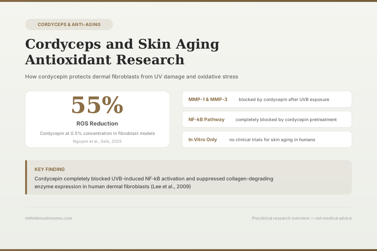
55% ROS Reduction
Cordycepin at 0.5% concentration reduced reactive oxygen species levels by 55% in fibroblast models over 24 hours, according to a 2025 review of Cordyceps militaris bioactive compounds for dermatology.
Nguyen et al., Gels, 2025
MMP-1 & MMP-3
collagen-degrading enzymes blocked by cordycepin after UVB exposure
NF-kB
inflammatory pathway suppressed by cordycepin in dermal fibroblasts
6 Studies
covering UV protection, oxidative stress, and cellular senescence
Nguyen et al., Gels, 2025
 View as image ↗
Skin aging is driven by two converging processes: intrinsic aging (cellular senescence, declining antioxidant defenses, reduced collagen synthesis) and extrinsic aging (UV radiation, pollution, oxidative stress). Both processes converge on the same molecular endpoints: increased matrix metalloproteinase (MMP) activity that degrades collagen and elastin, accumulated reactive oxygen species (ROS) that damage cellular structures, and accelerated cell senescence that reduces the skin's capacity for repair. Cordyceps species produce compounds, particularly cordycepin, that interact with several of these pathways in laboratory studies. For broader context on how cordyceps compounds affect cellular aging mechanisms, see cordyceps and cellular aging: oxidative stress protection. The research is predominantly in vitro, conducted on cultured human dermal fibroblasts, with limited translation to clinical outcomes.
Key Takeaways
- Cordycepin completely blocked UVB-induced NF-kB activation and suppressed MMP-1 and MMP-3 expression in a dose-dependent manner in human dermal fibroblasts, protecting the collagen matrix from UV-driven degradation (Lee et al., 2009)
- Cordyceps militaris extract (CME) increased antioxidant activity dose-dependently, improved fibroblast survival against hydrogen peroxide-induced oxidative stress, and prevented premature cellular senescence in vitro (Park et al., 2014)
- Cordycepin at 0.5% concentration reduced reactive oxygen species by 55% in fibroblast models over 24 hours, per a 2025 review of Cordyceps militaris bioactive gel formulations (Nguyen et al., 2025)
- Cordycepin also inhibited IL-1beta-induced MMP-1 and MMP-3 expression in synovial fibroblasts, demonstrating that MMP suppression is not limited to UV-driven pathways (Noh et al., 2009)
- All skin aging data come from cell culture studies; no published clinical trial has tested cordyceps or cordycepin specifically for skin aging outcomes in humans
How UV Radiation Ages Skin and Where Cordyceps Intervenes
Ultraviolet B radiation penetrates the epidermis and reaches dermal fibroblasts, the cells responsible for producing and maintaining the collagen and elastin matrix that gives skin its structure. UVB triggers a signaling cascade: it activates the NF-kB transcription factor, which upregulates expression of matrix metalloproteinases, particularly MMP-1 (collagenase) and MMP-3 (stromelysin). These enzymes break down the collagen framework. Over years, this process produces the wrinkles, loss of elasticity, and thinning that characterize photoaged skin.
Lee et al. (2009) tested cordycepin's ability to block this cascade in cultured human dermal fibroblasts. Using Western blot analysis and real-time PCR, they found that cordycepin inhibited UVB-induced MMP-1 and MMP-3 expression in a dose-dependent manner. The mechanism was direct: cordycepin pretreatment completely blocked UVB-induced NF-kB activation. Since NF-kB is the upstream trigger for MMP production in UV-exposed skin cells, blocking it at the source prevented the downstream collagen destruction.[1]
This finding is mechanistically clean. MMP-1 and MMP-3 levels are measurably higher in aged and photoaged skin compared to young, sun-protected skin. A compound that suppresses their expression in response to UV exposure addresses a well-established driver of skin aging. The limitation is that cell culture conditions do not replicate the complexity of in vivo skin: penetration through the stratum corneum, metabolism by keratinocytes, dilution across tissue layers, and the continuous nature of real-world UV exposure all affect whether a compound that works in a culture dish would work on a face.
Protecting Fibroblasts from Oxidative Stress and Premature Senescence
Beyond UV damage, oxidative stress from endogenous metabolism and environmental exposures drives skin aging by triggering apoptosis (cell death) and premature senescence (permanent growth arrest) in dermal fibroblasts. As fibroblasts senesce, they produce less collagen, secrete more inflammatory cytokines, and release additional MMPs, creating a self-reinforcing cycle of skin degradation.
Park et al. (2014) tested Cordyceps militaris extract (CME) against hydrogen peroxide-induced oxidative stress in human dermal fibroblasts. CME demonstrated dose-dependent antioxidant activity, scavenging free radicals measured by DPPH assay. At functionally relevant concentrations, CME improved fibroblast survival against H2O2-induced oxidative damage by inhibiting intracellular ROS production. The study also showed that CME prevented the premature cellular senescence that typically follows oxidative stress exposure, assessed by senescence-associated beta-galactosidase staining. Cells treated with CME before oxidative challenge maintained normal proliferative capacity rather than entering the senescent state.[2]
The anti-senescence finding is particularly relevant to skin aging because senescent cell accumulation in the dermis is a hallmark of aging skin. Senescent fibroblasts adopt a senescence-associated secretory phenotype (SASP) that degrades the surrounding tissue through inflammatory mediators and MMPs. Preventing fibroblasts from entering senescence, rather than just protecting them from acute death, addresses the chronic, progressive nature of skin aging. For research on how cordyceps compounds interact with broader cellular aging mechanisms including NAD+ metabolism, see cordyceps and NAD+ metabolism: the anti-aging energy connection.
MMP Suppression Beyond UV: Inflammation-Driven Collagen Loss
Skin aging is not driven exclusively by UV exposure. Inflammatory cytokines, particularly IL-1beta, also upregulate MMP expression in connective tissue cells. Chronic low-grade inflammation (sometimes called "inflammaging") is increasingly recognized as a driver of age-related tissue degradation, including in skin.
Noh et al. (2009) demonstrated that cordycepin inhibited IL-1beta-induced MMP-1 and MMP-3 expression in rheumatoid arthritis synovial fibroblasts. While synovial fibroblasts are not dermal fibroblasts, the signaling pathways are substantially shared: both cell types upregulate MMPs through NF-kB activation in response to inflammatory stimuli. Cordycepin blocked this pathway, reducing MMP production regardless of whether the trigger was UV radiation or inflammatory cytokines.[3]
This finding broadens the potential anti-aging mechanism from photoaging alone to inflammatory aging. The dual action, blocking both UV-triggered and cytokine-triggered MMP production through the same NF-kB suppression pathway, positions cordycepin as acting on a common node rather than a stimulus-specific endpoint.
Cordyceps Antioxidant Capacity and Free Radical Defense
The antioxidant properties of cordyceps extend beyond direct skin studies. Yan et al. (2013) demonstrated that extracts from the mycelia of Cordyceps sinensis exhibited antioxidant activity that contributed to cardiovascular protection, acting partially through adenosine receptors. The relevance to skin aging lies in the mechanism: adenosine receptor activation modulates cellular stress responses across tissue types, and cordycepin (3'-deoxyadenosine) is structurally related to adenosine, enabling it to interact with these receptors.[4]
The antioxidant activity of cordyceps compounds is not limited to a single mechanism. The Park et al. study identified direct free radical scavenging (measured by DPPH), the Lee et al. study identified NF-kB pathway suppression (which reduces secondary ROS generation from inflammatory cascades), and the Yan et al. study identified adenosine receptor-mediated stress protection. Multiple antioxidant mechanisms operating through different pathways could produce more robust protection than a single-pathway antioxidant, though this additive effect has not been tested in skin models. For comparison of how different mushroom species compare in antioxidant capacity, see chaga and antioxidants: the highest ORAC score in the mushroom kingdom.
Cordyceps in Topical Formulations
Nguyen et al. (2025) reviewed the emerging research on Cordyceps militaris-derived bioactive gels for dermatological applications, including anti-aging. The review consolidated data showing that cordycepin, polysaccharides, and adenosine from C. militaris provide robust antioxidant activity when incorporated into gel formulations. The reported 55% reduction in ROS levels in fibroblast models treated with 0.5% cordycepin over 24 hours was among the quantified findings. The review also described the moisturizing potential of cordyceps polysaccharides, which can promote hyaluronan synthesis in skin cells.[5]
Topical formulation represents the most direct pathway for cordyceps compounds to reach dermal target cells. Oral supplementation faces significant pharmacokinetic challenges: cordycepin is rapidly deaminated by adenosine deaminase in the bloodstream, and achieving therapeutic concentrations in dermal tissue through oral intake has not been demonstrated. Topical delivery bypasses systemic metabolism, though penetration through the stratum corneum remains a formulation challenge. The gel-based delivery systems described in the Nguyen review are designed to address this barrier through enhanced skin penetration characteristics.
Cellular Aging Mechanisms: Beyond Skin
Li et al. (2023) demonstrated that cordycepin delays postovulatory aging of oocytes through inhibition of maternal mRNA degradation via DCP1A polyadenylation suppression. While this study focused on reproductive biology rather than skin, the mechanism is relevant to broader aging research: cordycepin's ability to modulate poly(A) tail processing (a consequence of its structure as a 3'-deoxyadenosine analog) affects mRNA stability and thereby cellular function during aging across multiple cell types.[6]
The common thread across skin fibroblast studies, cardiovascular studies, and reproductive biology studies is that cordycepin operates through fundamental cellular mechanisms (NF-kB suppression, poly(A) processing interference, adenosine receptor signaling, free radical scavenging) rather than tissue-specific pathways. This makes the skin aging findings biologically plausible but also means they are not uniquely designed for skin. For the broader picture of how cordyceps compounds interact with aging at the cellular level, see cordyceps and anti-aging: research on longevity.
Evidence Boundaries
The cordyceps skin aging literature consists entirely of in vitro studies on cultured fibroblasts and review articles synthesizing preclinical data. No published randomized controlled trial has tested oral cordyceps supplementation or topical cordycepin application for skin aging endpoints (wrinkle depth, elasticity, hydration, collagen density) in human subjects.
Evidence Hierarchy
Cordyceps & Skin Aging — Clean in the Dish, Untested on a Face
The skin-aging case is strongest where it is most mechanistic: a named compound shutting down a named pathway in cultured human fibroblasts. It thins toward the top, where topical delivery is still emerging and a human trial on a skin-aging outcome does not exist. Bar width reflects how much direct evidence exists at each tier.
Molecular mechanism (cell)
evidenceLee 2009 · Noh 2009
In human dermal fibroblasts, cordycepin completely blocks UVB-induced NF-kB activation and suppresses MMP-1 and MMP-3 dose-dependently; it blocks the same MMPs when the trigger is IL-1β instead of UV
Fibroblast antioxidant & anti-senescence
evidencePark 2014 · Nguyen 2025
Cordyceps militaris extract scavenges free radicals dose-dependently, rescues fibroblasts from hydrogen-peroxide damage, and prevents premature senescence; cordycepin cut ROS by 55% at 0.5% over 24 h
Cross-tissue mechanism support
evidenceYan 2013 · Li 2023
The same core mechanisms — adenosine-receptor signaling and poly(A)-tail processing — protect heart tissue and delay oocyte aging, confirming the pathways are real but not specific to skin
Topical delivery to human skin
emergingNguyen 2025
Cordyceps gel formulations are being designed to carry cordycepin, polysaccharides, and adenosine across the stratum corneum, but human penetration and efficacy data are not yet published
Human skin-aging endpoint
gapDoes not yet exist
No trial has tested whether cordyceps or cordycepin improves wrinkles, elasticity, hydration, or collagen density in people
How to read it: the lower tiers are wide and solid because the mechanistic and fibroblast data converge on specific, repeatable effects. Topical formulations that could carry those compounds into living skin are only now being designed, and the trial that would test an actual skin-aging outcome in people has never been run. That top gap — not the quality of the mechanism — is the honest limit of the cordyceps skin-aging claim.
Sources: Lee 2009 [1]; Park 2014 [2]; Noh 2009 [3]; Yan 2013 [4]; Nguyen 2025 [5]; Li 2023 [6]. Bar widths are illustrative of evidence depth per tier, not quantitative effect sizes.
The in vitro findings are consistent and mechanistically sound: cordycepin blocks both UV-induced and inflammation-induced MMP expression through NF-kB suppression, cordyceps extract prevents oxidative stress-induced senescence in fibroblasts, and cordycepin reduces intracellular ROS in a dose-dependent manner. These are well-designed cell culture studies published in peer-reviewed journals.
The gap between "protects fibroblasts in a dish" and "reduces wrinkles on a face" is substantial. Cell culture studies cannot account for bioavailability, skin penetration, metabolism, dose-response in intact tissue, or long-term safety. Several specific challenges limit translation:
Bioavailability. Cordycepin is rapidly deaminated by adenosine deaminase in blood and tissues. Its half-life in circulation is measured in minutes. Oral supplementation would need to deliver sustained levels to dermal tissue, which requires either very high doses, adenosine deaminase inhibitors (which carry their own risks), or novel delivery systems. None of these approaches have been tested for skin aging endpoints.
Penetration. Topical application avoids systemic metabolism but faces the stratum corneum barrier. The molecular weight of cordycepin (251 Da) is within the range that can theoretically penetrate skin, but actual penetration depends on formulation, concentration, vehicle, and skin condition. The gel formulations described in the Nguyen review are designed to optimize this, but penetration data from human skin models are limited.
Dose translation. The concentrations used in cell culture (direct exposure of fibroblasts to cordycepin or CME in growth media) do not correspond to achievable tissue concentrations from either oral or topical administration. Effective in vitro concentrations represent an upper bound of potential activity, not a prediction of in vivo efficacy.
The cosmetics industry frequently markets ingredients based on in vitro MMP inhibition data without clinical proof of anti-aging efficacy. The cordyceps data should be evaluated by the same standard: promising mechanism, unproven outcome.
The Bottom Line
Cordycepin from Cordyceps species blocks UV-induced and inflammation-driven collagen degradation by suppressing NF-kB activation and downstream MMP-1/MMP-3 expression in human dermal fibroblasts. Cordyceps militaris extract prevents oxidative stress-induced premature senescence and reduces ROS levels dose-dependently. All data are from cell culture studies. No clinical trial has tested cordyceps for skin aging in humans.
Frequently Asked Questions
Sources & References
- 1RTHM-00160·Lee, Young Rae et al. (2009). “Cordycepin inhibits UVB-induced matrix metalloproteinase expression by suppressing the NF-kappaB pathway in human dermal fibroblasts..” Experimental & molecular medicine.Study breakdown →PubMed →↩
- 2RTHM-00633·Park, Jun Myoung et al. (2014). “Cordyceps militaris extract protects human dermal fibroblasts against oxidative stress-induced apoptosis and premature senescence..” Nutrients.Study breakdown →PubMed →↩
- 3RTHM-00167·Noh, E-M et al. (2009). “Cordycepin inhibits IL-1beta-induced MMP-1 and MMP-3 expression in rheumatoid arthritis synovial fibroblasts..” Rheumatology (Oxford.Study breakdown →PubMed →↩
- 4RTHM-00310·Yan, Xiao-Feng et al. (2013). “Cardiovascular protection and antioxidant activity of the extracts from the mycelia of Cordyceps sinensis act partially via adenosine receptors..” Phytotherapy research : PTR.Study breakdown →PubMed →↩
- 5RTHM-00947·Nguyen, Trung Quang et al. (2025). “Cordyceps militaris-Derived Bioactive Gels: Therapeutic and Anti-Aging Applications in Dermatology..” Gels (Basel.Study breakdown →PubMed →↩
- 6RTHM-00838·Li, Chong et al. (2023). “Cordycepin delays postovulatory aging of oocytes through inhibition of maternal mRNAs degradation via DCP1A polyadenylation suppression..” Cellular and molecular life sciences : CMLS.Study breakdown →PubMed →↩