Cordyceps and Liver Health
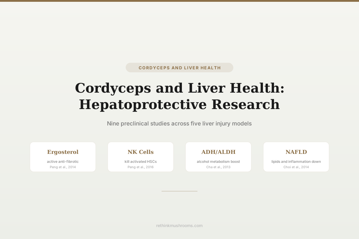
Ergosterol proposed as the anti-fibrotic compound
Peng Y. et al. (2014) traced the anti-liver-fibrosis activity of cultured Cordyceps sinensis mycelium to ergosterol, concluding it 'might be the efficacious antifibrotic substance' — a defined molecular candidate in an otherwise complex extract.
Peng Y. et al., Evidence-Based Complementary and Alternative Medicine, 2014
NK cell activation
drives anti-fibrotic effect in CCl4 mice
ADH and ALDH
enzymes boosted against alcohol injury
1 human report
a case of hepatoportal sclerosis, not a benefit
Peng Y. et al., Evidence-Based Complementary and Alternative Medicine, 2014
 View as image ↗
Liver disease is among the leading causes of death worldwide. Chronic insults from alcohol, viral hepatitis, drug toxicity, and metabolic dysfunction all converge on a common endpoint: inflammation, hepatocyte death, and progressive fibrosis that can advance to cirrhosis. Cordyceps sinensis and Cordyceps militaris have been studied across multiple liver injury models, with a body of preclinical evidence showing anti-fibrotic, anti-inflammatory, and antioxidant effects through increasingly well-defined mechanisms.[1]
The most precise finding in this field is the proposal of ergosterol as the anti-fibrotic compound in cultured Cordyceps sinensis mycelium. Other studies have mapped cordycepin's role in alcohol-induced liver damage, polysaccharide effects on drug-induced injury, and whole-extract protection in fulminant hepatic failure and NAFLD models. Every efficacy study is preclinical: no randomized controlled trial has tested Cordyceps for any human liver disease. The only published human liver data runs in the opposite direction — a 2023 case report attributing hepatoportal sclerosis to Cordyceps supplementation.[2] This article covers what the animal and cell studies show, which compounds drive the effects, what the single human report contains, and where the evidence stands. For Cordyceps effects on drug-specific liver injury, see Cordyceps and drug-induced liver injury: hepatoprotective mechanisms. For the liver fibrosis evidence specifically, see Cordyceps and liver fibrosis: can it help reverse damage?.
Key Takeaways
- Peng Y. et al. (2014) traced the anti-fibrotic activity of cultured Cordyceps sinensis mycelium to ergosterol, which inhibited activated hepatic stellate cells dose-dependently in cell culture and reproduced the whole-extract effect in CCl4-treated mice at a single tested dose; the authors concluded ergosterol "might be" the active substance
- Peng Y. et al. (2016) showed cultured Cordyceps sinensis mycelium alleviates CCl4-induced liver fibrosis partly by activating hepatic natural killer cells — antibody depletion of NK cells accelerated fibrosis, though the extract retained some effect even in NK-depleted mice
- Cha et al. (2013) found cordycepin-enriched Cordyceps militaris prevented alcohol-induced hepatotoxicity in rats by increasing alcohol dehydrogenase (ADH) and acetaldehyde dehydrogenase (ALDH) activity, alongside a silymarin positive control
- Choi et al. (2014) demonstrated that 1% Cordyceps militaris water extract for 10 weeks reduced hepatic lipids, triglycerides, TNF-alpha and IL-6 in ob/ob mice with NAFLD, while raising hepatic glutathione
- Cheng et al. (2014) showed Cordyceps sinensis attenuated apoptosis in D-galactosamine/LPS-induced fulminant hepatic failure via reduced caspase-3 and caspase-6 activity and a shifted cytokine response
- Liu et al. (2003) and Peng J. et al. (2013) independently established the TGF-beta1 axis as the core anti-fibrotic mechanism, the latter adding Smad2/3, MMP2/MMP9 and TIMP2 to the map
- Kaur et al. (2023) reported the only published human liver outcome: biopsy-confirmed hepatoportal sclerosis in an 81-year-old woman taking Cordyceps, which improved after she stopped it
Anti-Fibrotic Effects: From Whole Extract to Defined Compounds
Liver fibrosis is the accumulation of extracellular matrix (primarily collagen) in response to chronic injury. The central cellular driver is the hepatic stellate cell (HSC): quiescent in healthy liver, activated by injury into myofibroblast-like cells that deposit collagen. Reversing or preventing HSC activation is the primary therapeutic target.
Liu et al. (2003) provided the earliest mechanistic framework. Rats received CCl4 plus ethanol for nine weeks to induce fibrosis, with Cordyceps sinensis started 10 days after injury induction began — a treatment design rather than a pure pre-treatment design. Serum markers fell substantially in the CS group compared with untreated model animals: ALT 86.0 ± 34.4 versus 224.3 ± 178.9, and AST 146.7 ± 60.2 versus 272.6 ± 130.1, alongside reductions in the fibrosis markers hyaluronic acid and laminin. The proposed mechanism: CS suppressed TGF-beta1 expression, which downstream reduced PDGF, preventing HSC activation and lowering procollagen I and III deposition.[1]
A decade later, a separate group led by Jinghua Peng tested the isolated polysaccharide fraction. (Note that two unrelated first authors named Peng appear in this literature; they are distinguished here as Peng J. and Peng Y.) Peng J. et al. (2013) gave CCl4-treated rats a polysaccharide extracted from cultured Cordyceps sinensis mycelia (CS-PS), using the anti-fibrotic drug colchicine as a positive control. CS-PS lowered ALT, AST, total bilirubin and hepatic hydroxyproline, raised serum albumin, and improved histology to a degree the authors described as comparable to colchicine. Mechanistically it went further than Liu et al.: CS-PS downregulated alpha-smooth muscle actin, TGF-beta1, both TGF-beta receptors, phosphorylated Smad2 and Smad3, and TIMP2, while inhibiting MMP2 and MMP9 activity.[3]
That result matters for a practical reason: polysaccharides are the fraction most commercial Cordyceps products are standardized to, and this is the study that ties that specific fraction to the fibrosis endpoint.
Peng Y. et al. (2014) approached the same problem from the opposite direction, asking which small molecule carries the activity. Testing cultured mycelium Cordyceps sinensis (CMCS) at 120 mg/kg/day in CCl4-treated C57BL/6 mice over four weeks, the team found reduced serum liver enzymes, less hepatic inflammation, decreased collagen deposition, and lower alpha-SMA expression. They then detected ergosterol — a fungal sterol analogous to cholesterol in animals — by HPLC and screened it against activated HSCs in culture, where it inhibited the cells dose-dependently by increasing lysosomal membrane permeability and reducing EdU incorporation, F-actin, and alpha-SMA. Administered to mice at 50 mg/kg/day, ergosterol reproduced the anti-fibrotic effect of the whole extract.[4]
Two details are worth holding precisely, because they are easy to overstate. The dose-dependence was demonstrated in vitro, on cultured stellate cells; in the animals, each compound was given at a single dose. And the authors' own conclusion is hedged: ergosterol "might be the efficacious antifibrotic substance in CMCS." This is a strong candidate compound, not a settled attribution.
Still, the direction of travel is what makes the field interesting. It moves the research from "Cordyceps extract does something to fibrosis" toward "a specific, quantifiable compound may drive the effect." Ergosterol is present in measurable quantities in Cordyceps products, meaning its content could in principle be standardized across preparations.
Mechanism Split
Four Anti-Fibrosis Studies, Four Different Things in the Syringe
These four are often summarized as one result — “Cordyceps reduces liver fibrosis.” Each actually tested a different preparation and hit a different node of the fibrosis cascade. That is what makes them complementary rather than repeated confirmations of the same claim.
Liu et al., 2003[1]
CCl4 + ethanol rats, 9 weeksWhat was tested
Whole Cordyceps sinensis
Pathway node moved
TGF-β1 → PDGF → procollagen I/III
Treatment started 10 days after injury induction began. ALT fell to 86.0 ± 34.4 from 224.3 ± 178.9 and AST to 146.7 ± 60.2 from 272.6 ± 130.1, with hyaluronic acid and laminin down as well.
Peng J. et al., 2013[3]
CCl4 rats, colchicine controlWhat was tested
Isolated polysaccharide (CS-PS)
Pathway node moved
TGF-β1/Smad2/3 · MMP2, MMP9 · TIMP2
The fraction most commercial products standardize to. Lowered ALT, AST, bilirubin and hepatic hydroxyproline, raised albumin, and improved histology to a degree the authors called comparable to colchicine.
Peng Y. et al., 2014[4]
CCl4 mice, 4 weeksWhat was tested
Cultured mycelium, then isolated ergosterol
Pathway node moved
Direct hepatic stellate cell inhibition
Ergosterol detected by HPLC, then dose-dependently inhibited activated stellate cells in culture via lysosomal membrane permeability, EdU, F-actin and α-SMA. At 50 mg/kg/d it reproduced the whole-extract effect in mice — a single dose, not a dose curve.
Peng Y. et al., 2016[5]
CCl4 mice + NK depletion armWhat was tested
Cultured mycelium (CMCS)
Pathway node moved
Hepatic NK cell activation (NKG2D)
The only causally tested mechanism in the set: depleting NK cells with ASGM-1 accelerated fibrosis. But CMCS still slowed fibrosis in NK-depleted mice, so NK activation is part of the effect, not all of it.
How to read it: the recurring appearance of TGF-β1 across independent groups is the strongest signal here — two labs, two preparations, same axis. But “Cordyceps” is not one substance: a whole extract, an isolated polysaccharide, and a purified sterol are different products with different pharmacology, and a result obtained with one does not automatically transfer to another. All four studies are in rodents. Nothing here is medical advice.
Sources: Liu et al. 2003 [1]; Peng J. et al. 2013 [3]; Peng Y. et al. 2014 [4]; Peng Y. et al. 2016 [5]. Values quoted are as reported in the cited abstracts. Two unrelated first authors named Peng appear in this literature and are distinguished as Peng J. and Peng Y.
NK Cell Activation: An Immune Mechanism for Anti-Fibrosis
Peng Y. et al. (2016) added a layer to the anti-fibrotic story by investigating the immune component. C57BL/6 mice received 10% CCl4 three times weekly for four weeks with CMCS at 120 mg/kg/day. CMCS improved liver function, attenuated inflammation and fibrosis, and increased both the number of hepatic natural killer (NK) cells and their expression of NKG2D, an NK activating receptor. It also reduced desmin staining and increased TUNEL-positive signal adjacent to hepatic stellate cells — the signature of NK cells killing HSCs.[5]
The design's strength is that it tested the mechanism rather than merely correlating with it. A second arm depleted NK cells with the antibody ASGM-1. Depletion alone markedly accelerated liver inflammation and fibrosis, confirming that hepatic NK cells restrain fibrogenesis. In NK-depleted mice, CMCS still slowed fibrosis development and mildly restored NK numbers, but no longer changed NKG2D expression.
That last result is the reason for precision here. NK activation is a genuine and causally tested part of how CMCS works, but it is not the whole of it: the extract retained partial activity when NK cells were removed. Read alongside the TGF-beta1/Smad suppression documented by Liu et al. and Peng J. et al., and the ergosterol-mediated HSC inhibition of Peng Y. et al., Cordyceps appears to act on fibrosis through several parallel routes rather than one.
Alcohol-Induced Liver Injury
Cha et al. (2013) tested cordycepin-enriched Cordyceps militaris against alcohol-induced hepatotoxicity in Sprague-Dawley rats. Alcohol-fed animals received diets containing Paecilomyces japonica (CPJ), standard C. militaris (CCM), or cordycepin-enriched C. militaris (CCM-alpha) at 3% by weight, plus a silymarin arm at 0.1% as an established hepatoprotective comparator, for four weeks.[6]
Alcohol raised AST, ALT, gamma-GTP, ALP and LDH, along with blood alcohol and acetaldehyde levels. The cordycepin-enriched group was the preparation the authors credited with markedly preventing those elevations. The mechanistic finding: CCM-alpha increased hepatic activity of both alcohol dehydrogenase (ADH) and acetaldehyde dehydrogenase (ALDH), the two enzymes responsible for metabolizing ethanol and its toxic intermediate acetaldehyde. It also attenuated lipid droplet accumulation in hepatocytes, the defining lesion of alcoholic fatty liver.
The measured drop in circulating alcohol and acetaldehyde is what ties the enzyme finding to the injury finding. Acetaldehyde is the primary mediator of alcohol-induced liver damage, driving oxidative stress, mitochondrial dysfunction, and inflammatory signaling. Raising ALDH activity addresses the bottleneck in alcohol metabolism that allows acetaldehyde to accumulate — and in these rats, the accumulation was in fact reduced.
Non-Alcoholic Fatty Liver Disease (NAFLD)
Choi et al. (2014) tested C. militaris in the ob/ob mouse, a genetic obesity model that spontaneously develops NAFLD. Four-week-old males received either a control diet or one containing 1% C. militaris water extract for 10 weeks.[7]
C. militaris significantly decreased serum glucose and HOMA-IR, a measure of insulin resistance. Serum free fatty acids fell. Hepatic total lipids and triglyceride content dropped. Serum ALT declined, as did the proinflammatory cytokines TNF-alpha and IL-6, while hepatic glutathione rose and lipid peroxide levels fell — a coherent antioxidant signature rather than a single isolated marker.
The dual effect on lipid accumulation and on inflammation addresses both components of NAFLD progression. Fat accumulation (steatosis) alone is relatively benign; it is the addition of inflammation (steatohepatitis) that drives fibrosis and progression to cirrhosis. For NAFLD-specific evidence, see Cordyceps for fatty liver: NAFLD evidence.
Wang et al. (2015) extended the lipid story using a residue polysaccharide (RPS) recovered from the spent cultivation medium of Cordyceps militaris SU-12 — the material normally discarded after harvest. The polysaccharide was structurally characterized (glucose, arabinose and mannose in a 62:1.6:1 ratio) and, in mice, reduced blood and liver lipid levels, improved glutamate pyruvate transaminase and antioxidant activity, and attenuated liver cell injury on histopathology.[8]
Fulminant Hepatic Failure
Cheng et al. (2014) tested Cordyceps sinensis in one of the most severe liver injury models: D-galactosamine/LPS-induced fulminant hepatic failure in mice, which produces massive hepatocyte death within hours. Mice received C. sinensis by oral gavage at 20 mg or 40 mg, five days a week for four weeks, before being challenged with GalN (800 mg/kg) plus LPS (10 µg/kg).[9]
The challenge increased hepatocyte degeneration, circulating AST, TNF-alpha, IL-10, hepatic apoptosis and caspase activity. CS pre-treatment reduced AST, TNF-alpha and nitric oxide, and increased IL-10 and superoxide dismutase. Apoptosis was attenuated, as shown by TUNEL staining and by caspase-3 and caspase-6 activity assays. The authors summarized the mechanism as modulating the cytokine response and inhibiting apoptosis.
This is a severe, acute injury model rather than the chronic models used in the fibrosis studies. That CS protects in both chronic and acute contexts suggests its hepatoprotective effects span both long-term HSC activation and acute hepatocyte death pathways. It is also, notably, a pre-treatment design: the extract was given for four weeks before the insult, which does not test whether it helps once injury is underway.
Drug-Induced Liver Injury
Fan et al. (2018) investigated a combination approach: polysaccharides from Cordyceps sinensis (CSP) combined with polysaccharides from Ganoderma atrum (PSG) against cyclophosphamide-induced liver injury in mice. The combined fungal polysaccharides (CFP) reduced the toxicity markers AST, ALT and ALP.[10]
What the study actually resolved was not that the blend beat its parts on a head-to-head efficacy endpoint, but that the two polysaccharides act through different pathways. CSP inhibited Toll-like receptor 9 activation and the inflammatory signals downstream of it, including proinflammatory cytokines, inducible nitric oxide synthase, and COX-2. PSG worked through PPAR-alpha activation, restoring hepatic glutathione peroxidase and glutathione depleted by cyclophosphamide and preventing mitochondria-dependent apoptosis via the Bcl-2 family proteins Bad, Bax and Bcl-2. Both polysaccharides were structurally characterized, which makes the preparation reproducible. For more on drug-specific hepatoprotection, see Cordyceps and drug-induced liver injury: hepatoprotective mechanisms. For the viral hepatitis literature, see Cordyceps and hepatitis: antiviral liver research.
The Only Human Liver Data Points the Other Way
Nine of the ten studies above are animal or cell experiments reporting benefit. The tenth is the only published human liver outcome for Cordyceps, and it is a report of harm.
Kaur et al. (2023) described an 81-year-old woman taking a traditional Chinese herbal supplement, Cordyceps, who presented with splenomegaly and weight loss. After an extensive workup, liver biopsy confirmed loss of the small portal veins with obstruction at the level of both small and large portal veins — the histological signature of hepatoportal sclerosis, a form of non-cirrhotic portal hypertension. Other causes were excluded, the condition was attributed to Cordyceps use, and her features of hepatoportal sclerosis improved after she stopped taking it.[2]
A single case report cannot establish causation, incidence, or risk at any given dose, and no denominator exists for how many people take Cordyceps without incident. What gives this one more weight than an isolated coincidence is the combination of a biopsy-confirmed diagnosis, systematic exclusion of alternative causes, and improvement after withdrawal. That pattern is what clinicians call a positive dechallenge, and it is the standard signal that prompts closer attention rather than dismissal.
It also reframes the preclinical literature rather than cancelling it. The animal findings are real; they simply have not been tested in people, and the first human observation to reach publication described injury rather than protection. Anyone with existing liver disease, or taking Cordyceps alongside hepatically metabolized medication, should treat that asymmetry as a reason to involve a physician. Nothing in this article is medical advice.
Direction Inversion
Every Animal Study Says Protection. The Only Human Report Says Injury.
These two columns are almost never printed next to each other. Nine preclinical studies point one way; the single published human liver outcome points the other. Neither column cancels the other — but a reader who sees only the left one is reading half the evidence.
Preclinical — benefit
9 studiesLiu et al., 2003[1]
CCl4 + ethanol rats — fibrosis down
Peng J. et al., 2013[3]
CCl4 rats — polysaccharide, fibrosis down
Peng Y. et al., 2014[4]
CCl4 mice — ergosterol, fibrosis down
Peng Y. et al., 2016[5]
CCl4 mice — NK activation, fibrosis down
Cha et al., 2013[6]
Alcohol-fed rats — enzymes protected
Choi et al., 2014[7]
ob/ob mice — hepatic lipid, TNF-α down
Wang et al., 2015[8]
Mice — blood and liver lipid down
Cheng et al., 2014[9]
GalN/LPS mice — apoptosis attenuated
Fan et al., 2018[10]
Cyclophosphamide mice — AST/ALT/ALP down
All rodent or cell work. None tested a human liver endpoint.
Human — harm
1 case reportKaur et al., ACG Case Reports Journal, 2023[2]
81-year-old woman taking Cordyceps, presenting with splenomegaly and weight loss.
- •Liver biopsy confirmed loss of the small portal veins — hepatoportal sclerosis
- •Other causes excluded on extensive workup
- •Improved after stopping Cordyceps (positive dechallenge)
Human randomized controlled trial — does not exist for any Cordyceps preparation in any liver disease indication.
How to read it: the columns are not drawn to a shared scale, because none exists — nine rodent studies and one human case report are different kinds of evidence, not different amounts of it. A single case report cannot establish incidence, dose-dependence, or causation in general; what raises it above coincidence is the biopsy confirmation, the exclusion of other causes, and the improvement on withdrawal. The honest summary is that the benefit has never been tested in people and the first human liver outcome to be published described injury. Anyone with existing liver disease, or taking medication metabolized by the liver, should speak to a physician before using Cordyceps. Nothing here is medical advice.
Sources: Liu et al. 2003 [1]; Kaur et al. 2023 [2]; Peng J. et al. 2013 [3]; Peng Y. et al. 2014 [4]; Peng Y. et al. 2016 [5]; Cha et al. 2013 [6]; Choi et al. 2014 [7]; Wang et al. 2015 [8]; Cheng et al. 2014 [9]; Fan et al. 2018 [10]. Column headings describe the direction of each study’s reported finding, not its magnitude.
Converging Evidence and Open Questions
The ten studies in this article span multiple liver injury models (CCl4-chemical, alcohol, genetic obesity/NAFLD, drug-induced, acute galactosamine/LPS), multiple Cordyceps preparations (whole extract, cultured mycelium, cordycepin-enriched, isolated and residue polysaccharide fractions), and multiple mechanisms (TGF-beta1/PDGF and TGF-beta1/Smad suppression, MMP and TIMP modulation, NK cell activation, ergosterol-mediated HSC inhibition, ADH/ALDH enhancement, cytokine modulation, antioxidant defense).
The consistency across models is the strongest argument for the hepatoprotective hypothesis. Independent groups, working on different insults with different preparations, keep landing on the TGF-beta1 axis and on antioxidant restoration. The identification of candidate compounds — ergosterol for anti-fibrosis, cordycepin for alcohol metabolism enhancement — moves the field beyond "mushroom extract helps liver" toward testable pharmacological hypotheses.
The central gap is unchanged: no controlled human trial has tested any Cordyceps preparation for any liver disease indication. Dose equivalencies between animal models and human supplementation are undefined. Whether the ergosterol content of commercial Cordyceps products reaches the concentrations used in mice is unknown. Whether cordycepin from supplements achieves sufficient hepatic bioavailability to enhance ADH/ALDH activity has not been tested in humans. And the one human liver outcome on record is an adverse one, which means the translation question is not only whether the benefit carries over, but whether the safety profile in a healthy rodent liver carries over to an elderly human one.
Evidence Map
Ten Studies, Six Rungs — and What Each Model Can Actually Tell You
“Ten studies” sounds like ten confirmations. It is really coverage across six very different situations. Bar width reflects how closely each model corresponds to a human clinical scenario — the NAFLD models sit closest, the acute chemical challenges furthest. The dashed rung is the trial nobody has run; the red rung is the one human report, and it describes harm.
Human clinical trial
gapDoes not exist
No randomized or controlled trial has tested any Cordyceps preparation against any liver disease endpoint. Dose equivalence between the rodent studies below and human supplementation is undefined.
Human observation
harm signalKaur 2023
The only published human liver outcome: biopsy-confirmed hepatoportal sclerosis in an 81-year-old woman taking Cordyceps, with other causes excluded and improvement after she stopped. One case, adverse direction.
Chronic metabolic injury — NAFLD animal models
preclinicalChoi 2014 · Wang 2015
The closest preclinical match to the condition most readers are asking about. C. militaris water extract cut hepatic lipids, triglycerides, ALT, TNF-α and IL-6 while raising glutathione in ob/ob mice; a residue polysaccharide lowered blood and liver lipids in mice.
Chronic toxic fibrosis — CCl4 animal models
preclinicalLiu 2003 · Peng J. 2013 · Peng Y. 2014 · Peng Y. 2016
The densest cluster, and the one with the best-mapped mechanisms: TGF-β1/PDGF, TGF-β1/Smad2/3 with MMP and TIMP modulation, ergosterol-mediated stellate cell inhibition, and hepatic NK cell activation. Chemically induced fibrosis is a well-characterized model but not a human disease.
Chronic alcohol injury — animal model
preclinicalCha 2013
Cordycepin-enriched C. militaris prevented alcohol-driven rises in AST, ALT, γ-GTP, ALP and LDH in rats, raised ADH and ALDH activity, lowered circulating acetaldehyde, and reduced hepatocyte lipid droplets — benchmarked against a silymarin arm.
Acute chemical / drug insult — animal models
preclinicalFan 2018 · Cheng 2014
Furthest from ordinary human exposure: a cyclophosphamide hepatotoxicity model, and a D-galactosamine/LPS challenge that produces fulminant liver failure within hours. Both used pre-treatment, so neither tests whether Cordyceps helps once injury is underway.
How to read it: the consistency across independent labs, different insults and different Cordyceps preparations is a genuine reason to take the hepatoprotective hypothesis seriously — that is what a converging preclinical base looks like. It is also entirely rodent evidence. Animal liver models reproduce important features of human disease without reliably predicting human outcomes, and the two human rungs at the top are the ones that matter for anyone deciding whether to take a supplement: one is empty, and the other is a case of injury. Nothing here is medical advice.
Sources: Liu et al. 2003 [1]; Kaur et al. 2023 [2]; Peng J. et al. 2013 [3]; Peng Y. et al. 2014 [4]; Peng Y. et al. 2016 [5]; Cha et al. 2013 [6]; Choi et al. 2014 [7]; Wang et al. 2015 [8]; Cheng et al. 2014 [9]; Fan et al. 2018 [10]. Bar widths are illustrative of how closely each model corresponds to a human clinical scenario, not quantitative effect sizes.
The Bottom Line
Cordyceps sinensis and Cordyceps militaris show hepatoprotective effects across nine preclinical studies spanning chemical liver fibrosis, alcohol-induced injury, NAFLD, drug toxicity, and fulminant hepatic failure. The research has proposed specific active compounds (ergosterol for anti-fibrosis, cordycepin for alcohol metabolism enhancement) and mapped specific mechanisms (HSC inhibition via TGF-beta1/Smad suppression, NK cell-mediated killing of activated HSCs, antioxidant defense enhancement). No controlled human trial has been published for any liver disease indication, and the single published human liver outcome is a 2023 case report of biopsy-confirmed hepatoportal sclerosis attributed to Cordyceps that improved on withdrawal — making the translation from animal models to clinical application an entirely open question in both directions.
Frequently Asked Questions
Sources & References
- 1RTHM-00060·Liu, Yu-Kan et al. (2003). “Inhibitive effect of cordyceps sinensis on experimental hepatic fibrosis and its possible mechanism..” World journal of gastroenterology.Study breakdown →PubMed →↩
- 2RTHM-00834·Kaur, Bhupinder et al. (2023). “Traditional Chinese Medicine, Cordyceps, Related to Hepatoportal Sclerosis..” ACG case reports journal.Study breakdown →PubMed →↩
- 3RTHM-00303·Peng, Jinghua et al. (2013). “Anti-fibrotic effect of Cordyceps sinensis polysaccharide: Inhibiting HSC activation, TGF-β1/Smad signalling, MMPs and TIMPs..” Experimental biology and medicine (Maywood.Study breakdown →PubMed →↩
- 4RTHM-00344·Peng, Yuan et al. (2014). “Ergosterol Is the Active Compound of Cultured Mycelium Cordyceps sinensis on Antiliver Fibrosis..” Evidence-based complementary and alternative medicine : eCAM.Study breakdown →PubMed →↩
- 5RTHM-00440·Peng, Yuan et al. (2016). “Cultured Mycelium Cordyceps sinensis allevi¬ates CCl4-induced liver inflammation and fibrosis in mice by activating hepatic natural killer cells..” Acta pharmacologica Sinica.Study breakdown →PubMed →↩
- 6RTHM-00280·Cha, Jae-Young et al. (2013). “Protective effect of cordycepin-enriched Cordyceps militaris on alcoholic hepatotoxicity in Sprague-Dawley rats..” Food and chemical toxicology : an international journal published for the British Industrial Biological Research Association.Study breakdown →PubMed →↩
- 7RTHM-00316·Choi, Ha-Neul et al. (2014). “Cordyceps militaris alleviates non-alcoholic fatty liver disease in ob/ob mice..” Nutrition research and practice.Study breakdown →PubMed →↩
- 8RTHM-00407·Wang, Liqin et al. (2015). “Antihyperlipidemic and hepatoprotective activities of residue polysaccharide from Cordyceps militaris SU-12..” Carbohydrate polymers.Study breakdown →PubMed →↩
- 9RTHM-00315·Cheng, Yu-Jung et al. (2014). “Cordyceps sinensis prevents apoptosis in mouse liver with D-galactosamine/lipopolysaccharide-induced fulminant hepatic failure..” The American journal of Chinese medicine.Study breakdown →PubMed →↩
- 10RTHM-00507·Fan, Songtao et al. (2018). “Combinatorial usage of fungal polysaccharides from Cordyceps sinensis and Ganoderma atrum ameliorate drug-induced liver injury in mice..” Food and chemical toxicology : an international journal published for the British Industrial Biological Research Association.Study breakdown →PubMed →↩