Cordyceps and Liver Health
62% ALT Reduction
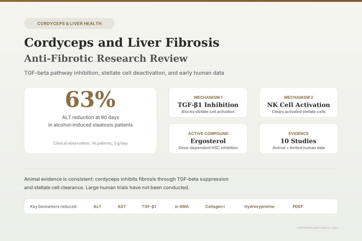
In a small clinical observation of 14 patients with alcohol-induced liver steatosis, 90 days of Cordyceps sinensis supplementation at 3 g/day reduced average ALT levels by 63.2%.
Liu & Bau, World Journal of Gastroenterology, 2003
TGF-β1 Pathway
primary anti-fibrotic mechanism identified
8+ Animal Studies
testing cordyceps against liver fibrosis models
CCl₄ + Alcohol
most common experimental fibrosis model used
Liu & Bau, World Journal of Gastroenterology, 2003
 View as image ↗
Liver fibrosis is the progressive accumulation of scar tissue that replaces functional hepatocytes after repeated injury. Left unchecked, it advances to cirrhosis. Cordyceps sinensis and Cordyceps militaris have both been tested against liver fibrosis in animal models, with multiple studies converging on a shared mechanism: suppression of the TGF-β1 signaling pathway that drives hepatic stellate cell activation and collagen deposition. A small body of human data exists but remains preliminary. For a broader overview of how cordyceps interacts with the liver beyond fibrosis, see Cordyceps and Liver Health: Hepatoprotective Research. If your interest is specifically in medication-related liver damage, Cordyceps and Drug-Induced Liver Injury covers that research separately.
Key Takeaways
- Cordyceps sinensis reduced TGF-β1 protein expression from 1.73 to 0.20 and collagen I mRNA from 2.64 to 1.10 in CCl₄-induced fibrosis in rats (Liu and Bau, World Journal of Gastroenterology, 2003)
- CS polysaccharide inhibited hepatic stellate cell activation and downregulated TGF-β1/Smad2/Smad3 signaling, MMP2, MMP9, and TIMP2 expression in fibrotic rat liver (Peng et al., Experimental Biology and Medicine, 2013)
- Ergosterol from cultured Cordyceps sinensis mycelium was identified as the specific compound responsible for anti-fibrotic activity in stellate cells (Peng et al., Evidence-Based Complementary and Alternative Medicine, 2014)
- Cultured mycelium Cordyceps sinensis alleviated CCl₄-induced liver fibrosis by activating hepatic natural killer cells that cleared activated stellate cells (Peng et al., Acta Pharmacologica Sinica, 2016)
- In 14 patients with alcohol-induced steatosis, 3 g/day of Cordyceps sinensis reduced ALT by 51.6% at 20 days and 63.2% at 90 days (clinical observation, not a controlled trial)
- A 2024 study found Cordyceps militaris protected against hepatocyte apoptosis and liver fibrosis induced by high palmitic acid diet in mice (Tan et al., Frontiers in Pharmacology, 2024)
The Central Mechanism: TGF-β1 and Stellate Cell Inhibition
Liver fibrosis follows a consistent biological cascade regardless of what caused the initial injury. Damaged hepatocytes release inflammatory signals that activate hepatic stellate cells (HSCs). Once activated, HSCs transform from quiescent vitamin A-storing cells into myofibroblasts that produce excessive collagen, forming scar tissue. The master regulator of this process is transforming growth factor beta-1 (TGF-β1).
Liu and Bau conducted the foundational study on cordyceps and hepatic fibrosis, published in the World Journal of Gastroenterology in 2003.[1] Using 66 male Wistar rats exposed to CCl₄ plus 5% ethanol to induce fibrosis, they administered Cordyceps sinensis suspension orally for 9 weeks. The cordyceps group showed marked reductions across every liver injury marker: ALT dropped from 224.3 to 86.0 U/L, AST from 272.6 to 146.7 U/L, hyaluronic acid from 316.5 to 202.0 μg/L, and laminin from 59.7 to 50.4 μg/L.
More telling than the biomarker improvements was what happened at the tissue level. TGF-β1 protein expression fell from 1.73 to 0.20 in the cordyceps group. TGF-β1 mRNA dropped from 3.17 to 1.68. Collagen I mRNA fell from 2.64 to 1.10. PDGF protein, which drives stellate cell proliferation, decreased from 1.91 to 0.87 (p < 0.01). The authors concluded that cordyceps inhibited fibrogenesis by suppressing TGF-β1, which in turn reduced PDGF expression and prevented stellate cell activation.
CS Polysaccharide: The Bioactive Fraction
Peng and colleagues followed up in 2013 by isolating the polysaccharide fraction from cultured Cordyceps sinensis mycelia (CS-PS) and testing it directly against liver fibrosis.[2] Using CCl₄-induced fibrosis in rats with colchicine as a positive control, they measured a comprehensive panel of fibrosis markers.
CS-PS reduced serum ALT, AST, and total bilirubin while increasing albumin levels. Hepatic hydroxyproline, a direct measure of collagen content in liver tissue, also decreased. At the molecular level, CS-PS downregulated hepatic alpha-smooth muscle actin (α-SMA, the hallmark of activated stellate cells), TGF-β1, TGF-β1 receptors (TβR-I and TβR-II), phosphorylated Smad2 and Smad3, and TIMP2. It also inhibited MMP2 and MMP9 activity. This matters because the MMP/TIMP balance determines whether scar tissue gets deposited or degraded. By shifting that balance, CS-PS did not just slow fibrosis progression; it created conditions for existing scar tissue remodeling.
The limitation: this study did not report specific percentage improvements or dose-response data. The results established that the polysaccharide fraction carries anti-fibrotic activity, but the magnitude remains poorly quantified. For context on how cordyceps interacts with drug metabolism in the liver, that research addresses a related but distinct mechanism.
Ergosterol: Identifying the Active Compound
In 2014, Peng and colleagues took the next logical step: identifying which specific compound within cultured mycelium Cordyceps sinensis (CMCS) was responsible for the anti-fibrotic effect.[3] Through bioactivity-guided fractionation, they identified ergosterol as the active compound.
Ergosterol showed dose-dependent inhibition of activated hepatic stellate cells in vitro. It upregulated lysosomal membrane permeability (promoting stellate cell death) while downregulating EdU (a proliferation marker), F-actin (cytoskeletal reorganization), and α-SMA (the activation marker). This was tested directly on isolated stellate cells, removing the ambiguity of whole-animal studies where multiple pathways interact.
This finding is relevant because ergosterol is present in measurable quantities in Cordyceps sinensis preparations and can be standardized in supplement manufacturing. It provides a clear molecular target for understanding how cordyceps acts against fibrosis.
NK Cell Activation: A Second Anti-Fibrotic Pathway
Peng and colleagues published a third study in 2016 in Acta Pharmacologica Sinica that identified a completely different anti-fibrotic mechanism.[4] Using CCl₄-induced liver fibrosis in mice, they found that cultured mycelium Cordyceps sinensis activated hepatic natural killer (NK) cells. These NK cells then directly killed activated stellate cells, reducing the population of collagen-producing myofibroblasts.
This dual mechanism, both direct inhibition of stellate cell activation (via TGF-β1 suppression) and indirect clearance of already-activated stellate cells (via NK cells), distinguishes cordyceps from single-pathway anti-fibrotic agents. Most pharmaceutical approaches target one or the other. For a look at cordyceps and broader anti-inflammatory mechanisms, that topic gets its own dedicated analysis.
Cordycepin and Acute Liver Injury
Cordycepin (3'-deoxyadenosine) is the other major bioactive compound in Cordyceps species, distinct from the polysaccharide and ergosterol fractions. Several studies have tested its hepatoprotective effects.
Cha and colleagues (2013) tested cordycepin-enriched Cordyceps militaris extract in Sprague-Dawley rats with alcohol-induced liver damage.[5] The extract protected against alcoholic hepatotoxicity, reducing markers of oxidative damage and inflammation in liver tissue.
Tan and colleagues published a 2024 study in Frontiers in Pharmacology examining both cordycepin alone and whole Cordyceps militaris extract against acute liver injury induced by lipopolysaccharide and D-galactosamine in mice.[6] Using transcriptomic and metabolomic analyses, they found that both cordycepin and the full extract attenuated liver injury, but through partially overlapping pathways. The whole extract showed broader activity than isolated cordycepin, suggesting synergy between bioactive compounds.
A 2024 study published in Frontiers in Pharmacology also tested Cordyceps militaris against liver fibrosis induced by a high palmitic acid diet. Treatment halted disease progression, reducing liver size and weight, fibrosis grade, and markers of mitochondrial dysfunction, endoplasmic reticulum stress, and oxidative stress. This model is relevant to non-alcoholic fatty liver disease (NAFLD), a growing clinical concern for which existing treatments are limited. For the NAFLD-specific evidence, see Cordyceps for Fatty Liver: NAFLD Evidence.
Supporting Evidence: Hepatoprotection Beyond Fibrosis
Several additional studies extend the picture of how cordyceps protects liver tissue.
Wang and colleagues (2015) found that residue polysaccharide from Cordyceps militaris SU-12 exhibited both antihyperlipidemic and hepatoprotective activities in mice, reducing lipid accumulation and liver damage markers.[7] This is relevant because lipid overload is a primary driver of NAFLD-related fibrosis.
Fan and colleagues (2018) tested a combination of polysaccharides from Cordyceps sinensis and Ganoderma atrum against drug-induced liver injury in mice.[8] The combination ameliorated liver damage, though isolating the cordyceps-specific contribution from the combination is not possible.
Cheng and colleagues (2014) demonstrated that Cordyceps sinensis prevented apoptosis in mouse liver after D-galactosamine/lipopolysaccharide-induced fulminant hepatic failure.[9] This addresses acute liver failure rather than chronic fibrosis, but shares the underlying hepatoprotective mechanism.
Li and colleagues (2022) found that cordyceps inhibited ceramide biosynthesis, improving insulin resistance and hepatic steatosis.[10] Since insulin resistance drives fatty liver disease, which drives fibrosis, this represents an upstream intervention point. For the broader context of cordyceps and viral liver disease, see Cordyceps and Hepatitis.
The Human Data: Small and Uncontrolled
The most frequently cited human observation involved 14 patients with alcohol-induced liver steatosis who received 3.0 g/day of Cordyceps sinensis. After 20 days, average ALT levels decreased by 51.6%. After 90 days, ALT decreased by 63.2%. These numbers are striking, but this was a clinical observation, not a randomized controlled trial. There was no placebo group, no blinding, and the sample size of 14 is too small for statistical reliability.
A clinical trial reportedly examined cordyceps in patients with chronic hepatitis B-related liver fibrosis (referenced in the Mycology Research literature), but the full data from controlled human trials on cordyceps for liver fibrosis specifically remain sparse. Most systematic reviews on cordyceps and liver disease note the absence of large, well-designed human trials.
This gap between robust animal evidence and limited human data is the central limitation of this entire research area. The animal models consistently show anti-fibrotic effects across multiple species, injury models, and Cordyceps preparations. But animal models of liver fibrosis (particularly CCl₄-induced models) do not perfectly replicate human fibrosis progression, and drug effects frequently fail to translate from rodents to humans.
What the Evidence Supports and What It Does Not
Supported by multiple animal studies:
- Cordyceps sinensis and its polysaccharide fraction inhibit TGF-β1/Smad signaling in fibrotic liver tissue
- Ergosterol is a specific anti-fibrotic compound that directly inhibits hepatic stellate cell activation
- Cordyceps activates hepatic NK cells that clear activated stellate cells
- Cordycepin protects against acute liver injury from alcohol, toxins, and lipopolysaccharide
Not supported by current evidence:
- Cordyceps can reverse established cirrhosis in humans
- Specific dosing protocols for anti-fibrotic benefit in human patients
- Cordyceps can replace standard treatments for chronic liver disease
- Long-term safety data for cordyceps supplementation in patients with existing liver damage
The Bottom Line
Animal research on cordyceps and liver fibrosis is consistent and mechanistically coherent. Multiple studies from independent groups identify TGF-β1 pathway inhibition, stellate cell deactivation, NK cell activation, and ergosterol as the active anti-fibrotic compound. A small clinical observation showed ALT reductions exceeding 60% in patients with alcoholic steatosis. The gap between animal evidence and human proof remains substantial: no large, placebo-controlled trial has tested cordyceps for liver fibrosis in human patients.
Frequently Asked Questions
Sources & References
- 1RTHM-00060·Liu, Yu-Kan et al. (2003). “Inhibitive effect of cordyceps sinensis on experimental hepatic fibrosis and its possible mechanism..” World journal of gastroenterology.Study breakdown →PubMed →↩
- 2RTHM-00303·Peng, Jinghua et al. (2013). “Anti-fibrotic effect of Cordyceps sinensis polysaccharide: Inhibiting HSC activation, TGF-β1/Smad signalling, MMPs and TIMPs..” Experimental biology and medicine (Maywood.Study breakdown →PubMed →↩
- 3RTHM-00344·Peng, Yuan et al. (2014). “Ergosterol Is the Active Compound of Cultured Mycelium Cordyceps sinensis on Antiliver Fibrosis..” Evidence-based complementary and alternative medicine : eCAM.Study breakdown →PubMed →↩
- 4RTHM-00440·Peng, Yuan et al. (2016). “Cultured Mycelium Cordyceps sinensis allevi¬ates CCl4-induced liver inflammation and fibrosis in mice by activating hepatic natural killer cells..” Acta pharmacologica Sinica.Study breakdown →PubMed →↩
- 5RTHM-00280·Cha, Jae-Young et al. (2013). “Protective effect of cordycepin-enriched Cordyceps militaris on alcoholic hepatotoxicity in Sprague-Dawley rats..” Food and chemical toxicology : an international journal published for the British Industrial Biological Research Association.Study breakdown →PubMed →↩
- 6RTHM-00912·Tan, Ding et al. (2024). “Transcriptomic and Metabolomic Analyses Reveal the Attenuating Role of Cordycepin and Cordyceps militaris Extract on Acute Liver Injury Induced by LPS in Piglets..” Animals : an open access journal from MDPI.Study breakdown →PubMed →↩
- 7RTHM-00407·Wang, Liqin et al. (2015). “Antihyperlipidemic and hepatoprotective activities of residue polysaccharide from Cordyceps militaris SU-12..” Carbohydrate polymers.Study breakdown →PubMed →↩
- 8RTHM-00507·Fan, Songtao et al. (2018). “Combinatorial usage of fungal polysaccharides from Cordyceps sinensis and Ganoderma atrum ameliorate drug-induced liver injury in mice..” Food and chemical toxicology : an international journal published for the British Industrial Biological Research Association.Study breakdown →PubMed →↩
- 9RTHM-00315·Cheng, Yu-Jung et al. (2014). “Cordyceps sinensis prevents apoptosis in mouse liver with D-galactosamine/lipopolysaccharide-induced fulminant hepatic failure..” The American journal of Chinese medicine.Study breakdown →PubMed →↩
- 10RTHM-00779·Li, Ying et al. (2022). “Cordyceps inhibits ceramide biosynthesis and improves insulin resistance and hepatic steatosis..” Scientific reports.Study breakdown →PubMed →↩