Cordyceps and Liver Health
TLR9 pathway inhibited
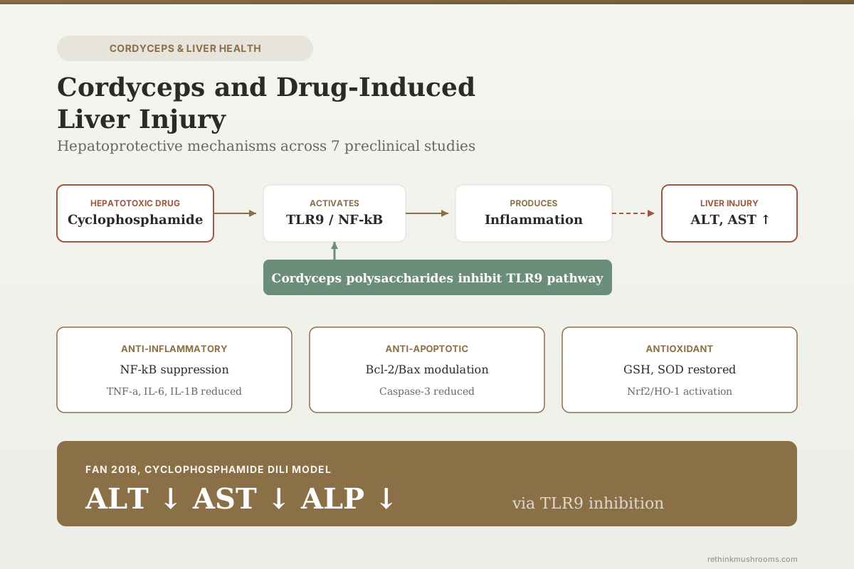
Cordyceps sinensis polysaccharides reduced cyclophosphamide-induced liver injury in mice by inhibiting TLR9 activation and downstream inflammatory signaling, decreasing ALT, AST, and ALP.
Fan et al., Food and Chemical Toxicology, 2018
Cyclophosphamide
the only drug-specific DILI model tested with cordyceps
NF-κB ↓ / Nrf2 ↑
convergent mechanism across 7 independent studies
0 Human Trials
no clinical trial of cordyceps for DILI has been published
Fan et al., Food and Chemical Toxicology, 2018
 View as image ↗
Drug-induced liver injury (DILI) is the leading cause of acute liver failure in the United States, responsible for roughly half of all cases. It occurs when a medication or its metabolites damage hepatocytes directly (dose-dependent, like acetaminophen) or trigger an immune-mediated reaction (idiosyncratic, like isoniazid or amoxicillin-clavulanate). Cordyceps species have been tested in several animal models of toxicant-induced liver damage, including one study using a specific pharmaceutical agent. For the broader picture of cordyceps and liver biology, see the cordyceps and liver health overview. This article focuses specifically on what happens when cordyceps compounds meet drug- and chemical-induced hepatotoxicity in controlled experiments.
Only one published study has tested cordyceps against a pharmaceutical drug causing liver injury: Fan and colleagues' 2018 work using cyclophosphamide.[1] The remaining evidence comes from chemical hepatotoxicity models (CCl4, D-galactosamine/lipopolysaccharide, alcohol, ischemia/reperfusion) that share mechanistic overlap with clinical DILI but are not direct equivalents. That distinction matters throughout this article.
Key Takeaways
- Cordyceps sinensis polysaccharides combined with Ganoderma atrum polysaccharides reduced cyclophosphamide-induced ALT, AST, and ALP in mice, with C. sinensis acting specifically through TLR9 pathway inhibition (Fan et al., Food and Chemical Toxicology, 2018)
- Cordycepin at 200 mg/kg elevated survival rate and reduced ALT, AST, hepatocyte apoptosis, and necrosis in mice with GalN/LPS-induced fulminant hepatic failure by suppressing TLR4 and NF-κB (Li et al., Mediators of Inflammation, 2017)
- Cordyceps sinensis mycelium prevented apoptosis via Bcl-2 upregulation and reduced TNF-α, IL-6, IL-1β, and caspase-3 in GalN/LPS-induced fulminant hepatic failure mice (Cheng et al., American Journal of Chinese Medicine, 2014)
- Cultured mycelium Cordyceps sinensis alleviated CCl4-induced liver inflammation and fibrosis in mice by activating hepatic natural killer cells and increasing NKG2D expression (Peng et al., Acta Pharmacologica Sinica, 2016)
- Cordycepin at 25 and 50 mg/kg decreased ALT, AST, TNF-α, IL-6, and IL-1β in hepatic ischemia/reperfusion injury via MAPK/NF-κB pathway inhibition (Ding et al., Mediators of Inflammation, 2022)
- One case report documented hepatoportal sclerosis in an 81-year-old woman after 5 years of daily cordyceps tablets, resolving after discontinuation (Kaur et al., ACG Case Reports Journal, 2023)
What Drug-Induced Liver Injury Actually Is
DILI falls into two categories. Intrinsic DILI is dose-dependent and predictable: acetaminophen overwhelms glutathione conjugation at high doses, producing NAPQI, which binds covalently to mitochondrial proteins and triggers hepatocyte necrosis. Idiosyncratic DILI is unpredictable, dose-independent, and involves immune-mediated mechanisms or metabolic idiosyncrasies that vary between individuals. Drugs like isoniazid, amoxicillin-clavulanate, and diclofenac are common causes.
The preclinical models used to study hepatoprotective compounds do not replicate either category precisely. Cyclophosphamide hepatotoxicity (used in Fan 2018) is the closest to clinical DILI: cyclophosphamide is an actual pharmaceutical that causes liver damage through its acrolein metabolite. GalN/LPS produces fulminant hepatic failure that resembles viral or drug-induced acute liver failure in its immune activation pattern. CCl4, while mechanistically useful, is a chemical toxicant rather than a drug. These distinctions determine how much weight each study carries when evaluating cordyceps for DILI specifically.
Fan 2018: The Only Pharmaceutical DILI Study
Fan and colleagues tested combined fungal polysaccharides (CFP) from Cordyceps sinensis (CSP) and Ganoderma atrum (PSG) against cyclophosphamide-induced liver injury in mice.[1] Cyclophosphamide is an alkylating chemotherapy agent whose hepatotoxicity involves oxidative stress, mitochondrial dysfunction, and inflammatory cytokine release via its metabolite acrolein.
The combined polysaccharide treatment reduced serum ALT, AST, and ALP. Mechanistically, the two fungal polysaccharides acted through complementary pathways:
- Cordyceps sinensis polysaccharides inhibited TLR9 activation and its downstream inflammatory cascade, including pro-inflammatory cytokines, inducible nitric oxide synthase (iNOS), and cyclooxygenase-2 (COX-2)
- Ganoderma atrum polysaccharides activated PPARα (peroxisome proliferator-activated receptor alpha), increasing hepatic glutathione peroxidase and glutathione content depleted by cyclophosphamide, and prevented mitochondria-dependent apoptosis via Bcl-2 family protein regulation
The TLR9 finding is mechanistically specific. TLR9 recognizes unmethylated CpG DNA released from damaged cells, and cyclophosphamide's cytotoxicity generates abundant cellular debris containing these danger-associated molecular patterns. Cordyceps sinensis polysaccharides blocking this specific receptor is a targeted anti-inflammatory mechanism rather than a generic antioxidant effect.
The limitation: this study used cordyceps polysaccharides combined with ganoderma polysaccharides, making it impossible to attribute the full hepatoprotective effect to cordyceps alone. The complementary mechanism (cordyceps on TLR9/inflammation, ganoderma on PPARα/antioxidant) suggests both components contributed, but the individual dose-response for cordyceps was not isolated. For how cordyceps interacts with liver fibrosis pathways specifically, see the cordyceps and liver fibrosis article.
GalN/LPS Fulminant Hepatic Failure: Two Independent Studies
D-galactosamine sensitizes hepatocytes to LPS-induced TNF-α damage by depleting UTP, disrupting RNA and protein synthesis. The result is fulminant hepatic failure with massive hepatocyte apoptosis and necrosis. This model is considered relevant to clinical acute liver failure because it reproduces the TNF-α-driven immune activation pattern seen in severe drug reactions and viral hepatitis.
Cheng 2014: Cordyceps sinensis Mycelium
Cheng and colleagues pretreated mice with Cordyceps sinensis mycelium extract before GalN/LPS challenge.[2] The extract prevented hepatocyte apoptosis by upregulating Bcl-2 (anti-apoptotic) and downregulating Bax (pro-apoptotic), reduced TNF-α, IL-6, and IL-1β, decreased caspase-3 activation, and preserved liver architecture on histology.
The anti-apoptotic mechanism is relevant to DILI because many drug-induced liver injuries involve both necrosis and apoptosis. Acetaminophen toxicity, for instance, produces primarily necrosis, while idiosyncratic DILI from drugs like diclofenac involves significant apoptotic signaling. A compound that modulates both pathways has broader theoretical applicability.
Li 2017: Cordycepin (3'-Deoxyadenosine)
Li and colleagues tested cordycepin, the primary bioactive nucleoside from Cordyceps militaris, at 200 mg/kg for 3 weeks before GalN/LPS injection.[3] Results:
- Elevated survival rate compared to untreated GalN/LPS controls
- Reduced serum ALT and AST
- Suppressed hepatocyte apoptosis and necrosis on histology
- Inhibited neutrophil and macrophage infiltration into liver tissue
- Decreased TNF-α, IL-6, and IL-1β through TLR4 and NF-κB suppression
- Reduced reactive oxygen species and lipid peroxidation via decreased NAD(P)H oxidase activity
- Prevented excessive autophagy in hepatic tissue
The survival rate finding is a hard endpoint that animal studies on liver protection rarely report. Most hepatoprotective studies measure only biochemical markers (ALT, AST) and histological scores. A survival benefit in a fulminant hepatic failure model is a stronger signal than enzyme reduction alone. For fatty liver connections involving some of these same pathways, see cordyceps for fatty liver.
Ding 2022: Hepatic Ischemia/Reperfusion Injury
Hepatic ischemia/reperfusion (I/R) injury occurs during liver surgery, transplantation, and hemorrhagic shock. It is not DILI in the traditional sense, but it shares the same downstream damage pathways: oxidative stress, inflammatory cytokine release, NF-κB activation, and hepatocyte apoptosis. It is included here because surgical liver injury is a clinical scenario where hepatoprotective compounds have translational potential.
Ding and colleagues tested cordycepin at 25 mg/kg (low dose) and 50 mg/kg (high dose) in BALB/c mice for 7 days before surgical hepatic I/R.[4] At both 6 and 24 hours post-injury:
- Serum ALT and AST decreased in cordycepin-treated groups
- Hepatocyte apoptosis was reduced
- TNF-α, IL-6, and IL-1β were suppressed
- The MAPK/NF-κB signaling pathway was inhibited at both the phosphorylation and transcription levels
The 25 mg/kg dose is considerably lower than the 200 mg/kg used in Li 2017's GalN/LPS study, suggesting dose-response relationships may vary substantially across injury models. Whether the lower dose would be effective in a chemical DILI model has not been tested.
Peng 2016: CCl4 Liver Inflammation and Fibrosis
Peng and colleagues administered cultured mycelium Cordyceps sinensis (CMCS) to mice with CCl4-induced chronic liver injury.[5] CCl4 is a chemical toxicant rather than a drug, but its CYP2E1-mediated activation to the trichloromethyl radical mirrors the metabolic activation step in many DILI cases (acetaminophen via CYP2E1 to NAPQI, for instance).
CMCS treatment produced:
- Improved liver function markers
- Reduced liver inflammation
- Attenuated liver fibrosis
- Increased numbers of hepatic natural killer (NK) cells
- Elevated expression of NKG2D (an activating receptor on NK cells)
The NK cell mechanism distinguishes this study from the others. Hepatic NK cells are part of the innate immune system's response to liver injury, and their activation by NKG2D ligands on stressed hepatocytes can kill activated hepatic stellate cells, which are responsible for fibrosis. CMCS promoting NK cell-mediated killing of stellate cells is a distinct anti-fibrotic mechanism that operates through immune regulation rather than direct antioxidant or anti-inflammatory effects. For the broader picture of how mushrooms including chaga and antrodia approach liver protection, those species-specific articles provide comparative context.
Alcohol and Lipid-Related Hepatotoxicity
Cha 2013: Cordycepin-Enriched C. militaris and Alcohol
Cha and colleagues fed rats cordycepin-enriched Cordyceps militaris at 3% (w/w) of diet for 4 weeks alongside alcohol administration, using silymarin as a positive control.[6] The cordycepin-enriched preparation prevented alcohol-induced ALT and AST elevations and reduced lipid droplet accumulation in hepatocytes.
The specific mechanism involved increased alcohol dehydrogenase (ADH) and acetaldehyde dehydrogenase (ALDH) activities. These enzymes convert ethanol to acetaldehyde and then to acetate, meaning cordycepin enhanced the liver's capacity to metabolize alcohol rather than just mopping up downstream damage. This is mechanistically relevant to DILI because many drug hepatotoxicities involve impaired drug metabolism or accumulation of toxic metabolites.
Wang 2015: C. militaris Residue Polysaccharide
Wang and colleagues isolated residue polysaccharide (RPS) from Cordyceps militaris SU-12 and tested its hepatoprotective activity.[7] The polysaccharide, composed primarily of glucose with minor arabinose and mannose, reduced blood and liver lipid levels, improved glutamate pyruvate transaminase activity, and enhanced antioxidant capacity. Histological examination confirmed reduced liver cell injury. This study extends the hepatoprotective evidence to the polysaccharide fraction remaining after primary extraction, suggesting that waste products from cordyceps processing retain bioactivity.
Tan 2024: Transcriptomic Evidence in Piglets
Tan and colleagues moved beyond rodents, testing cordycepin and Cordyceps militaris extract in LPS-challenged weaned piglets and using transcriptomic and metabolomic profiling to map the response.[8] Both treatments reduced C-reactive protein and ALT/AST levels. Transcriptomic analysis revealed that cordycepin suppressed cell adhesion molecule pathways, while the whole extract downregulated complement cascades and cytokine-receptor interactions.
The pig model is relevant because porcine liver physiology is closer to human liver physiology than rodent models. Pigs metabolize drugs through similar CYP450 pathways, have comparable liver-to-body-weight ratios, and share hepatic immune cell populations with humans. Protection in piglets is a step closer to translational relevance than protection in mice, though still preclinical.
The Convergent Mechanism Across All Studies
Seven independent studies using six different injury models converge on the same signaling profile:
- NF-κB suppression: documented in Fan 2018, Li 2017, Ding 2022, Peng 2016, and Tan 2024
- TLR pathway inhibition: TLR9 in Fan 2018, TLR4 in Li 2017
- Anti-apoptotic signaling: Bcl-2/Bax ratio modulation in Cheng 2014 and Fan 2018
- Antioxidant enzyme restoration: GSH, SOD, catalase in Wang 2015, Cha 2013
- Pro-inflammatory cytokine reduction: TNF-α, IL-6, IL-1β across all studies
- Nrf2/HO-1 activation: implied by antioxidant enzyme induction patterns
The consistency across different cordyceps preparations (whole mycelium, polysaccharide fractions, purified cordycepin), different injury types (cyclophosphamide, GalN/LPS, CCl4, alcohol, I/R, LPS), and different species (mice, rats, piglets) strengthens the biological plausibility. It does not substitute for testing in the specific clinical scenarios where DILI occurs in humans. For how the related species cordyceps interacts with viral hepatitis, that intersection is covered separately.
The Safety Counterweight
Cordyceps is rated "E" (unlikely cause) for clinically apparent liver injury by LiverTox, and no serum aminotransferase elevations during therapy have been reported in clinical trials. One case report complicates the otherwise clean safety profile.
Kaur and colleagues documented hepatoportal sclerosis in an 81-year-old woman who had taken cordyceps tablets daily for 5 years.[9] She presented with splenomegaly (21.5 cm), thrombocytopenia (76 x 10^3/uL), and weight loss. Liver biopsy showed loss of small portal veins with features of portal vein obstruction. After discontinuing cordyceps, spleen size decreased to 17.8 cm within 2 years and platelet count improved to 106 x 10^3/uL.
Hepatoportal sclerosis is a distinct entity from DILI. It involves obliteration of small portal veins leading to non-cirrhotic portal hypertension, and its mechanism is vascular rather than hepatocellular. Whether the cordyceps caused, contributed to, or coincided with this condition cannot be determined from a single case. The temporal relationship (improvement after discontinuation) is suggestive but not definitive, given the patient's age and the possibility of undiagnosed pre-existing vascular disease.
For other liver-related safety information across mushroom species, the chaga and liver protection article discusses reishi hepatotoxicity case reports.
What Would Confirm the Effect in Humans
A clinically meaningful trial would require: a standardized cordyceps preparation with characterized cordycepin and polysaccharide content; a patient population at high DILI risk (chemotherapy patients receiving hepatotoxic regimens, tuberculosis patients on isoniazid, or patients recovering from acetaminophen overdose); baseline and serial liver function tests (ALT, AST, GGT, bilirubin, INR); and an active comparator or placebo arm with adequate sample size for the expected DILI incidence. Such a trial has not been published, registered, or announced at the time of writing.
The preclinical mechanistic convergence, with Fan 2018's cyclophosphamide data as the most directly relevant study, provides a biological rationale for designing one. It is not a substitute for having results from one. The clinical question is whether cordyceps polysaccharides or cordycepin can reduce the incidence or severity of DILI in patients receiving hepatotoxic drugs, and that question remains entirely unanswered.
The Bottom Line
Cordyceps compounds have been tested against drug- and chemical-induced liver injury in seven independent animal studies. Only one (Fan 2018) used a pharmaceutical drug (cyclophosphamide) as the hepatotoxicant, finding that Cordyceps sinensis polysaccharides reduced ALT, AST, and ALP through TLR9 pathway inhibition. The remaining studies used GalN/LPS, CCl4, alcohol, LPS, and ischemia/reperfusion models, consistently showing NF-κB suppression, anti-apoptotic signaling, and antioxidant enzyme restoration. Cordycepin at 200 mg/kg improved survival in fulminant hepatic failure mice (Li 2017). One case report documents hepatoportal sclerosis after 5 years of daily cordyceps, resolved by discontinuation. No human trial of cordyceps for DILI prevention or treatment has been published.
Frequently Asked Questions
Sources & References
- 1RTHM-00507·Fan, Songtao et al. (2018). “Combinatorial usage of fungal polysaccharides from Cordyceps sinensis and Ganoderma atrum ameliorate drug-induced liver injury in mice..” Food and chemical toxicology : an international journal published for the British Industrial Biological Research Association.Study breakdown →PubMed →↩
- 2RTHM-00315·Cheng, Yu-Jung et al. (2014). “Cordyceps sinensis prevents apoptosis in mouse liver with D-galactosamine/lipopolysaccharide-induced fulminant hepatic failure..” The American journal of Chinese medicine.Study breakdown →PubMed →↩
- 3RTHM-00478·Li, Jin et al. (2017). “The Protective Effect of Cordycepin on D-Galactosamine/Lipopolysaccharide-Induced Acute Liver Injury..” Mediators of inflammation.Study breakdown →PubMed →↩
- 4RTHM-00754·Ding, Jiameng et al. (2022). “Cordycepin Protects against Hepatic Ischemia/Reperfusion Injury via Inhibiting MAPK/NF-κB Pathway..” Mediators of inflammation.Study breakdown →PubMed →↩
- 5RTHM-00440·Peng, Yuan et al. (2016). “Cultured Mycelium Cordyceps sinensis allevi¬ates CCl4-induced liver inflammation and fibrosis in mice by activating hepatic natural killer cells..” Acta pharmacologica Sinica.Study breakdown →PubMed →↩
- 6RTHM-00280·Cha, Jae-Young et al. (2013). “Protective effect of cordycepin-enriched Cordyceps militaris on alcoholic hepatotoxicity in Sprague-Dawley rats..” Food and chemical toxicology : an international journal published for the British Industrial Biological Research Association.Study breakdown →PubMed →↩
- 7RTHM-00407·Wang, Liqin et al. (2015). “Antihyperlipidemic and hepatoprotective activities of residue polysaccharide from Cordyceps militaris SU-12..” Carbohydrate polymers.Study breakdown →PubMed →↩
- 8RTHM-00912·Tan, Ding et al. (2024). “Transcriptomic and Metabolomic Analyses Reveal the Attenuating Role of Cordycepin and Cordyceps militaris Extract on Acute Liver Injury Induced by LPS in Piglets..” Animals : an open access journal from MDPI.Study breakdown →PubMed →↩
- 9RTHM-00834·Kaur, Bhupinder et al. (2023). “Traditional Chinese Medicine, Cordyceps, Related to Hepatoportal Sclerosis..” ACG case reports journal.Study breakdown →PubMed →↩