Cordyceps Liver Health
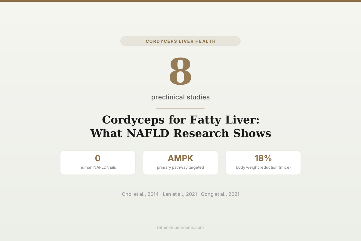
8 preclinical studies
Eight animal studies have tested cordyceps or cordycepin against NAFLD and hepatic steatosis. Most report reduced liver triglycerides, lower inflammatory markers, and improved insulin sensitivity. Zero human trials have been published.
Choi et al., Nutr Res Pract, 2014; Lan et al., Hepatology, 2021
0
human NAFLD trials
AMPK
primary pathway targeted
Cordycepin
most-studied active compound
Choi et al., Nutr Res Pract, 2014; Lan et al., Hepatology, 2021
 View as image ↗
Non-alcoholic fatty liver disease affects roughly 30% of adults globally, making it the most common chronic liver condition. Cordyceps, particularly Cordyceps militaris and its primary bioactive compound cordycepin, has been tested against NAFLD in multiple animal models over the past decade. The findings are consistent: reduced liver fat, lower inflammatory cytokines, and improved insulin resistance markers. One study published in Hepatology, a top-tier liver journal, mapped the specific molecular pathway through which cordycepin activates AMPK to reverse steatohepatitis in mice.[1] But the evidence remains entirely preclinical, no human trial has tested cordyceps specifically for NAFLD. This article reviews what eight studies found, the mechanisms they identified, and one contradictory result that complicates the picture. For the broader view of cordyceps and liver health, that article covers the full hepatoprotective evidence.
Key Takeaways
- In ob/ob mice fed 1% C. militaris extract for 10 weeks, hepatic triglyceride content decreased and serum ALT, TNF-alpha, and IL-6 all dropped compared to controls (Choi et al., 2014)
- Cordycepin activated AMPK signaling in a NASH mouse model, reducing hepatic steatosis, inflammation, and fibrosis markers in a study published in Hepatology (Lan et al., 2021)
- Cordycepin reduced body weight by 18% and liver weight in HFD-fed mice while downregulating lipid anabolism proteins and upregulating beta-oxidation pathways (Gong et al., 2021)
- Fermented C. militaris extract reduced hepatic lipid droplets and triglycerides by upregulating fatty acid oxidation genes including CPT1A and ACOX1 in HFD-fed mice (Tran et al., 2019)
- Cordyceps extract inhibited ceramide biosynthesis by suppressing serine palmitoyltransferase, improving insulin resistance and hepatic steatosis in obese mice (Li et al., 2022)
- One contradictory study found C. sinensis polysaccharides aggravated liver fibrosis and steatosis in HFD-fed mice by altering gut microbiota composition (Chen et al., PLoS ONE, 2020)
Why Cordyceps Is Studied for Fatty Liver Disease
NAFLD describes fat accumulation in liver cells without significant alcohol consumption. It ranges from simple steatosis (fat buildup with minimal inflammation) to non-alcoholic steatohepatitis (NASH), which adds inflammation and cell damage. NASH can progress to fibrosis, cirrhosis, and liver failure. The primary drivers are insulin resistance, dyslipidemia, and chronic low-grade inflammation, all conditions where cordyceps compounds have shown activity in other contexts.
The interest in cordyceps for NAFLD comes from three observations in the broader literature. First, cordycepin (3'-deoxyadenosine) activates AMP-activated protein kinase (AMPK), a central regulator of lipid metabolism in hepatocytes. Second, C. militaris extracts reduce inflammatory cytokines like TNF-alpha and IL-6 in multiple disease models. Third, cordyceps polysaccharides have shown antioxidant activity in liver tissue. Whether these properties translate to meaningful NAFLD improvement is what the studies below tested.
The distinction between NAFLD and NASH matters for evaluating cordyceps research. NAFLD alone (simple steatosis) is reversible with lifestyle changes and may not cause lasting liver damage. NASH, which affects an estimated 3-5% of the global population, involves active inflammation and hepatocyte injury that can progress to cirrhosis. Most cordyceps studies model HFD-induced steatosis, but the Lan 2021 study specifically targeted NASH, making its findings more clinically relevant for the more serious form of the disease.
The Foundational Study: C. militaris in ob/ob Mice
The first direct test of cordyceps against NAFLD came from Choi et al. (2014), using genetically obese (ob/ob) mice, a standard NAFLD model due to their leptin deficiency and spontaneous fatty liver development.[2]
Four-week-old male ob/ob mice received either a standard AIN-93G diet or the same diet supplemented with 1% C. militaris water extract for 10 weeks. The study measured serum glucose, insulin, free fatty acids (FFA), alanine transaminase (ALT), proinflammatory cytokines, and hepatic lipid content.
The cordyceps group showed reduced serum glucose and improved HOMA-IR (a measure of insulin resistance). Serum FFA levels dropped. Hepatic total lipids and triglyceride contents both decreased. Serum ALT activity (a marker of liver cell damage) fell, as did TNF-alpha and IL-6 levels. On the antioxidant side, hepatic glutathione (GSH) increased and lipid peroxide levels decreased.
The study's strengths: it used a well-characterized obesity model and measured multiple relevant endpoints. Its weaknesses: the sample size was small, it used a single dose level (1% of diet), and it could not identify which specific compounds drove the effects. Cordyceps extract contains cordycepin, polysaccharides, adenosine, and other bioactives. Separating their contributions required follow-up work.
Cordycepin and AMPK: The Hepatology Study
The most rigorous investigation of cordycepin's mechanism against NASH was published in Hepatology by Lan et al. (2021).[1] This is notable because Hepatology is among the highest-impact journals in liver research, reflecting the significance the editors assigned to the findings.
The study tested cordycepin in mice with diet-induced NASH and found that it ameliorated steatohepatitis through activation of the AMPK signaling pathway. AMPK is sometimes called the cell's "energy sensor." When activated, it shifts hepatic metabolism away from fat synthesis (lipogenesis) and toward fat breakdown (beta-oxidation). In NAFLD, AMPK activity is typically suppressed.
Lan et al. demonstrated that cordycepin directly activated AMPK in hepatocytes, which led to reduced expression of lipogenic enzymes (SREBP-1c, FAS, ACC) and increased expression of fatty acid oxidation genes. The result was reduced hepatic steatosis, lower inflammation markers, and less fibrosis in the NASH model.
This pathway matters for understanding how cordycepin differs from whole cordyceps extract. Where the Choi 2014 study showed broad improvements without identifying a mechanism, the Lan 2021 study pinpointed AMPK activation as a primary driver.
For how cordyceps affects liver fibrosis specifically, see cordyceps and liver fibrosis.
Cordycepin Reduces Body Weight and Liver Fat in HFD Models
Gong et al. (2021) tested cordycepin directly in mice fed a high-fat diet (HFD), the most common method for inducing NAFLD experimentally.[3]
Cordycepin treatment reduced body weight by approximately 18%, decreased liver weight, and relieved liver injury as measured by histopathological examination and liver enzyme markers. At the molecular level, cordycepin downregulated proteins involved in lipid anabolism (fat-building pathways) and upregulated proteins involved in beta-oxidation (fat-burning pathways).
The study also tracked inflammatory responses. Cordycepin-treated mice showed reduced hepatic inflammation, consistent with the anti-inflammatory effects seen in the Choi 2014 and Lan 2021 studies. The dual action, reducing both fat accumulation and inflammation, is relevant because NAFLD progression to NASH depends on both factors (the "two-hit hypothesis").
Autophagy: A Third Mechanism
Li et al. (2019) identified a different pathway through which cordycepin reduces liver fat: protective autophagy.[4]
Autophagy is the cell's cleanup process, breaking down and recycling damaged organelles and accumulated lipids. In NAFLD, hepatic autophagy is impaired, contributing to lipid droplet accumulation. Li et al. found that cordycepin induced autophagy in hepatocytes via the PKA/mTOR pathway, which reduced intracellular lipid accumulation.
This is a distinct mechanism from AMPK activation (Lan 2021), though the two pathways interact. AMPK activation can itself promote autophagy by inhibiting mTOR. The Li 2019 study suggests cordycepin may engage multiple overlapping pathways to clear hepatic fat.
Fermented Extracts and Fatty Acid Oxidation
Tran et al. (2019) took a different approach, testing a fermented C. militaris extract rather than isolated cordycepin.[5]
The study used HFD-fed mice and found that fermented extract reduced body weight, lowered plasma liver enzyme activities (AST, ALT), and decreased hepatic lipid droplets and triglyceride levels. The mechanism was upregulation of fatty acid oxidizing genes, specifically CPT1A and ACOX1, with activation of fatty acid oxidation via SPHK2 (sphingosine kinase 2) in the liver.
This study is relevant because fermented extracts are closer to commercial cordyceps supplements than purified cordycepin. The fact that a whole fermented preparation showed hepatoprotective effects through a defined mechanism adds practical relevance, though the dose used in mice does not translate directly to human supplementation.
Ceramide Biosynthesis: A Newer Target
Li et al. (2022) found that cordyceps extract inhibited ceramide biosynthesis, a pathway linking obesity to insulin resistance and liver fat accumulation.[6]
Ceramides are lipid molecules that accumulate in liver and muscle tissue during obesity, where they impair insulin signaling and promote hepatic steatosis. Li et al. showed that cordyceps suppressed serine palmitoyltransferase (SPT), the first enzyme in the ceramide biosynthesis pathway. This reduced tissue ceramide levels, improved insulin sensitivity, and decreased hepatic fat content in obese mice.
This is a different angle from the AMPK and autophagy mechanisms described above. It suggests cordyceps may affect NAFLD through at least three independent pathways: AMPK-mediated fat metabolism, autophagy-mediated lipid clearance, and ceramide-mediated insulin sensitization.
Protection Against Palmitic Acid-Induced Liver Damage
Tsai et al. (2024) tested C. militaris against liver damage caused by a high palmitic acid diet, a model that specifically mimics the lipotoxicity seen in advanced NAFLD.[7]
Palmitic acid is the most abundant saturated fatty acid in Western diets, and excess palmitic acid is directly toxic to hepatocytes. Tsai et al. found that C. militaris extract protected against hepatocyte apoptosis (programmed cell death) and reduced liver fibrosis markers in their model. Published in Frontiers in Pharmacology, this 2024 study represents the most recent addition to the cordyceps-NAFLD literature.
The antihyperlipidemic activity of cordyceps polysaccharides has also been demonstrated separately. Wang et al. (2015) showed that a residue polysaccharide from C. militaris SU-12 had both antihyperlipidemic and hepatoprotective effects in animal models.[8]
For related hepatoprotective research on other mushrooms, Antrodia for liver health covers Taiwan's most-studied liver mushroom, and Agaricus blazei liver protection examines a different species with hepatoprotective properties.
The Contradictory Evidence
Not all cordyceps research on liver fat shows benefit. Chen et al. (2020) published a study in PLoS ONE (PMID: 32407370) that found polysaccharides isolated from Cordyceps sinensis actually aggravated liver fibrosis and steatosis in HFD-fed mice.
The mechanism was unexpected: C. sinensis polysaccharides altered gut microbiota composition, increasing the relative abundance of the Actinobacteria phylum. The high Actinobacteria abundance disrupted intestinal flora and contributed to NASH progression. The polysaccharide supplementation prevented body weight gain (a positive effect), but simultaneously worsened liver outcomes as measured by increased ALT, inflammation markers, and lipid metabolism markers.
This study matters for several reasons. First, it used C. sinensis polysaccharides, not cordycepin. Most of the positive NAFLD studies used either whole C. militaris extract or purified cordycepin. The species and the specific compound fraction may determine whether cordyceps helps or harms the fatty liver. Second, the gut microbiota mechanism highlights that the liver does not respond to cordyceps in isolation. The gut-liver axis mediates many effects, and altering gut bacteria can produce counterintuitive outcomes.
For other ways cordyceps affects the liver, see cordyceps and drug-induced liver injury and cordyceps and hepatitis research.
What the Evidence Does and Does Not Show
The preclinical case for cordycepin against NAFLD is mechanistically detailed. Multiple studies identify specific pathways (AMPK, autophagy, ceramide biosynthesis) through which the compound reduces liver fat and inflammation. The consistency across different research groups and animal models (ob/ob mice, HFD-fed mice, palmitic acid models) adds weight.
The limitations are significant. Every study reviewed here used animal models. No randomized controlled trial has tested cordyceps or cordycepin for NAFLD in humans. The doses used in animal studies do not have established human equivalents for NAFLD. The one contradictory study (Chen 2020) raises questions about whether certain cordyceps preparations could worsen liver outcomes, particularly through gut microbiota disruption. The distinction between C. militaris cordycepin and C. sinensis polysaccharides appears meaningful but is not fully characterized.
There is also a publication bias concern. Negative results are less likely to be published, meaning there may be additional failed experiments that never reached journals. The studies also use different cordyceps species (C. militaris vs. C. sinensis), different preparations (whole extract, fermented extract, purified cordycepin, isolated polysaccharides), and different dosing regimens, making direct comparisons difficult. A compound that works as purified cordycepin in a mouse model may not produce the same effect in a commercially available supplement containing a different concentration of active ingredients.
The Bottom Line
Cordycepin, the primary bioactive compound in Cordyceps militaris, reduces hepatic steatosis, inflammation, and insulin resistance through AMPK activation, autophagy induction, and ceramide biosynthesis inhibition in animal NAFLD models. The evidence is mechanistically detailed and consistent across multiple research groups. However, zero human trials exist for this specific indication, one study found C. sinensis polysaccharides worsened liver outcomes via gut microbiota disruption, and the clinical translation of these animal findings remains entirely hypothetical.
Frequently Asked Questions
Sources & References
- 1RTHM-01379·Lan, Tian et al. (2021). “Cordycepin Ameliorates Nonalcoholic Steatohepatitis by Activation of the AMP-Activated Protein Kinase Signaling Pathway..” Hepatology (Baltimore.Study breakdown →PubMed →↩
- 2RTHM-00316·Choi, Ha-Neul et al. (2014). “Cordyceps militaris alleviates non-alcoholic fatty liver disease in ob/ob mice..” Nutrition research and practice.Study breakdown →PubMed →↩
- 3RTHM-01356·Gong, Xiaobao et al. (2021). “Cordycepin attenuates high-fat diet-induced non-alcoholic fatty liver disease via down-regulation of lipid metabolism and inflammatory responses..” International immunopharmacology.Study breakdown →PubMed →↩
- 4RTHM-01086·Li, Tianjiao et al. (2019). “Cordycepin alleviates hepatic lipid accumulation by inducing protective autophagy via PKA/mTOR pathway..” Biochemical and biophysical research communications.Study breakdown →PubMed →↩
- 5RTHM-01127·Tran, Nguyen Khoi Song et al. (2019). “Fermented Cordyceps militaris Extract Prevents Hepatosteatosis and Adipocyte Hypertrophy in High Fat Diet-Fed Mice..” Nutrients.Study breakdown →PubMed →↩
- 6RTHM-00779·Li, Ying et al. (2022). “Cordyceps inhibits ceramide biosynthesis and improves insulin resistance and hepatic steatosis..” Scientific reports.Study breakdown →PubMed →↩
- 7RTHM-02006·Tsai, Wan-Ting et al. (2024). “Protective effects of Cordyceps militaris against hepatocyte apoptosis and liver fibrosis induced by high palmitic acid diet..” Frontiers in pharmacology.Study breakdown →PubMed →↩
- 8RTHM-00407·Wang, Liqin et al. (2015). “Antihyperlipidemic and hepatoprotective activities of residue polysaccharide from Cordyceps militaris SU-12..” Carbohydrate polymers.Study breakdown →PubMed →↩