Cordyceps and Sexual Health
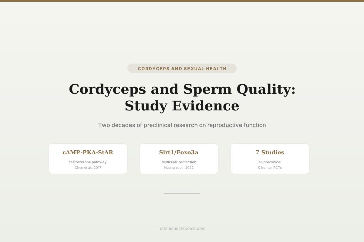
cAMP-PKA-StAR pathway activated
A 2017 review by Chen et al. established that cordycepin stimulates testosterone production through the cAMP-PKA-StAR steroidogenesis pathway in Leydig cells, providing a defined molecular mechanism for the traditional aphrodisiac claim.
Chen et al., Journal of Food and Drug Analysis, 2017
Testosterone
stimulated in vivo and in vitro
Sperm motility
improved in supplemented rats
Sirt1/Foxo3a
pathway protects testes in diabetic models
Chen et al., Journal of Food and Drug Analysis, 2017
 View as image ↗
Cordyceps sinensis has been used in traditional Chinese medicine as an aphrodisiac and reproductive tonic for centuries. The formal research testing these claims began in the early 2000s with Leydig cell studies, progressed through animal reproductive function trials, and has been synthesized in a 2017 review that established cordycepin's mechanism of action on testosterone biosynthesis. The evidence is entirely preclinical: mouse Leydig cells, rat supplementation studies, and diabetic animal models. No human randomized controlled trial has directly measured cordyceps effects on sperm parameters.[1]
The research base is narrow but mechanistically coherent. Multiple studies from independent labs converge on the same steroidogenic pathway. This article covers the testosterone production evidence, sperm quality outcomes in animal models, the diabetic fertility connection, and the specific molecular pathways identified. For testosterone-specific data, see Cordyceps and testosterone: does it increase male hormones?. For the broader sexual function picture, see Cordyceps and libido: research on sexual function.
Key Takeaways
- Chen et al. (2017) reviewed the evidence and established that cordycepin stimulates testosterone production through the cAMP-PKA-StAR steroidogenesis pathway in mouse Leydig cells, providing a defined molecular mechanism for the traditional reproductive tonic claim
- Hsu et al. (2003) demonstrated that Cordyceps sinensis, its water-soluble protein fraction (F2), and its polysaccharide/protein fraction (F3) all stimulated testosterone production in vitro in purified mouse Leydig cells (the water-soluble polysaccharide F1 had no effect), and each significantly elevated plasma testosterone in living mice
- Chang et al. (2008) supplemented Sprague-Dawley rats with dietary Cordyceps militaris mycelium (1% and 5%) for 6 weeks and found increased epididymal sperm count and motility alongside elevated serum testosterone and estradiol, while FSH, luteinizing hormone, and prolactin were unchanged
- Huang et al. (2004) showed that oral Cordyceps sinensis mycelium and its F2 and F3 fractions significantly raised plasma testosterone in both immature and mature mice within 3 to 7 days, without changing the weights of the reproductive organs
- Nguyen et al. (2021) found Cordyceps militaris at 100 mg/kg for 21 days improved epididymal sperm parameters, serum testosterone, and copulatory behavior in streptozotocin-induced diabetic rats
- Huang et al. (2022) identified cordycepin protection against diabetes-induced testicular damage through the Sirt1/Foxo3a pathway, reducing oxidative stress markers in testicular tissue
Testosterone Production: The Leydig Cell Evidence
Leydig cells in the testes produce testosterone in response to luteinizing hormone (LH) stimulation. The biosynthetic pathway involves cholesterol transport into mitochondria via the StAR protein (steroidogenic acute regulatory protein), followed by enzymatic conversion through the steroidogenic cascade.
Huang et al. (2001) provided the first evidence in normal (non-tumor) Leydig cells. Testing Cordyceps sinensis across concentrations from 0.1 to 10 mg/mL on purified mouse Leydig cells, the team found dose-dependent increases in testosterone production, reaching statistical significance at 3 mg/mL. The effect required new protein synthesis, and the data indicated that CS acts on the steroidogenic signaling pathway downstream of cyclic AMP formation.[2]
Hsu et al. (2003) extended this work with both in vitro and in vivo experiments. In purified mouse Leydig cells, whole Cordyceps sinensis extract, the water-soluble protein fraction (F2), and the poorly water-soluble polysaccharide/protein fraction (F3) all stimulated testosterone production, with maximal responses near 3 mg/mL; the water-soluble polysaccharide fraction (F1) had no effect. In living mice, CS, F2, and F3 each significantly elevated plasma testosterone.[3]
The in vivo confirmation was critical: it demonstrated that the testosterone-stimulating effect observed in isolated cells translates to measurable hormone changes in living animals, suggesting adequate bioavailability of the active compounds after oral administration.
Identifying the Pathway: cAMP-PKA-StAR
Chen et al. (2017) published a comprehensive review synthesizing a decade of Leydig cell studies and identifying the specific molecular pathway. Cordycepin, as an adenosine analogue, stimulates cAMP production. Elevated cAMP activates protein kinase A (PKA), which phosphorylates and activates the StAR protein. StAR transports cholesterol into mitochondria, the rate-limiting step in testosterone biosynthesis.[1]
This cAMP-PKA-StAR mechanism is the canonical pathway for LH-stimulated testosterone production. Cordycepin appears to activate the same pathway through its adenosine-like activity, essentially mimicking or amplifying the natural hormonal signal. The review also noted that CS promoted the expression of multiple steroidogenic enzymes downstream of StAR, including 3-beta-HSD and 17-beta-HSD, suggesting effects at multiple points in the testosterone biosynthetic cascade rather than a single bottleneck.
Sperm Production and Motility in Animal Models
Chang et al. (2008) directly measured sperm parameters in Sprague-Dawley rats supplemented with Cordyceps militaris mycelium. Ninety rats were divided into three groups of 30 — a control diet and diets supplemented with 1% or 5% CM mycelium — for 6 weeks. Epididymal sperm count rose significantly by the fifth week and peaked at the sixth, and the increase persisted for two weeks after supplementation stopped. The proportion of motile sperm also increased. Serum testosterone and estradiol rose, while FSH, luteinizing hormone (LH), and prolactin were unchanged.[4]
The hormonal pattern is informative. Testosterone rose without any accompanying increase in LH, the pituitary signal that normally drives Leydig cells. That dissociation points to CM acting directly on the testis rather than upstream on the hypothalamic-pituitary axis, consistent with the direct Leydig cell stimulation documented in the cell studies above. The parallel rise in estradiol is expected, since estradiol is produced by aromatization of testosterone.
Huang et al. (2004) tested oral CS mycelium and its fractions in male mice over shorter timeframes. Feeding CS, F2, or F3 at 0.02 and 0.2 mg per gram of body weight significantly raised plasma testosterone in both immature and mature mice within 3 to 7 days, without a meaningful change in the weights of the reproductive organs. This in vivo hormonal effect complements Chang's sperm-parameter data: the two studies together connect short-term testosterone elevation in mice to longer-term gains in sperm output in rats.[5]
Timeframe matters for interpreting these results. A full spermatogenic cycle takes roughly seven weeks in the rat, so Chang's six-week supplementation is long enough for a genuine effect on sperm production to emerge, which fits the observed rise between weeks five and six. The mouse studies, by contrast, captured hormonal changes within days — too brief to reflect new sperm production, and indeed they measured plasma testosterone rather than sperm counts.
Diabetic Models: Fertility Under Metabolic Stress
Diabetes impairs male fertility through oxidative stress damage to testicular tissue, reduced testosterone production, and altered sperm parameters. Two studies tested Cordyceps in this specific context.
Nguyen et al. (2021) treated streptozotocin-induced diabetic rats with Cordyceps militaris at 100 mg/kg daily for 21 days. The CM-treated diabetic rats showed improved epididymal sperm parameters (count, motility, morphology), elevated serum testosterone, and improved copulatory behavior compared to untreated diabetic controls. Antioxidant parameters in testicular tissue also improved.[6]
Huang et al. (2022) focused specifically on cordycepin and identified a molecular pathway for testicular protection. In diabetic mice, cordycepin ameliorated testicular damage through the Sirt1/Foxo3a pathway. Sirt1 is a NAD+-dependent deacetylase that regulates cellular stress responses; Foxo3a is a downstream transcription factor that controls antioxidant gene expression. Cordycepin activated this pathway, reducing oxidative stress markers and protecting testicular tissue architecture.[7]
The Sirt1/Foxo3a mechanism is distinct from the cAMP-PKA-StAR pathway identified for testosterone production. Cordycepin appears to have dual roles in male reproduction: stimulating testosterone biosynthesis through one pathway and protecting testicular tissue from oxidative damage through another. For erectile function evidence specifically, see Cordyceps for erectile dysfunction: evidence and mechanisms. For related research on female fertility, see Cordyceps and female sexual health: fertility and hormone research.
Species Differences: C. sinensis vs. C. militaris
The testosterone and steroidogenesis studies primarily used Cordyceps sinensis (Hsu 2003, Huang 2001, Huang 2004). The sperm parameter and diabetic model studies primarily used Cordyceps militaris (Chang 2008, Nguyen 2021, Huang 2022). Both species contain cordycepin, though C. militaris typically has higher cordycepin concentrations.
The Chen et al. (2017) review treated the two species' evidence as convergent, with cordycepin identified as the common active compound. Wild C. sinensis is prohibitively expensive, while cultured C. militaris is commercially accessible. If cordycepin is indeed the primary mediator of reproductive effects, then C. militaris products may be equivalent or superior to C. sinensis for this application, given their typically higher cordycepin content.
However, the Hsu et al. (2003) finding that the water-soluble protein fraction (F2) of C. sinensis also stimulated testosterone suggests that cordycepin is not the only active compound. Protein-based active compounds may differ between species, making direct species substitution an assumption rather than a demonstrated equivalence.
What the Evidence Does Not Show
Evidence Hierarchy
Cordyceps & Sperm Quality — An Upside-Down Evidence Pyramid
A normal evidence pyramid is widest at the bottom (mechanisms) and narrows to a single, decisive human trial at the top. Here it is inverted: the cell and animal tiers are crowded with studies, while the tier that would guide a real decision — a controlled trial of sperm parameters in men — is empty. Bar width reflects how much direct evidence exists at each tier, not the size of any effect.
Controlled human trial (sperm parameters)
gapDoes not yet exist
The evidence that would actually guide a man deciding whether to take cordyceps — a randomised, controlled trial measuring sperm count, motility, or morphology in people — has never been run
Whole-animal reproductive outcomes
evidenceChang 2008 · Nguyen 2021
The tier closest to a real fertility outcome: Cordyceps militaris raised epididymal sperm count and motility in rats over six weeks, and improved sperm parameters and copulatory behaviour in diabetic rats — but these are rodents, at doses far above ordinary supplementation
In vivo hormone & tissue effects (animals)
evidenceHsu 2003 · Huang 2004 · Huang 2022
Feeding Cordyceps sinensis raised plasma testosterone in mice within days, and cordycepin protected testicular tissue from oxidative damage in diabetic mice through the Sirt1/Foxo3a pathway — hormonal and protective signals, not fertility outcomes
Isolated Leydig cell steroidogenesis
evidenceHuang 2001 · Hsu 2003 · Chen 2017
The deepest, cleanest tier: in purified mouse Leydig cells, cordyceps stimulated testosterone dose-dependently, and a 2017 review mapped the mechanism to cordycepin driving the cAMP-PKA-StAR steroidogenesis pathway — the same signal luteinizing hormone uses
How to read it: the bars grow wider toward the bottom because the mechanistic and animal literature is deep and converges on a coherent steroidogenic pathway. But every step upward toward a human man loses evidence — the whole-animal sperm data is limited to rodents at high doses, and the controlled human trial that would settle the question has never been run. The gap at the top, not the quality of the mechanisms, is the honest limit of the cordyceps–sperm claim.
Sources: Chen 2017 [1]; Huang 2001 [2]; Hsu 2003 [3]; Chang 2008 [4]; Huang 2004 [5]; Nguyen 2021 [6]; Huang 2022 [7]. Every tier is preclinical — mouse Leydig cells, rat and mouse feeding studies, and diabetic-animal models. Bar widths are illustrative of evidence depth per tier, not quantitative effect sizes.
Every study in this article is preclinical: mouse Leydig cells, rat feeding studies, or diabetic animal models. No randomized controlled trial has measured sperm parameters (count, motility, morphology) in men taking Cordyceps supplements. No trial has evaluated time-to-pregnancy, fertilization rates, or any clinical fertility outcome.
The testosterone increases demonstrated in mice may not translate proportionally to humans. Leydig cell physiology differs between species, and the doses used in animal studies — whether high dietary fractions (1 to 5 percent of the diet) or daily doses of 100 mg/kg and above — may not correspond to achievable human supplementation levels once adjusted for body size.
The diabetic model studies show protective effects under metabolic stress, which is a different question from whether Cordyceps improves fertility in otherwise healthy men. Protecting damaged tissue from further harm is not the same as enhancing normal function above baseline.
The Bottom Line
Research on cordyceps and sperm quality spans two decades and converges on a defined mechanism: cordycepin stimulates testosterone biosynthesis through the cAMP-PKA-StAR pathway in Leydig cells, confirmed both in vitro and in vivo. Animal supplementation studies show improved sperm count, motility, and hormonal profiles. Diabetic models add a second mechanism: cordycepin protects testicular tissue via the Sirt1/Foxo3a antioxidant pathway. All evidence is preclinical. No human trial has measured sperm parameters or clinical fertility outcomes with Cordyceps supplementation, and the translation from animal doses to human supplementation levels remains undefined.
Frequently Asked Questions
Sources & References
- 1RTHM-00466·Chen, Yung-Chia et al. (2017). “Functional study of Cordyceps sinensis and cordycepin in male reproduction: A review..” Journal of food and drug analysis.Study breakdown →PubMed →↩
- 2RTHM-00037·Huang, B M et al. (2001). “Effects of Cordyceps sinensis on testosterone production in normal mouse Leydig cells..” Life sciences.Study breakdown →PubMed →↩
- 3RTHM-00056·Hsu, Chih Chao et al. (2003). “In vivo and in vitro stimulatory effects of Cordyceps sinensis on testosterone production in mouse Leydig cells..” Life sciences.Study breakdown →PubMed →↩
- 4RTHM-00127·Chang, Ying et al. (2008). “Effect of Cordyceps militaris supplementation on sperm production, sperm motility and hormones in Sprague-Dawley rats..” The American journal of Chinese medicine.Study breakdown →PubMed →↩
- 5RTHM-00070·Huang, Yuan-Li et al. (2004). “In vivo stimulatory effect of Cordyceps sinensis mycelium and its fractions on reproductive functions in male mouse..” Life sciences.Study breakdown →PubMed →↩
- 6RTHM-00710·Nguyen, Toan Van et al. (2021). “A study of the aphrodisiac properties of Cordyceps militaris in streptozotocin-induced diabetic male rats..” Veterinary world.Study breakdown →PubMed →↩
- 7RTHM-00767·Huang, Tao et al. (2022). “Cordycepin, a major bioactive component of Cordyceps militaris, ameliorates diabetes-induced testicular damage through the Sirt1/Foxo3a pathway..” Andrologia.Study breakdown →PubMed →↩