Mushrooms and Cardiac Protection
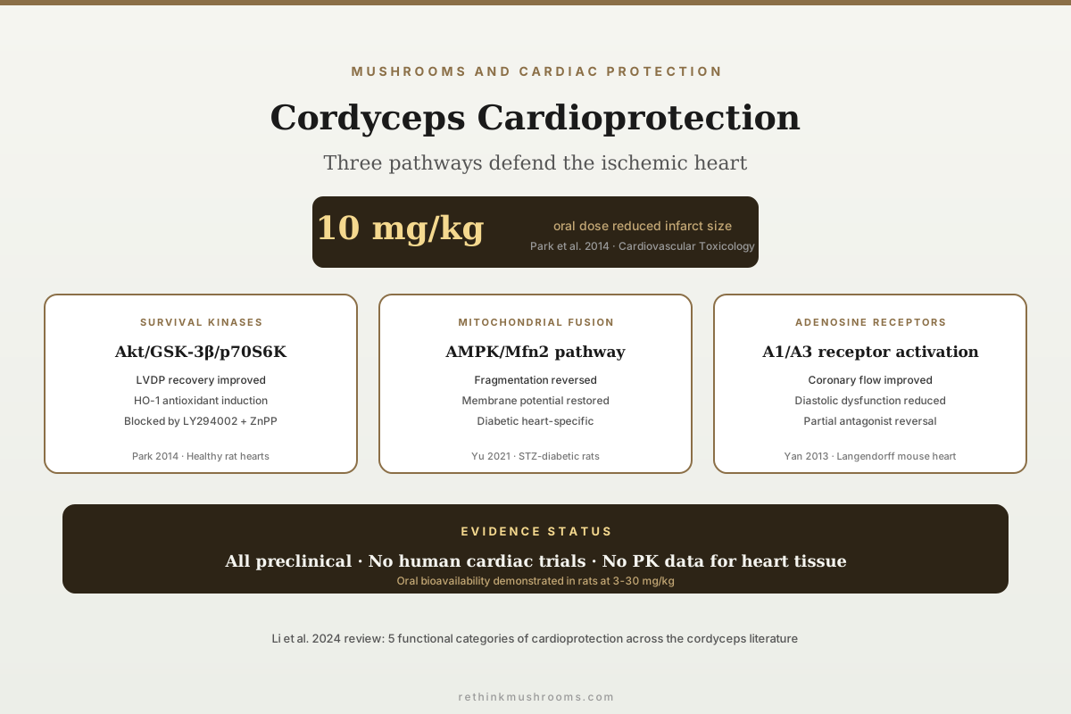
10 mg/kg Cardioprotective Dose
Oral cordycepin at 10 mg/kg significantly increased left ventricular developed pressure during reperfusion and reduced infarct size in rat hearts subjected to ischemia-reperfusion injury, acting through Akt/GSK-3β/p70S6K pathway activation and HO-1 induction (Park et al., Cardiovascular Toxicology, 2014).
Park et al., Cardiovascular Toxicology, 2014
AMPK/Mfn2
Cordycepin restored mitochondrial fusion and reduced infarct size in diabetic hearts through AMPK/Mfn2-dependent signalling (Yu et al., 2021)
Adenosine receptors
Cordyceps sinensis mycelia extract improved post-ischemic ventricular function partially via adenosine receptor activation (Yan et al., 2013)
5 pathways
A 2024 comprehensive review catalogued cordyceps cardioprotection across perfusion, ROS, inflammation, apoptosis, and regeneration (Li et al., 2024)
Park et al., Cardiovascular Toxicology, 2014
 View as image ↗
Myocardial ischemia-reperfusion (I/R) injury occurs when blood flow returns to heart tissue after a period of oxygen deprivation, paradoxically causing damage through reactive oxygen species, calcium overload, and inflammatory cascades. Cordyceps preparations and isolated cordycepin have been tested against this injury model across multiple preclinical systems, from isolated perfused hearts to intact diabetic animals. The evidence converges on at least three distinct cardioprotective mechanisms: survival kinase pathway activation, mitochondrial dynamics regulation, and adenosine receptor-mediated protection.[1][2] For the broader context of mushroom cardiac protection, see mushrooms and cardiac protection: mechanisms of heart defense; for the Antrodia parallel, see Antrodia camphorata and heart protection: Taiwan's research-backed fungus.
Key Takeaways
- Park et al. (2014) in Cardiovascular Toxicology showed oral cordycepin at 3-30 mg/kg dose-dependently increased left ventricular developed pressure and reduced infarct size in rat I/R models through activation of the Akt/GSK-3β/p70S6K signalling pathway and induction of heme oxygenase-1 (HO-1)
- Yu et al. (2021) in Frontiers in Pharmacology demonstrated cordycepin reduced I/R injury specifically in diabetic hearts by upregulating AMPK/Mfn2-dependent mitochondrial fusion, restoring mitochondrial membrane potential and reducing cardiomyocyte apoptosis in both STZ-induced diabetic rats and high-glucose H9c2 cells
- Yan et al. (2013) in Phytotherapy Research found Cordyceps sinensis mycelia extracts reduced post-ischemic diastolic dysfunction and improved ventricular pressure development and coronary flow in mouse Langendorff hearts, with effects partially blocked by adenosine receptor antagonists
- Li et al. (2024) in the Journal of Ethnopharmacology published a comprehensive review cataloguing cordyceps cardioprotection across five pathways: improving blood perfusion, mitigating ROS damage, suppressing inflammation, preventing apoptosis, and promoting tissue regeneration
- Liu et al. (2014) showed Cordyceps sinensis at 4 mg/kg/day for 8 weeks protected against heart injury in a 5/6-nephrectomy CKD rat model, with metabolomic analysis revealing normalisation of cardiac metabolite profiles disrupted by chronic kidney disease
- Cordycepin crosses the blood-heart barrier when administered orally, with pharmacological effects detectable in cardiac tissue at doses as low as 3 mg/kg in rats, though human pharmacokinetic data for cardiac tissue exposure are absent
The Survival Kinase Pathway: Akt/GSK-3β/p70S6K
The RISK (Reperfusion Injury Salvage Kinase) pathway is the best-characterised endogenous cardioprotective cascade. When activated at the onset of reperfusion, it reduces mitochondrial permeability transition pore opening and limits infarct size. Park et al. (2014) tested whether cordycepin activates this pathway.
Sprague-Dawley rats received cordycepin orally at 3, 10, or 30 mg/kg, then underwent either isolated heart perfusion (Langendorff model) or in vivo regional ischemia (left anterior descending artery occlusion). In the Langendorff model, cordycepin at 10 mg/kg significantly increased left ventricular developed pressure (LVDP) during the reperfusion period compared to controls. In the in vivo model, cordycepin reduced myocardial infarct size dose-dependently.[1]
The molecular mechanism was mapped through Western blot analysis. Cordycepin increased phosphorylation of Akt, GSK-3β (at the inhibitory Ser9 site, preventing its pro-apoptotic activity), and p70S6K (a downstream effector of mTOR signalling). It also induced heme oxygenase-1 (HO-1), an enzyme that generates carbon monoxide and biliverdin with antioxidant and anti-inflammatory properties. The cardioprotective effect was abolished by LY294002 (a PI3K/Akt inhibitor) and ZnPP (an HO-1 inhibitor), confirming these are required rather than incidental pathways.
The dose-response across a 10-fold range (3-30 mg/kg) and the pharmacological blockade experiments make this the most mechanistically rigorous of the cordyceps cardiac studies. The oral administration route is relevant because it demonstrates bioavailability: cordycepin or its metabolites reach cardiac tissue at effective concentrations when given by mouth, not just by direct perfusion.
Diabetic Hearts: AMPK/Mfn2 Mitochondrial Fusion
Diabetic cardiomyopathy increases vulnerability to I/R injury through mitochondrial fragmentation, impaired fatty acid oxidation, and elevated oxidative stress. Yu et al. (2021) addressed this specifically high-risk population.
In STZ-induced diabetic rats, cordycepin reduced myocardial infarct size and improved cardiac function after I/R. In high-glucose-treated H9c2 cardiomyocytes (an in vitro model of diabetic cardiac stress), cordycepin restored mitochondrial membrane potential, reduced reactive oxygen species production, and inhibited cardiomyocyte apoptosis.[2]
The novel mechanistic contribution was identifying AMPK/Mfn2-dependent mitochondrial fusion as the cardioprotective pathway. Mfn2 (mitofusin 2) is a GTPase on the outer mitochondrial membrane that promotes fusion of fragmented mitochondria into interconnected networks. In diabetic hearts, Mfn2 expression is suppressed, leading to mitochondrial fragmentation and impaired energy production. Cordycepin upregulated AMPK phosphorylation, which in turn increased Mfn2 expression and restored the balance between mitochondrial fusion and fission.
AMPK activation by cordycepin has also been documented in the blood sugar literature (see cordyceps and blood sugar: hypoglycemic research), where it drives glucose metabolism improvements. The cardiac application shows the same upstream kinase activation produces tissue-specific downstream effects: glucose regulation in skeletal muscle, mitochondrial dynamics in cardiomyocytes. This shared AMPK node may explain why cordyceps preparations show pleiotropic benefits across metabolic and cardiovascular models.
Adenosine Receptor-Mediated Protection
Cordycepin (3'-deoxyadenosine) is a structural analogue of adenosine, the endogenous nucleoside that mediates cardioprotection through A1 and A3 adenosine receptors. Adenosine released during ischemia activates these receptors on cardiomyocytes, triggering protective signalling cascades that reduce calcium overload, preserve mitochondrial integrity, and limit cell death. Yan et al. (2013) tested whether Cordyceps sinensis extracts exploit this structural similarity to provide exogenous cardioprotection.
Using the Langendorff mouse heart perfusion model, they subjected isolated hearts to 30 minutes of global ischemia followed by 40 minutes of reperfusion. Hearts pre-treated with C. sinensis mycelia extracts showed reduced post-ischemic diastolic dysfunction (contracture during reperfusion, a hallmark of intracellular calcium overload) and improved recovery of LVDP and coronary flow. Oxidative stress markers in cardiac tissue homogenates, including malondialdehyde (a lipid peroxidation product) and superoxide dismutase activity, were significantly improved compared to untreated ischemic controls.[3]
The adenosine receptor contribution was established using pharmacological antagonists. When adenosine receptor blockers were co-administered with the C. sinensis extract, the cardioprotective effects were partially (but not completely) reversed. This indicates that adenosine receptor activation contributes to but does not fully account for the cardioprotection, consistent with the multi-pathway model where additional mechanisms (Akt activation, direct antioxidant activity of polysaccharide fractions) operate in parallel. The incomplete reversal by receptor antagonists suggests that adenosine receptor activation may account for roughly 40-60% of the cardioprotective effect, with the remainder operating through receptor-independent mechanisms.
This study used whole mycelia extract rather than isolated cordycepin, making it more representative of commercial C. sinensis preparations (including the Cs-4 fermentation product widely available as a supplement). Mycelia extracts contain not only cordycepin but also polysaccharides, adenosine itself, ergosterol, and mannitol, each of which has independent bioactivity. The partial adenosine receptor dependence suggests that the structural similarity between cordycepin and adenosine is pharmacologically functional in cardiac tissue, while the non-receptor-dependent component likely reflects contributions from other extract constituents.
The Comprehensive Mechanistic Picture
Li et al. (2024) published a comprehensive review in the Journal of Ethnopharmacology that catalogued cordyceps cardioprotective mechanisms across the published literature. They organised the evidence into five functional categories:[4]
Improving blood perfusion: cordyceps preparations increase coronary flow and reduce vascular resistance in perfused heart models, improving oxygen and nutrient delivery to ischemic myocardium.
Mitigating ROS damage: both whole extracts and cordycepin reduce superoxide, hydrogen peroxide, and lipid peroxidation products in cardiac tissue, primarily through HO-1 induction and direct antioxidant activity of polysaccharide fractions.
Suppressing inflammation: NF-kB pathway inhibition reduces TNF-α, IL-1β, and IL-6 production in cardiac tissue, limiting neutrophil infiltration and secondary damage during reperfusion.
Preventing apoptosis: Akt activation, Bcl-2 upregulation, and caspase-3/9 suppression reduce cardiomyocyte death in the peri-infarct zone, potentially limiting infarct expansion.
Promoting tissue regeneration: emerging evidence suggests cordyceps polysaccharides may support angiogenesis and cardiac progenitor cell activation, though this evidence is preliminary and limited to in vitro models.
Heart Protection in Chronic Kidney Disease
Liu et al. (2014) approached cardiac protection from a different angle: the cardiorenal syndrome. In chronic kidney disease, uremic toxins and metabolic derangements damage the heart. Using a 5/6-nephrectomy CKD rat model, they administered Cordyceps sinensis at 4 mg/kg/day for 8 weeks and performed metabolomic analysis of cardiac tissue.[5]
C. sinensis treatment normalised cardiac metabolite profiles disrupted by CKD, including restoration of energy metabolism intermediates and reduction of oxidative stress markers. This study bridges the cordyceps kidney literature (see cordyceps and diabetic kidney disease: nephroprotective effects) with the cardiac literature, showing that the same preparation protects both organs in a model of systemic metabolic disease.
Why Diabetic Hearts Matter More
The Yu et al. (2021) study deserves additional emphasis because diabetic patients face disproportionately worse outcomes from myocardial infarction. Diabetes impairs endogenous cardioprotective mechanisms: ischemic preconditioning (the heart's natural ability to protect itself through brief ischemic episodes) is blunted in diabetic myocardium. Mitochondrial fragmentation, elevated baseline oxidative stress, and impaired autophagy all reduce the diabetic heart's tolerance for ischemic insults.
The finding that cordycepin restores AMPK/Mfn2-dependent mitochondrial fusion specifically addresses one of the core vulnerabilities of the diabetic heart. Mfn2 downregulation in diabetes leads to small, fragmented mitochondria with reduced respiratory capacity and increased susceptibility to permeability transition pore opening during reperfusion. By rescuing Mfn2 expression through AMPK activation, cordycepin may restore a protective mechanism that diabetes has disabled. This positions cordycepin differently from conventional cardioprotective agents, most of which were developed and tested in non-diabetic models. However, the gap between this mechanistic rationale and clinical application remains large: no dosing, formulation, or delivery strategy has been optimised for human diabetic cardiac protection.
Limitations
All cardioprotective evidence is preclinical. No clinical trial has tested cordyceps or cordycepin for myocardial ischemia, angina, heart failure, or post-infarction recovery in human patients. The approved Chinese indication for C. sinensis preparations includes arrhythmia, but this is based on traditional use classification rather than contemporary randomised trial evidence.
Doses used in animal models (3-30 mg/kg cordycepin in rats) translate to approximate human equivalent doses of 0.5-5 mg/kg using body surface area scaling, but actual human cardiac tissue penetration after oral cordyceps supplementation has not been measured. Cordycepin's short plasma half-life (due to rapid deamination by adenosine deaminase) raises questions about whether sustained cardiac exposure is achievable with standard oral dosing regimens. Co-administration with adenosine deaminase inhibitors (such as pentostatin) extends cordycepin's half-life in oncology settings, but this approach has not been tested for cardioprotection.
The studies used different models (isolated perfused hearts, in vivo coronary occlusion, CKD-associated cardiac injury), different species (C. sinensis mycelia extract, isolated cordycepin, whole C. sinensis), and different animal models (healthy rats, diabetic rats, CKD rats). While this diversity strengthens the aggregate mechanistic picture, it means no single preparation-dose-population combination has been replicated across independent laboratories.
The 2024 comprehensive review (Li et al.) is a narrative review synthesising heterogeneous evidence; it is not a systematic review with meta-analytic pooling. The regenerative mechanism category (cardiac progenitor cell activation) has particularly thin evidence and relies primarily on in vitro observations. For how mushroom polysaccharides may contribute to cardiac remodelling, see mushroom polysaccharides and cardiac remodeling after injury.
The Bottom Line
Cordycepin demonstrates cardioprotective activity against myocardial ischemia-reperfusion injury through three established preclinical pathways: Akt/GSK-3β survival kinase activation (reducing infarct size in healthy rat hearts), AMPK/Mfn2-dependent mitochondrial fusion restoration (specifically effective in diabetic hearts), and adenosine receptor-mediated vascular and functional protection. Oral bioavailability to cardiac tissue has been demonstrated in rats at doses as low as 3 mg/kg. No human clinical trial has tested these effects, and pharmacokinetic data for human cardiac tissue exposure are absent.
Frequently Asked Questions
Sources & References
- 1RTHM-00343·Park, Eun-Seok et al. (2014). “Cordycepin, 3'-deoxyadenosine, prevents rat hearts from ischemia/reperfusion injury via activation of Akt/GSK-3β/p70S6K signaling pathway and HO-1 expression..” Cardiovascular toxicology.Study breakdown →PubMed →↩
- 2RTHM-00733·Yu, Houyou et al. (2021). “Cordycepin Decreases Ischemia/Reperfusion Injury in Diabetic Hearts via Upregulating AMPK/Mfn2-dependent Mitochondrial Fusion..” Frontiers in pharmacology.Study breakdown →PubMed →↩
- 3RTHM-00310·Yan, Xiao-Feng et al. (2013). “Cardiovascular protection and antioxidant activity of the extracts from the mycelia of Cordyceps sinensis act partially via adenosine receptors..” Phytotherapy research : PTR.Study breakdown →PubMed →↩
- 4RTHM-00891·Li, Yong et al. (2024). “Cordyceps: Alleviating ischemic cardiovascular and cerebrovascular injury - A comprehensive review..” Journal of ethnopharmacology.Study breakdown →PubMed →↩
- 5RTHM-00337·Liu, Xia et al. (2014). “Cordyceps sinensis protects against liver and heart injuries in a rat model of chronic kidney disease: a metabolomic analysis..” Acta pharmacologica Sinica.Study breakdown →PubMed →↩