Cordyceps for Diabetes
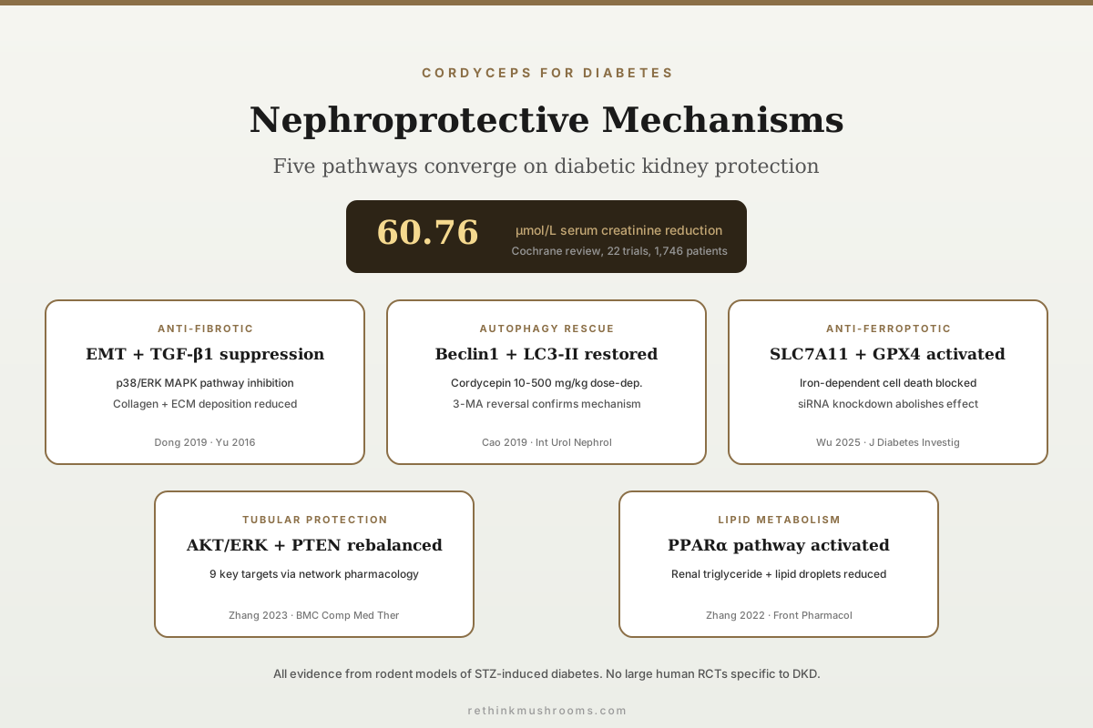
60.76 µmol/L SCr reduction
A Cochrane systematic review of 22 randomised trials (1,746 CKD patients) found Cordyceps sinensis preparations reduced serum creatinine by a mean of 60.76 µmol/L versus controls, though all included trials had high or unclear risk of bias (Zhang et al., Cochrane Database Syst Rev, 2014).
Zhang et al., Cochrane Database of Systematic Reviews, 2014
Anti-fibrotic
Cordyceps nucleoside extract inhibited epithelial-mesenchymal transition and ECM deposition via p38/ERK pathways in STZ-diabetic mice (Dong et al., 2019)
Autophagy rescue
Cordycepin at 10-500 mg/kg dose-dependently suppressed apoptosis and restored autophagy markers in DN rat kidneys (Cao et al., 2019)
PPARα activation
Bailing Capsule reduced renal triglyceride accumulation and lipid droplet formation through PPARα upregulation in diabetic rats (Zhang et al., 2022)
Zhang et al., Cochrane Database of Systematic Reviews, 2014
 View as image ↗
Diabetic kidney disease (DKD) is the leading cause of end-stage renal failure worldwide, and standard treatments targeting hyperglycemia, proteinuria, and hypertension slow but do not stop its progression. Cordyceps sinensis and its bioactive constituent cordycepin have accumulated a body of preclinical evidence suggesting nephroprotective activity across multiple molecular pathways: anti-fibrotic, anti-inflammatory, anti-apoptotic, lipid-metabolic, and, most recently, anti-ferroptotic.[1] A 2014 Cochrane systematic review of 22 randomised trials involving 1,746 chronic kidney disease patients found statistically significant improvements in serum creatinine, creatinine clearance, and proteinuria with cordyceps preparations as adjuvant therapy, but rated all included trials as high or unclear risk of bias.[2] For the broader glucose-lowering evidence, see cordyceps and blood sugar: hypoglycemic research; for the insulin-signalling mechanism, see cordyceps and insulin sensitivity: mechanisms and evidence.
Key Takeaways
- Zhang et al. (2014) in the Cochrane Database of Systematic Reviews pooled 22 randomised trials (1,746 CKD patients) and found Cordyceps sinensis preparations decreased serum creatinine by 60.76 µmol/L (95% CI: -85.82 to -35.71), increased creatinine clearance by 9.22 mL/min, and reduced 24-hour proteinuria by 0.15 g, though evidence quality was low across all outcomes
- Dong et al. (2019) in Molecules showed a nucleoside/nucleobase-rich Cordyceps sinensis extract inhibited the epithelial-mesenchymal transition and extracellular matrix deposition in STZ-diabetic mice and high-glucose HK-2 cells by suppressing p38 and ERK signalling pathways
- Cao et al. (2019) in International Urology and Nephrology demonstrated cordycepin (10-500 mg/kg) dose-dependently suppressed renal apoptosis and fibrosis while restoring autophagy markers (Beclin1, LC3-II) in diabetic nephropathy rats; the autophagy inhibitor 3-MA reversed these effects
- Wu et al. (2025) in the Journal of Diabetes Investigation identified a novel ferroptosis mechanism: cordycepin elevated SLC7A11 and GPX4 expression in high-glucose podocytes, suppressing iron-dependent cell death, and silencing either gene abolished these protective effects
- Yu et al. (2016) in PLoS ONE found 8 weeks of Cordyceps militaris treatment (fruiting body + mycelium combination) significantly reduced serum creatinine, kidney-to-body weight ratio, and TGF-β1 levels in type 2 diabetic nephropathy mice while attenuating collagen and advanced glycation end-product accumulation
- Zhang et al. (2022) in Frontiers in Pharmacology showed Bailing Capsule activated PPARα and downstream fatty acid oxidation genes (ACOX1, SCD) while suppressing FASN, reducing renal triglyceride content and lipid droplet formation in diabetic rats
- Zheng et al. (2023) in Chinese Medicine independently replicated cordycepin's protective effect through a different route again — reversing high-glucose induction of miR-193b-5p and restoring its target MCL-1 in DN mice and HK-2 cells — bringing the count of separately demonstrated mechanisms to seven
- The seven mechanisms were produced by five different substances: a purified nucleoside fraction, a C. militaris powder blend, isolated cordycepin, fermented C. sinensis, and a pharmaceutical Bailing Capsule. Isolated cordycepin accounts for three of the seven — genuine convergence across three independent groups — while each of the other four preparations was used exactly once, and the assorted consumer-grade C. sinensis products behind the human trials account for none of them
Why Diabetic Kidneys Fail: The Pathways Cordyceps Targets
DKD progresses through several interconnected molecular cascades. Chronic hyperglycemia drives advanced glycation end-product (AGE) formation, activates transforming growth factor-beta 1 (TGF-β1) signalling, triggers oxidative stress, and promotes a fibrotic remodelling programme that replaces functional nephrons with scar tissue. Podocytes (the specialised cells wrapping glomerular capillaries) undergo apoptosis, tubular epithelial cells shift toward a mesenchymal phenotype (epithelial-mesenchymal transition, EMT), and lipid accumulates in tubular cells through dysregulated fatty acid metabolism.
Cordyceps preparations have been tested against each of these cascades in animal models, with each study typically isolating one or two pathways. The aggregate picture from Liu et al.'s 2022 mechanistic review is that cordyceps does not act through a single molecular target. Instead, its multiple bioactive fractions (whole aqueous extract, nucleoside-rich fraction, polysaccharides, isolated cordycepin) each engage different nodes in the DKD progression network.[1]
Anti-Fibrotic Effects: Blocking the Scar Tissue Programme
Renal fibrosis is the final common pathway of DKD. Once the kidney's functional tissue is replaced by collagen and extracellular matrix (ECM), the damage is irreversible. Two studies directly address how cordyceps preparations interfere with this process.
Dong et al. (2019) isolated a nucleoside/nucleobase-rich extract from Cordyceps sinensis (CS-N) containing adenosine, cordycepin, and related nucleosides, then tested it in STZ-induced diabetic mice and high-glucose-exposed HK-2 (human kidney proximal tubular) cells. CS-N significantly attenuated renal functional abnormalities, ameliorated histopathological changes, and inhibited both EMT and ECM accumulation. The mechanism mapped to suppression of p38 and ERK MAPK signalling pathways, both of which are upstream activators of the fibrotic transcription programme.[3]
Yu et al. (2016) tested CmNo1, a novel combination of Cordyceps militaris fruiting bodies and mycelia, in a type 2 diabetic nephropathy mouse model (STZ-nicotinamide plus high-fat diet). After 8 weeks, CmNo1 significantly reduced carboxymethyl lysine (an AGE), attenuated collagen deposition, and dampened TGF-β1 expression in glomeruli. The same treatment reduced serum creatinine, kidney-to-body weight ratio, triglycerides, and cholesterol.[4] This study is relevant because it used C. militaris rather than the more commonly studied C. sinensis — a species widely sold in consumer products — and because TGF-β1 is the master regulator of renal fibrogenesis. It should be read as a result about CmNo1 specifically, a defined blend of fruiting body and mycelium, rather than about C. militaris products generally. For more on how cordyceps creatinine-level effects have been measured, see cordyceps and creatinine levels: what the studies show.
Autophagy Rescue: Restarting the Cellular Cleanup System
Autophagy is the cell's internal recycling programme, essential for podocyte survival under metabolic stress. In DKD, autophagy becomes impaired, allowing damaged organelles and misfolded proteins to accumulate and drive apoptosis. Cao et al. (2019) directly tested whether cordycepin could restore this process.
Using Wistar rats with STZ-induced diabetes, they administered cordycepin at 10 mg/kg (low dose), 100 mg/kg (medium dose), and 500 mg/kg (high dose). Compared to diabetic controls, cordycepin treatment dose-dependently decreased cholesterol, blood glucose, triglycerides, creatinine, and urine protein. Histopathological injury was visibly reduced. At the molecular level, cordycepin suppressed apoptosis markers (cleaved caspase-3, Bax) while rescuing autophagy markers (Beclin1, LC3-II upregulation, p62 downregulation) and suppressing fibrosis markers (α-SMA, t-TG, TIMP-1).[5]
The critical control in this study was 3-methyladenine (3-MA), a specific autophagy inhibitor. When 3-MA was co-administered, it abolished cordycepin's protective effects, re-inducing apoptosis and fibrosis. This confirms that autophagy induction is mechanistically required for the nephroprotective effect, not merely correlated with it. The dose-dependent response across a 50-fold concentration range (10-500 mg/kg) adds pharmacological credibility, though the effective doses in rats do not translate directly to human dosing.
Cao et al. (2019) · International Urology and Nephrology
- Population
- Wistar rats with streptozotocin-induced diabetes, randomised to normal control, diabetic control, and three cordycepin dose groups, plus a parallel in vitro arm
- Intervention
- Isolated cordycepin at 10 mg/kg, 100 mg/kg or 500 mg/kg
- Comparator
- Untreated diabetic rats; and, in vitro, cordycepin co-administered with the autophagy inhibitor 3-methyladenine
- Finding
- Cordycepin dose-dependently lowered cholesterol, blood glucose, triglycerides, creatinine and urine protein, and reduced histopathological injury. At the protein level it suppressed the apoptosis markers cleaved caspase-3 and Bax, suppressed the fibrosis markers α-SMA, t-TG and TIMP-1, and rescued the autophagy markers Beclin1 and LC3-II while lowering p62. Adding 3-methyladenine abolished the protection and re-induced apoptosis and fibrosis.
positive Positive and unusually well controlled — the inhibitor arm establishes that autophagy induction is required rather than merely coincident, which is the experiment most mechanism papers in this field skip. It remains a rodent study using a purified compound, and the authors themselves frame it as a first demonstration.
Cao et al., International Urology and Nephrology, 2019
Ferroptosis Inhibition: A New Mechanism from 2025
Ferroptosis is an iron-dependent form of regulated cell death distinct from apoptosis. In DKD, high glucose conditions deplete glutathione and suppress antioxidant defence systems, leaving podocytes vulnerable to lipid peroxidation and iron-catalysed cell death. Wu et al. (2025) identified that cordycepin protects podocytes through this newly characterised pathway.
In high-glucose-treated mouse podocytes (MPC5 cells), cordycepin elevated the expression of SLC7A11 (the cystine/glutamate antiporter that maintains glutathione synthesis) and GPX4 (glutathione peroxidase 4, the enzyme that detoxifies lipid hydroperoxides). This suppressed high-glucose-induced apoptosis, inflammatory cytokine release, and ferroptosis markers (elevated Fe2+, ROS, malondialdehyde; depleted glutathione). Overexpressing either SLC7A11 or GPX4 reproduced the protection on its own; when either gene was silenced instead, cordycepin's protective effects were abolished.[6]
In the in vivo component, cordycepin ameliorated renal structural injury and inflammation in STZ-induced diabetic mice while modulating SLC7A11 and GPX4 expression. Ferroptosis inhibition represents a mechanistically distinct target from the anti-fibrotic and autophagy pathways documented earlier, expanding the range of cordycepin's demonstrated nephroprotective activity. It is also, at the time of writing, a single paper: the gene-silencing arm makes the mechanism internally convincing, but no independent group has yet reproduced it.
Wu et al. (2025) · Journal of Diabetes Investigation
- Population
- High-glucose-treated MPC5 mouse podocytes in vitro, plus streptozotocin-induced diabetic mice in vivo
- Intervention
- Cordycepin, with parallel arms overexpressing or silencing SLC7A11 and GPX4 by siRNA
- Comparator
- High-glucose-exposed podocytes without cordycepin; untreated diabetic mice
- Finding
- Cordycepin suppressed high-glucose-induced apoptosis, inflammation and ferroptosis — measured as Fe2+, glutathione, reactive oxygen species and malondialdehyde — and raised SLC7A11 and GPX4 expression. Overexpressing either gene alone reproduced the protection; silencing either one abolished cordycepin's effect, placing the compound upstream of both. In vivo, cordycepin reduced renal structural injury and inflammation while modulating the same two genes.
preliminary The loss-of-function and gain-of-function arms make this a genuinely well-constructed mechanism paper rather than a correlation. What holds it at preliminary is replication: it is one group, one publication, one compound, and no human has been given cordycepin for a kidney endpoint in any trial.
Wu et al., Journal of Diabetes Investigation, 2025
Proximal Tubular Cell Protection: The AKT/ERK Axis
DKD has traditionally been characterised as a glomerular disease, but accumulating evidence points to proximal tubular dysfunction as equally important for disease progression. Zhang et al. (2023) investigated fermented Cordyceps sinensis in high-glucose-exposed HK-2 cells (human kidney proximal tubular cells) using network pharmacology to identify therapeutic targets, then validated them experimentally.
Database analysis and molecular docking identified 9 key therapeutic targets: RELA, JNK1, PTEN, VEGFA, EGF, ERK2, CASP3, AKT1, and MMP9. Gene ontology enrichment showed these targets clustered around positive regulation of cell proliferation and negative regulation of apoptosis. In vitro, fermented C. sinensis significantly attenuated high-glucose cytotoxicity, promoted HK-2 cell proliferation, reduced apoptosis, and downregulated Bax, Caspase-3, VEGFA, P-AKT, and P-ERK while upregulating PTEN.[7]
The PTEN upregulation is particularly notable. PTEN is a negative regulator of the PI3K/AKT pathway; its loss in DKD contributes to uncontrolled cell growth and fibrosis. By restoring PTEN expression while simultaneously suppressing AKT and ERK phosphorylation, fermented C. sinensis may rebalance the proliferation-versus-fibrosis axis in tubular cells. The network pharmacology approach identifies multiple simultaneous targets rather than a single mechanism, consistent with the multi-pathway picture emerging from the broader cordyceps-DKD literature.
Lipid Accumulation: The PPARα Pathway
Ectopic lipid deposition in renal tubular cells (lipotoxicity) is an increasingly recognised driver of DKD progression. Triglycerides accumulate in tubular epithelial cells, disrupting mitochondrial function and promoting inflammation. Zhang et al. (2022) tested Bailing Capsule, a Chinese-approved medicine whose active ingredient is Cordyceps sinensis, in STZ-induced diabetic rats over 12 weeks.
Bailing Capsule at both low and high doses significantly improved renal function markers (serum creatinine, blood urea nitrogen, 24-hour urinary protein, urinary albumin), reduced glomerulosclerosis and tubulointerstitial injury, and attenuated renal fibrosis. The distinct contribution of this study was demonstrating that Bailing Capsule markedly reduced kidney triglyceride content and lipid droplet formation. Renal transcriptomics revealed 498 upregulated and 448 downregulated genes in treated kidneys, enriched in the PPAR pathway and fatty acid metabolism. Specifically, Bailing Capsule activated PPARα and its downstream target ACOX1 (acyl-CoA oxidase 1, the rate-limiting enzyme in peroxisomal fatty acid β-oxidation) and SCD (stearoyl-CoA desaturase), while suppressing FASN (fatty acid synthase).[8]
This lipid-metabolic mechanism is distinct from the anti-fibrotic and anti-apoptotic pathways documented by the other studies. Renal lipotoxicity has gained recognition as a therapeutic target in DKD only in recent years. The transcriptomic approach used here provides genome-wide evidence rather than a candidate-gene hypothesis, strengthening the mechanistic case. For how cordyceps blood sugar effects connect to downstream kidney complications, see cordyceps for type 2 diabetes: clinical trial findings.
Seven Mechanisms, Five Different Substances
There is one more mechanism to add before stepping back. Zheng et al. (2023), publishing in Chinese Medicine, found that high glucose raised the microRNA miR-193b-5p in HK-2 cells while suppressing its target MCL-1, an anti-apoptotic protein, and that cordycepin reversed both movements. A luciferase reporter assay confirmed MCL-1 as a direct target of the microRNA, and cordycepin attenuated high-glucose-induced cell damage, inflammation and abnormal energy metabolism in DN mice as well as in culture.[9] That is a seventh distinct route, and it matters mostly as an independent replication: a different group, a different molecular axis, the same compound, the same direction of effect.
Seven mechanisms sounds like a convergent body of evidence, and in one respect it genuinely is. Laid side by side, though, the picture is more awkward than the summary suggests: those seven mechanisms were produced by five different substances.
Preparation Audit
Seven Mechanisms, Five Different Substances
The middle column is the one usually dropped from summaries of this literature. Every mechanism below is real and was demonstrated competently. But they were not produced by one thing. Isolated cordycepin accounts for three of the seven — real convergence, from three separate groups — and each of the remaining four preparations was used exactly once. “Cordyceps has seven nephroprotective mechanisms” is therefore a claim about five substances that share a genus name, not about a product.
Anti-fibrotic — blocks EMT and matrix deposition via p38/ERK
Dong 2019 [3]Purified fractionCS-N, a nucleoside/nucleobase-rich fraction isolated on resin and glucan columns
· STZ-diabetic mice + high-glucose HK-2 cells
Anti-fibrotic — lowers TGF-β1, carboxymethyl lysine and collagen
Yu 2016 [4]Whole mushroomCmNo1, a powder blend of C. militaris fruiting bodies and mycelia
· STZ-nicotinamide + high-fat-diet mice, 8 weeks
Autophagy restoration — Beclin1 and LC3-II up, p62 down
Cao 2019 [5]Isolated cordycepinIsolated cordycepin, 10 / 100 / 500 mg/kg
· STZ-diabetic Wistar rats, with 3-MA reversal control
Ferroptosis inhibition — SLC7A11 and GPX4 activation
Wu 2025 [6]Isolated cordycepinIsolated cordycepin
· High-glucose MPC5 podocytes + STZ-diabetic mice
miR-193b-5p / MCL-1 axis — oxidative stress and apoptosis
Zheng 2023 [9]Isolated cordycepinIsolated cordycepin
· DN mice + high-glucose HK-2 cells
Tubular survival — AKT/ERK suppression with PTEN restoration
Zhang 2023 [7]Fermented myceliumFermented C. sinensis, a cultivated substitute for the wild fungus
△ High-glucose HK-2 cells only — no animal arm
Anti-lipotoxic — PPARα, ACOX1 and SCD up, FASN down
Zhang 2022 [8]Pharmaceutical productBailing Capsule, an approved Chinese pharmaceutical
· STZ-diabetic rats, 12 weeks
Human renal endpoints — creatinine, clearance, proteinuria
Zhang 2014 [2]Assorted, human trialsAssorted C. sinensis preparations, differing across all 22 trials
△ Human CKD patients not on dialysis
What this means at the shelf: The substance with the most mechanistic support here is isolated cordycepin — three mechanisms, three independent groups. It is also the substance furthest from anything on a shelf: a purified secondary metabolite of Cordyceps militaris [9], given as a compound rather than as a mushroom. Two of the remaining mechanisms required a laboratory fraction purified over resin and glucan columns or an approved pharmaceutical capsule. And the one preparation class with any human data — the assorted C. sinensis products used across the 22 trials in the Cochrane review — is the class that demonstrated none of the seven. No consumer product in this table has been shown to produce any of these mechanisms.
Sources: Zhang et al., Cochrane Database of Systematic Reviews (2014) [2]; Dong et al., Molecules (2019) [3]; Yu et al., PLoS ONE (2016) [4]; Cao et al., International Urology and Nephrology (2019) [5]; Wu et al., Journal of Diabetes Investigation (2025) [6]; Zhang et al., BMC Complementary Medicine and Therapies (2023) [7]; Zhang et al., Frontiers in Pharmacology (2022) [8]; Zheng et al., Chinese Medicine (2023) [9]. Preparation descriptions are taken from each study’s own methods section. Ordering follows the article, not effect size.
Read that middle column carefully, because it is the part almost always lost in summary. Three of the seven mechanisms — autophagy restoration, ferroptosis inhibition, and the miR-193b-5p axis — were produced by isolated cordycepin, described in Zheng's own paper as a secondary metabolite of Cordyceps militaris, given as a purified compound rather than as any mushroom preparation.[9] That is the strongest single thread in this literature: three independent groups, three different molecular axes, one compound, a consistent direction of effect. It is also the thread furthest removed from anything a person can buy.
Each of the remaining four preparations was used exactly once. The EMT result came from a nucleoside fraction purified over resin and glucan columns; the TGF-β1 and AGE result from a C. militaris fruiting body and mycelium blend; the tubular AKT/ERK/PTEN result from fermented C. sinensis in cell culture with no animal arm at all; the PPARα result from an approved pharmaceutical capsule. And the one preparation class with any human data — the assorted C. sinensis products used across the Cochrane trials — accounts for none of these mechanisms in any model.
The honest way to state the position is therefore not "cordyceps protects the diabetic kidney through seven mechanisms." It is that a purified C. militaris metabolite has three times been shown to influence diabetic kidney injury in rodents and cell culture, and that four other cordyceps-derived preparations have each been shown once to influence a different node of the disease. Those are compatible claims, but they license very different expectations of a capsule.
What the Human Evidence Actually Shows
The strongest human-level evidence comes from the 2014 Cochrane systematic review by Zhang et al. This review searched the Cochrane Renal Group's specialised register to 14 April 2014, along with CINAHL, AMED, Current Controlled Trials, OpenSIGLE and a set of Chinese-language databases including CBM, CMCC, TCMLARS and the Chinese Dissertation Database. It included 22 randomised or quasi-randomised trials involving 1,746 participants with CKD, all using Cordyceps sinensis preparations as adjuvant therapy alongside conventional medicine, and reported results for the subgroup not receiving dialysis.[2] Two features of the included population matter for anyone reading this article for diabetic kidney disease specifically: the trials were identified predominantly through Chinese-language sources and conducted in Chinese clinical settings, and the review's inclusion criterion was chronic kidney disease of any cause, not diabetic kidney disease.
The pooled results showed that cordyceps preparations, compared to conventional treatment alone:
- Decreased serum creatinine: MD -60.76 µmol/L (95% CI: -85.82 to -35.71; 14 studies, 987 participants)
- Increased creatinine clearance: MD 9.22 mL/min (95% CI: 3.10 to 15.34; 6 studies, 362 participants)
- Reduced 24-hour proteinuria: MD -0.15 g/24h (95% CI: -0.24 to -0.05; 4 studies, 211 participants)
The review also reported improvements in haemoglobin and serum albumin, both markers of CKD-associated complications.
However, the Cochrane reviewers rated risk of bias as high in 4 studies and unclear in 18 studies. Common methodological problems included inadequate randomisation, lack of blinding, incomplete outcome reporting, and small sample sizes. No study was rated as low risk of bias across all domains. The review's conclusion explicitly stated that "definitive conclusions could not be made because of the low quality of evidence."
Evidence Foundation Audit
What “22 Trials, 1,746 Patients” Actually Rests On
Both halves of this panel come from the same paragraph of the 2014 Cochrane review. The top half is the quality of the inputs. The bottom half is how much of the advertised sample stood behind each individual number.
Risk of bias across the 22 included trials
- 0 Low risk of bias
- 4 High risk of bias
- 18 Unclear risk of bias
Not one trial was rated low risk of bias in every domain. Meta-analysis inherits the bias of the studies it pools — it narrows the confidence interval without adding validity, so the tight interval on the creatinine result below is a measure of precision, not of trustworthiness.
How much of the 22 trials / 1,746 participants backed each result
Serum creatinine
−60.76 µmol/L95% CI −85.82 to −35.71
14 of 22 trials · 987 of 1,746 participants = 56.5% of the headline sample
Creatinine clearance
+9.22 mL/min95% CI 3.10 to 15.34
6 of 22 trials · 362 of 1,746 participants = 20.7% of the headline sample
24-hour proteinuria
−0.15 g/24 h95% CI −0.24 to −0.05
4 of 22 trials · 211 of 1,746 participants = 12.1% of the headline sample
The number worth carrying: the proteinuria result — the outcome closest to what a person with diabetic kidney disease is actually monitored on — came from 4 trials and 211 people, roughly one eighth of the sample the review is usually cited for, none of them rated low risk of bias, and none of them restricted to diabetic kidney disease specifically. The review pooled chronic kidney disease of any cause. Its own authors concluded that “definitive conclusions could not be made because of the low quality of evidence.”
Source: Zhang HW, Lin ZX, Tung YS, Kwan TH, Mok CK. Cordyceps sinensis (a traditional Chinese medicine) for treating chronic kidney disease. Cochrane Database of Systematic Reviews 2014, CD008353 [2]. All counts, effect estimates and confidence intervals are quoted from the review’s Main Results and its risk-of-bias assessment.
Zhang et al. (2014) · Cochrane Database of Systematic Reviews
- Population
- 1,746 participants with chronic kidney disease of any cause, across 22 randomised or quasi-randomised trials identified largely through Chinese-language databases
- Intervention
- Cordyceps sinensis preparations as adjuvant therapy, added to conventional medicine
- Comparator
- Conventional treatment alone, placebo, or no treatment
- Finding
- Among participants not receiving dialysis, cordyceps preparations decreased serum creatinine by 60.76 µmol/L (95% CI −85.82 to −35.71; 14 trials, 987 participants), increased creatinine clearance by 9.22 mL/min (95% CI 3.10 to 15.34; 6 trials, 362 participants) and reduced 24-hour proteinuria by 0.15 g (95% CI −0.24 to −0.05; 4 trials, 211 participants). Haemoglobin and serum albumin also improved. Risk of bias was high in 4 trials and unclear in 18.
mixed Every pooled estimate crossed statistical significance, and every one of them rests on trials the reviewers could not rate as low risk of bias in any single case. The review's own authors declined to draw a conclusion, and the enrolled population was chronic kidney disease generally rather than diabetic kidney disease.
Zhang et al., Cochrane Database of Systematic Reviews, 2014
- Cell studiescompleted
High-glucose HK-2 tubular cells and MPC5 podocytes across at least five papers, with gene-silencing and pathway-inhibitor controls in the strongest of them. Refs 3, 5, 6, 7, 9.
- Animal studiescompleted
Streptozotocin rats and mice, streptozotocin-nicotinamide plus high-fat-diet mice, treatment periods of 8 to 12 weeks, functional and histological endpoints. Six of the seven mechanisms reached an animal model, using four different preparations between them. Refs 3, 4, 5, 6, 8, 9.
- Small human trialscurrent
Twenty-two randomised or quasi-randomised trials exist, but 4 were rated high risk of bias and 18 unclear, none low, and they enrolled chronic kidney disease of any cause rather than diabetic kidney disease. This is where genuine primary human evidence tops out. Ref 2.
- Adequately powered, low-bias RCT in diabetic kidney diseasemissing
This is the empty rung. No blinded, adequately powered, multi-centre trial has tested a defined cordyceps preparation in a DKD-specific population with a hard renal endpoint. Nothing above it can be firmer than this.
- Meta-analysiscompleted
A 2014 Cochrane review and a 2024 PROSPERO-registered meta-analysis both exist and both report benefit — but each pools the same underlying pool of Chinese trials, so both inherit rather than resolve the bias beneath them. Refs 2, 10.
This ladder is unusual: the top rung is occupied and the rung below it is empty. Two meta-analyses have been published, which normally signals a mature evidence base, but both pool trials that no reviewer could rate as low risk of bias, and neither is DKD-specific. Pooling biased trials produces a narrower confidence interval, not a more reliable answer. Until a properly blinded, adequately powered trial of a defined preparation in diabetic kidney disease exists, the correct description is a strong preclinical case with an unresolved clinical one — not an established therapy.
Refs 2, 3, 4, 5, 6, 7, 8, 9, 10
This gap between consistent preclinical findings and weak clinical evidence defines the current state of cordyceps-DKD research. The animal data converge on real molecular mechanisms through rigorous mechanistic work. The human data show signals of benefit that have not been confirmed by adequately powered, well-blinded, multi-centre trials. For the related intersection of chaga and diabetic kidney complications, see chaga and diabetic complications: kidney and eye protection.
Limitations of the Current Evidence
Every preclinical study cited here used rodent models of diabetes, primarily STZ-induced. These models replicate some features of human DKD (hyperglycemia, proteinuria, mesangial expansion) but not the full metabolic syndrome context of human type 2 diabetes. Doses used in animal studies (e.g., cordycepin at 10-500 mg/kg in rats) have unclear translational relevance to human supplementation.
The cordyceps preparations studied vary substantially: whole aqueous extracts, nucleoside-rich fractions, isolated cordycepin, polysaccharide fractions, and commercial Bailing Capsules. Effects seen with one preparation do not automatically extend to others. Consumer cordyceps supplements are rarely standardised to the same fractions used in research.
The Cochrane review's trials were identified predominantly through Chinese-language databases and used preparations available in the Chinese pharmaceutical market. Western clinical trials on cordyceps for kidney disease are largely absent. The 2014 review is now 12 years old, and newer meta-analyses draw from a similar pool of trials with similar methodological limitations.
The most recent of these deserves specific comment, because a reader who encounters it first will come away with a very different impression. Wu et al. (2024), publishing in Frontiers in Medicine under a registered PROSPERO protocol, pooled 15 studies and 1,310 patients with renal dysfunction — with an emphasis on acute kidney injury rather than DKD — and reported pooled sensitivity of 0.89 (95% CI 0.84-0.93), specificity of 0.69 (95% CI 0.59-0.77), a diagnostic odds ratio of 18.0, and an SROC area under the curve of 0.88. Its subgroup analysis compared northern with southern Chinese patients, which itself indicates the geographic concentration of the underlying trials. The authors concluded that C. sinensis "can be considered a dependable clinical treatment" for renal dysfunction.[10]
That conclusion is markedly more confident than Cochrane's, and the difference is not explained by new randomised evidence of higher quality appearing between 2014 and 2024. It is largely the same literature, differently framed, with a different condition emphasis and a different analytic approach. When two systematic reviews of an overlapping trial pool reach "definitive conclusions could not be made" and "a dependable clinical treatment," the divergence is a fact about the reviews rather than about cordyceps, and the more cautious of the two is the one that assessed risk of bias trial by trial and reported that none was low.
The ferroptosis data (Wu et al., 2025) and transcriptomic data (Zhang et al., 2022) represent single studies that have not been independently replicated. Both report promising mechanisms, but single-study findings in in vitro and rodent models have a high attrition rate when subjected to independent replication.
Fact check
Does the evidence show cordyceps repairs diabetic kidney damage?
The claim
“Cordyceps has been proven to protect and repair the kidneys in diabetes, with a Cochrane review confirming it lowers creatinine in over 1,700 patients.”
Two true facts fused into a false one. The Cochrane review does report a pooled creatinine reduction across 1,746 enrolled participants, and the preclinical literature does document real anti-fibrotic and anti-apoptotic mechanisms. Supplement marketing joins them with the word 'proven' and the mechanism claims silently borrow the human review's sample size.
What research shows
OverstatedThe creatinine result came from 14 of the 22 trials and 987 of the 1,746 participants, in chronic kidney disease of any cause rather than diabetic kidney disease, with 4 trials at high risk of bias, 18 unclear and none low. The mechanistic work is separate and was done in rodents and cell culture, using purified compounds and fractions that no consumer product supplies. Nothing in either body of work measured repair; the endpoints were slowed decline and reduced scarring.
This is not a debunking. The preclinical mechanisms are real, are unusually well controlled by the standards of mushroom research, and include loss-of-function experiments that most supplement studies never attempt. The problem is exclusively one of reach: no adequately powered blinded trial has tested a defined preparation in a DKD population, and the substances that produced the mechanisms are not the substances sold.
Zhang et al., 2014; Cao et al., 2019; Wu et al., 2025; Zheng et al., 2023
Research dose vs. what people take
Isolated cordycepin — the Cao 2019 protocol, the most-cited dosing in this literature: Research vs. commercial doses
What research used
10, 100 or 500 mg/kg body weight
Oral administration to rats
Daily · Study treatment period following streptozotocin induction
Wistar rats with STZ-induced diabetes; dose-dependent effects across the full 50-fold range
What supplements sell
1–3 g of cordyceps product per day
Oral capsule or powder
Once or twice daily
C. sinensis mycelium (Cs-4-type) fermentation products, C. militaris fruiting body extracts, and blended mushroom formulas that rarely state cordycepin content at all
The rat-to-human conversion is not the main problem here, though it is real — applying the standard body-surface-area factor of 6.2 to even the low 10 mg/kg arm gives roughly 1.6 mg/kg in humans, about 113 mg of pure cordycepin daily for a 70 kg adult. The larger problem is that this is a dose of cordycepin, not of cordyceps. Consumer products are dosed by total mushroom or extract weight, and most do not disclose cordycepin content, so there is no way to convert a capsule label into the quantity that was actually tested.
- *Cordycepin is a secondary metabolite of C. militaris. Products based on C. sinensis or its fermented mycelium are a different starting material, and the three cordycepin mechanisms in this article say nothing about them.
- *Cao's effects were dose-dependent across 10–500 mg/kg, so the low arm cannot be assumed to be the minimum effective dose in any other model or species.
- *Dong 2019 used a nucleoside fraction purified over resin and glucan columns; Zhang 2022 used an approved pharmaceutical capsule. Neither dose is expressible as a quantity of any commercial supplement.
- *No human dose-finding study for any cordyceps preparation with a renal endpoint has been published, so no dose in this table has ever been shown to do anything to a human kidney.
Cao et al., 2019; Dong et al., 2019; Zhang et al., 2022; Zheng et al., 2023
Research gaps
What would it take?
What would it take to establish that cordyceps protects the diabetic kidney in people?
A defined, characterised preparation
Trials would have to fix the species, the extraction method, and the assayed content of cordycepin, nucleosides or polysaccharides. Across the seven mechanisms described here, five different substances were used, and four of the five were used exactly once, so 'cordyceps' does not currently name a single testable intervention.
A DKD-specific population
The 22 Cochrane trials enrolled chronic kidney disease of any cause, and the 2024 meta-analysis emphasised acute kidney injury. Neither answers the question this article asks.
Blinding and adequate randomisation
No included trial was rated low risk of bias in every domain. Inadequate randomisation, absent blinding and incomplete outcome reporting were the recurring failures.
Hard renal endpoints over a meaningful horizon
Existing trials report serum creatinine, clearance and proteinuria — reasonable surrogates. What is absent is eGFR slope, progression to end-stage renal disease or dialysis initiation, measured over years rather than weeks.
Replication outside China
Effectively the entire clinical base is Chinese, and the 2024 meta-analysis found different efficacy in northern versus southern Chinese patients — a result that itself needs explaining before the literature can be generalised.
Independent replication of the newest mechanisms
Cordycepin's protective effect has now been shown by separate groups via autophagy, ferroptosis and the miR-193b-5p/MCL-1 axis, which is meaningful convergence. The ferroptosis and transcriptomic findings individually remain single papers.
Refs 2, 3, 4, 5, 6, 7, 8, 9, 10
Before adding cordyceps to kidney care
ModerateImpaired kidneys are the organ least able to tolerate an unquantified supplement
Concern
Nothing in this article is medical advice, and nothing in it supports using cordyceps to treat diabetic kidney disease or replace any part of prescribed care. The specific hazard is structural rather than toxicological: the kidney is the main route of clearance for most compounds, so reduced kidney function means anything taken accumulates for longer than it would in a healthy person. Supplement products are not dose-characterised for that population, heavy-metal and adulteration screening is inconsistent in this category, and several of these preparations lowered blood glucose in animals — an effect that would stack with insulin, sulfonylureas or alpha-glucosidase inhibitors rather than complement them. Protein and potassium restrictions are also common in CKD care, and mushroom powders are not free of either.
What the research says
Treat cordyceps as an unquantified addition to a regimen that is being actively titrated, not as a kidney therapy. The decision belongs to the nephrologist or prescriber managing the case, before starting rather than after a change in laboratory values, because the person interpreting a rising creatinine or a hypoglycaemic episode needs to know what else is being taken.
Particularly relevant for: Anyone with reduced eGFR or diagnosed CKD; people taking insulin, sulfonylureas, meglitinides or alpha-glucosidase inhibitors; transplant recipients and anyone on immunosuppressants, given cordyceps' documented immunomodulatory activity; people on potassium or protein restrictions; and people who are pregnant or breastfeeding, for whom no data exist
What to do
Do not adjust, delay or discontinue prescribed diabetes or kidney medication on the basis of preclinical mushroom research. Raise cordyceps with the clinician managing your kidney care before starting, and be aware that the Cochrane review's reassuring adverse-event record comes from small, short trials with incomplete adverse-event reporting — an absence of recorded harm, not a demonstration of long-term safety.
Zhang et al., 2014; Zhang et al., 2022; Wu et al., 2024
The Bottom Line
Cordyceps preparations demonstrate nephroprotective activity through at least seven distinct preclinical pathways: anti-fibrotic EMT suppression via p38/ERK, TGF-β1 and AGE reduction, autophagy restoration, anti-ferroptotic SLC7A11/GPX4 activation, the miR-193b-5p/MCL-1 axis, proximal tubular AKT/ERK/PTEN rebalancing, and anti-lipotoxic PPARα-mediated triglyceride clearance. The mechanistic work is unusually well controlled for this field, including autophagy-inhibitor and gene-silencing arms that establish necessity rather than correlation. The complication is that those seven mechanisms were produced by five different substances — a purified nucleoside fraction, a C. militaris powder blend, isolated cordycepin, fermented C. sinensis, and a pharmaceutical Bailing Capsule. Isolated cordycepin accounts for three of the seven, which is real convergence across three independent groups, but it is a purified C. militaris metabolite rather than a mushroom product; the other four preparations were each used exactly once. On the human side, a Cochrane review of 22 trials found significant improvements in serum creatinine, creatinine clearance and proteinuria, but 4 trials were rated high risk of bias, 18 unclear and none low, the population was chronic kidney disease of any cause rather than DKD specifically, and each pooled result rested on a fraction of the headline sample. A more bullish 2024 meta-analysis draws on the same trial pool. The preclinical case is strong and the clinical one is unresolved; the gap between them is not a matter of degree but of substance, population and study quality all at once.
Frequently Asked Questions
Sources & References
- 1RTHM-00783·Liu, Wu et al. (2022). “Mechanism of Cordyceps sinensis and its Extracts in the Treatment of Diabetic Kidney Disease: A Review..” Frontiers in pharmacology.Study breakdown →PubMed →↩
- 2RTHM-00360·Zhang, Hong Wei et al. (2014). “Cordyceps sinensis (a traditional Chinese medicine) for treating chronic kidney disease..” The Cochrane database of systematic reviews.Study breakdown →PubMed →↩
- 3RTHM-00552·Dong, Zhonghua et al. (2019). “A Nucleoside/Nucleobase-Rich Extract from Cordyceps Sinensis Inhibits the Epithelial-Mesenchymal Transition and Protects against Renal Fibrosis in Diabetic Nephropathy..” Molecules (Basel.Study breakdown →PubMed →↩
- 4RTHM-00458·Yu, Sung-Hsun et al. (2016). “Cordyceps militaris Treatment Preserves Renal Function in Type 2 Diabetic Nephropathy Mice..” PloS one.Study breakdown →PubMed →↩
- 5RTHM-00547·Cao, Tao et al. (2019). “The protective effect of Cordycepin on diabetic nephropathy through autophagy induction in vivo and in vitro..” International urology and nephrology.Study breakdown →PubMed →↩
- 6RTHM-00954·Wu, Bing et al. (2025). “Cordycepin ameliorates diabetic nephropathy injury by activating the SLC7A11/GPX4 pathway..” Journal of diabetes investigation.Study breakdown →PubMed →↩
- 7RTHM-00861·Zhang, Yuhan et al. (2023). “Protective effect of Cordyceps sinensis against diabetic kidney disease through promoting proliferation and inhibiting apoptosis of renal proximal tubular cells..” BMC complementary medicine and therapies.Study breakdown →PubMed →↩
- 8RTHM-00807·Zhang, Qian et al. (2022). “Bailing capsule (Cordyceps sinensis) ameliorates renal triglyceride accumulation through the PPARα pathway in diabetic rats..” Frontiers in pharmacology.Study breakdown →PubMed →↩
- 9RTHM-00862·Zheng, Rong et al. (2023). “Cordycepin from Cordyceps militaris ameliorates diabetic nephropathy via the miR-193b-5p/MCL-1 axis..” Chinese medicine.Study breakdown →PubMed →↩
- 10RTHM-02021·Wu, Fenfang et al. (2024). “Efficacy of traditional Chinese medicine Cordyceps sinensis as an adjunctive treatment in patients with renal dysfunction: a systematic-review and meta-analysis..” Frontiers in medicine.Study breakdown →PubMed →↩