Cordyceps and Blood Sugar
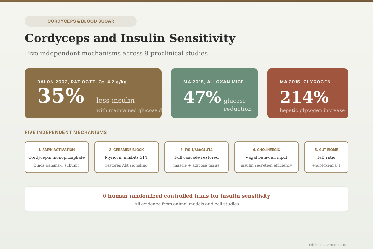
35% less insulin needed
Rats given Cordyceps sinensis fermentation product (Cs-4) at 2 g/kg daily for 30 days cleared an oral glucose load with 35% less insulin, measured as insulin area under the curve at day 20. In the same study, skeletal muscle glucose transport and muscle GLUT-4 did not change.
Balon et al., Journal of Alternative and Complementary Medicine, 2002
AMPK Activator
cordycepin converts to a monophosphate that binds AMPK's gamma-1 subunit at the same site as AMP
Ceramide ↓
cordyceps extract containing myriocin blocks ceramide-driven insulin resistance in obese mice
0 Human RCTs
no randomized controlled trial has tested cordyceps for insulin sensitivity in people
Balon et al., Journal of Alternative and Complementary Medicine, 2002
 View as image ↗
Insulin sensitivity determines how efficiently cells respond to insulin's signal to take up glucose. When it declines, the pancreas compensates by producing more insulin, blood sugar drifts upward, and eventually the system fails into type 2 diabetes. Cordyceps species and their bioactive compounds have been tested in this pathway at multiple points: the AMPK energy sensor, the ceramide lipotoxicity cascade, the insulin receptor signaling chain (IRS-1/Akt/GLUT4), and cholinergic neural inputs to the pancreas. For the broader picture of cordyceps and glucose regulation, see the cordyceps and blood sugar overview. This article isolates the insulin sensitivity question specifically.
The strongest mechanistic finding is that cordycepin functions as a pro-drug that converts to cordycepin monophosphate, a direct AMP analog that activates AMPK through the same gamma-subunit binding site as AMP itself.[1] AMPK activation promotes glucose uptake independently of insulin, making cordycepin a compound that can theoretically bypass insulin resistance rather than just treat its downstream consequences. Every study below is an animal or cell experiment. None is a human trial.
Key Takeaways
- Cordycepin activates AMPK by converting to cordycepin monophosphate, which binds the gamma-1 subunit at the same site as AMP; activation correlates with monophosphate content, not di- or triphosphate (Hawley et al., Cell Chemical Biology, 2020)
- Cordyceps extract containing myriocin reduced ceramide accrual, improved glucose homeostasis, raised liver pAKT (Ser473), and prevented diet-induced obesity in mice on high-fat diets (Li et al., Scientific Reports, 2022)
- The same study screened eight commercial cordyceps products and found myriocin content ranging from zero to 4–6 nmol/g; the myriocin-free product did not reproduce any benefit and impaired insulin tolerance (Li et al., 2022)
- Cs-4 (Cordyceps sinensis fermentation product) at 2 g/kg for 30 days cut insulin area under the curve by 35% at day 20 in rats — but glucose area under the curve, muscle glucose transport, and muscle GLUT-4 were all unchanged (Balon et al., Journal of Alternative and Complementary Medicine, 2002)
- C. militaris fruiting body and mycelia combination (360 mg/kg for 8 weeks) increased IRS-1, pIRS-1, AKT, pAKT, and GLUT-4 protein in skeletal muscle and adipose tissue of high-fat-diet diabetic mice (Yu et al., Journal of Diabetes Research, 2015)
- Cordycepin given by intraperitoneal injection — not orally — reduced blood glucose by 47% and increased hepatic glycogen by 214% in alloxan-induced diabetic mice (Ma et al., Nutrition Research, 2015)
- C. militaris extracts and cordycepin improved glucose and lipid parameters in diabetic mice alongside gut microbiota shifts, with KEGG enrichment of metabolite profiles pointing predominantly to the PI3K/Akt/mTOR pathway (Liu et al., Frontiers in Pharmacology, 2023)
How Cordycepin Activates AMPK
AMPK (AMP-activated protein kinase) is the cell's master energy sensor. When cellular energy drops and AMP levels rise, AMPK activates and triggers glucose uptake, fatty acid oxidation, and mitochondrial biogenesis. Pharmacological AMPK activators like metformin are first-line diabetes drugs. Cordycepin turns out to be another, though it reaches the target by a different route.
Hawley and colleagues demonstrated the mechanism in 2020.[1] Cordycepin (3'-deoxyadenosine) enters cells via adenosine transporters and is converted by cellular metabolism into mono-, di-, and triphosphate forms. The critical finding: AMPK activation in intact cells correlates with the content of cordycepin monophosphate and not with other cordycepin or adenine nucleotides. In cell-free assays, cordycepin monophosphate mimics all three effects of AMP on the AMPK complex: allosteric activation, promotion of Thr172 phosphorylation, and inhibition of Thr172 dephosphorylation. A gamma-subunit mutation that prevents activation by AMP also blocks activation by cordycepin in cells, confirming the two molecules use the same site.
Two details from the same paper are usually left out when this finding gets repeated. First, at high cordycepin concentrations the cordycepin nucleotides can almost entirely replace the cell's own adenine nucleotide pool — this is a compound that substitutes for the cell's energy currency, not merely a signal that nudges it. Second, genetic knockout of AMPK made cells more sensitive to cordycepin's cytotoxic effects, meaning AMPK activation is partly protecting cells against cordycepin rather than being a purely beneficial downstream effect. Both facts belong in any honest reading of "cordycepin activates AMPK."
Hawley et al. (2020) · Cell Chemical Biology
- Population
- Cultured cell lines, plus cell-free AMPK assays
- Intervention
- Cordycepin exposure with measurement of intracellular cordycepin mono-, di- and triphosphate content, AMPK activation, and a gamma-subunit mutation that blocks AMP binding
- Finding
- AMPK activation tracked cordycepin monophosphate content specifically. Cordycepin monophosphate reproduced all three of AMP's effects on AMPK in cell-free assay, and the gamma-subunit mutation that blocks AMP activation also blocked cordycepin activation — establishing a shared binding site. At high concentrations cordycepin nucleotides nearly replaced the cellular adenine nucleotide pool, and AMPK knockout increased cordycepin cytotoxicity.
positive Mechanistically decisive for the question it asked, and entirely in vitro. It establishes how cordycepin activates AMPK; it says nothing about whether an oral cordyceps supplement reaches intracellular concentrations high enough to do this in a person.
Hawley et al., Cell Chemical Biology, 2020
Ceramide Inhibition: The Lipotoxicity Pathway
Ceramides are sphingolipid metabolites that accumulate in obesity and directly cause insulin resistance by inhibiting Akt phosphorylation and blocking GLUT4 translocation. Li and colleagues published a finding in 2022 that reframes how cordyceps might affect insulin sensitivity — and, unusually for this literature, tested products people can actually buy.[2]
Cordyceps naturally contains myriocin, a potent inhibitor of serine palmitoyltransferase (SPT), the enzyme catalyzing the first step of ceramide biosynthesis. Myriocin was originally isolated from the fungus Isaria sinclairii and is abundant in closely related species including Cordyceps. It is not approved for human use as an isolated compound.
The researchers acquired eight commercially available cordyceps products — capsules bought on Amazon.com in the US, capsules and liquid from Singapore, and a wild mushroom from Qinghai, China — and measured myriocin content by mass spectrometry. The results are the most practically useful finding in this entire article:
Product Variability
Eight Cordyceps Products People Actually Buy, Measured for Myriocin
Myriocin is the compound that blocks ceramide synthesis — the mechanism behind the insulin-sensitivity benefit in this study. Li and colleagues bought eight cordyceps products from US, Singaporean and Chinese sources and quantified myriocin by LC/MS. Bar width reflects measured myriocin content. The label on the bottle predicted none of it.
Sample 1 · Amazon.com, U.S. · Capsule
4–6 nmol/gFed to obese mice at 19.7 mg/kg for 12 weeks: reduced circulating ceramides from week 4, lowered fed and fasted glucose, improved glucose and insulin tolerance, raised liver pAKT (S473), increased energy expenditure, prevented diet-induced obesity and resolved hepatic steatosis.
Sample 8 · Qinghai, China · Wild mushroom
4–6 nmol/gSample 2 · Amazon.com, U.S. · Capsule
0.5–2 nmol/gSample 3 · Amazon.com, U.S. · Capsule
0.5–2 nmol/gSample 4 · Amazon.com, U.S. · Capsule
0.5–2 nmol/gSample 5 · Amazon.com, U.S. · Capsule
0.5–2 nmol/gSample 7 · Eu Yan Sang, Singapore · Liquid
0.5–2 nmol/gSample 6 · Eu Yan Sang, Singapore · Capsule
None detectedno myriocin detected
Fed to obese mice as the negative control: no change in ceramides, energy expenditure or body weight — and on insulin tolerance testing it performed worse than control, with elevated serum insulin.
How to read it: only two of eight products carried myriocin in the 4–6 nmol/g band, five carried a quarter of that or less, and one carried none. Both of the products the study actually fed to animals are on this list — and they produced opposite results on the same insulin endpoint. “Cordyceps” on a label does not identify a dose, an extract type, or in this case even the presence of the active compound.
Source: Li et al., Scientific Reports 12:7273 (2022) [ref 2] — Table 1 and Figures 1, 3 and 5. Myriocin values are the bands reported by the authors, not point estimates; bar width uses each band’s midpoint. A higher dose of Sample 1 (39.5 mg/kg) caused intestinal inflammation and edema and that study arm was discontinued. The senior author discloses consulting and shareholding in a ceramide-therapeutics company.
They then fed obese mice a high-fat diet supplemented with Sample 1 (one of the two highest-myriocin products) at 19.7 mg/kg — a dose back-calculated from the manufacturer's own recommended human serving. Over 12 weeks, that treatment reduced circulating ceramides and related sphingolipids from four weeks onward, lowered fed and fasted blood glucose, improved glucose and insulin tolerance testing, raised liver pAKT (Ser473) on Western blot, increased energy expenditure, prevented diet-induced weight gain, and resolved hepatic steatosis. Mechanistically the authors attributed the effect to increased adipose tissue browning, improved brown adipose function, and improved hepatic insulin sensitivity, alongside shifts in gut bacteria including Clostridium and Bilophila.
The control that matters most was Sample 6 — a commercial cordyceps capsule from Singapore containing no detectable myriocin. It did not reduce ceramides. It did not change energy expenditure or body weight. On insulin tolerance testing it performed worse than control, with elevated serum insulin. That is a within-study demonstration that "cordyceps" is not a single intervention: two products sold for the same purpose produced opposite results on the study's primary insulin endpoint.
The ceramide mechanism is also distinct from the AMPK pathway. Ceramides block Akt activation through PP2A and PKC-zeta, sabotaging the insulin signal at a step between the insulin receptor and GLUT4 translocation. Removing ceramides with an SPT inhibitor restores that chain. Two caveats travel with the finding: a higher dose arm (39.5 mg/kg) produced intestinal inflammation and edema and had to be discontinued, and the senior author discloses that he is a consultant and shareholder in a company developing ceramide-targeting therapeutics.
For the intersection of cordyceps and cellular energy production that connects to AMPK biology, see cordyceps and ATP.
Li et al. (2022) · Scientific Reports
- Population
- Eight commercially available cordyceps products screened by mass spectrometry; 4-week-old C57BL/6J mice on a 12-week high-fat diet (N = 5–7 per group for tolerance testing, N = 3–4 for tissue endpoints)
- Intervention
- High-fat diet supplemented with Sample 1 (4–6 nmol/g myriocin) at 19.7 mg/kg, versus starch control and versus Sample 6 (no detectable myriocin)
- Comparator
- Rice-starch high-fat diet, and a myriocin-free commercial cordyceps product
- Finding
- Sample 1 reduced circulating ceramides from week 4, lowered fed and fasted glucose, improved glucose and insulin tolerance, increased liver pAKT (S473), raised energy expenditure, prevented diet-induced obesity and resolved hepatic steatosis. Sample 6 — a real product sold to consumers — reproduced none of it and showed impaired insulin tolerance with elevated serum insulin. A higher Sample 1 dose (39.5 mg/kg) caused intestinal inflammation and edema; that arm was discontinued.
mixed Positive for the mechanism and genuinely rigorous about product variability, but two things temper it: a dose only twice the effective dose was overtly toxic to the gut, and the senior author is a consultant and shareholder in a ceramide-therapeutics company. Group sizes are small (N = 3–7).
Li et al., Scientific Reports, 2022
Balon 2002: The Direct Insulin Sensitivity Measurement
Balon and colleagues published the study most often cited as evidence that cordyceps improves insulin sensitivity.[3] They administered CordyMax Cs-4 — a standardized, branded Cordyceps sinensis mycelial fermentation product — orally to rats at 2 g/kg body weight daily for 30 days.
After 10 days, the C-peptide response to an oral glucose load fell from 70,696 to 52,802 pM × 120 min (p < 0.05). C-peptide is secreted in equimolar amounts with insulin, so a lower C-peptide response with unchanged glucose clearance means the same glucose was handled with less insulin. After 20 days, integrated insulin area under the curve fell from 82.1 to 53.3 ng/mL × 120 min (p < 0.01) and the glucose-insulin index fell from 9.9 to 6.6 units. The insulin AUC change is the 35% figure that headlines this article.
What almost never gets quoted is the rest of the results paragraph. Integrated glucose area under the curve was unchanged on either glucose tolerance test. Basal and insulin-stimulated 2-deoxyglucose transport into skeletal muscle was unchanged. Skeletal muscle GLUT-4 concentration was unchanged. Swim time to exhaustion did not differ between groups. The authors' own conclusion is deliberately hedged: Cs-4 "may have potential beneficial effects by maintaining whole-body glucose disposal with a less pronounced increase in insulin secretion after a carbohydrate challenge."
That distinction is the whole story of this study. The systemic readout moved: less insulin was needed for the same glucose handling. The tissue-level readouts that would explain why — muscle glucose uptake, muscle GLUT-4 — did not move at all. So the effect is real at the level of whole-body insulin economy, and unexplained at the level of the muscle, which is where most insulin-mediated glucose disposal happens. It is equally consistent with reduced insulin secretion, altered hepatic insulin clearance, or slowed gut absorption. Calling it "increased insulin sensitivity" (as the paper's own title does) runs ahead of what the muscle data supports.
The rats were not diabetic, so this describes modulation of normal physiology rather than rescue of a diseased state. And 2 g/kg of a branded fermentation product is an enormous exposure, addressed below.
Balon et al. (2002) · Journal of Alternative and Complementary Medicine
- Population
- Non-diabetic rats
- Intervention
- Oral CordyMax Cs-4, 2 g/kg body weight daily for 30 days
- Comparator
- Vehicle-treated rats
- Finding
- Insulin AUC on OGTT fell 82.1 → 53.3 ng/mL × 120 min (p < 0.01, a 35% reduction) and the glucose-insulin index fell 9.9 → 6.6 units at day 20; C-peptide response fell at day 10 (p < 0.05). Glucose AUC, basal and insulin-stimulated 2-deoxyglucose transport in skeletal muscle, muscle GLUT-4 concentration, and swim time to exhaustion were all unchanged.
mixed The most-cited cordyceps insulin result, and the null findings sit in the same paragraph as the positive one. A whole-body insulin-sparing effect with no detectable change in muscle glucose transport leaves the mechanism unidentified.
Balon et al., Journal of Alternative and Complementary Medicine, 2002
Yu 2015: The Insulin Signaling Chain
Yu and colleagues tested CmNo1, a combination of Cordyceps militaris fruiting body and mycelia powder, in C57BL/6J mice made type 2 diabetic by six months of high-fat diet.[4] Treatment was 360 mg/kg/day orally for eight consecutive weeks, with weekly oral glucose tolerance testing. This study went beyond blood glucose to map the molecular cascade:
- IRS-1 and pIRS-1 increased in skeletal muscle and adipose tissue
- AKT and pAKT (the critical kinase downstream of the insulin receptor) increased
- GLUT-4 protein expression increased in both tissues
- PPAR-gamma expression in adipose tissue restored triglyceride and cholesterol levels
- Blood glucose fell, with no change in body weight
The IRS-1/Akt/GLUT4 cascade is the canonical insulin signaling pathway. IRS-1 is phosphorylated by the insulin receptor, activates PI3K, which activates Akt, which phosphorylates TBC1D4/AS160, releasing GLUT4-containing vesicles to fuse with the plasma membrane. Showing that CmNo1 raised every step of this chain in the two tissues where insulin resistance manifests is a systematic mechanistic map — and it is exactly the tissue-level evidence Balon 2002 failed to find, in a different species with a different preparation.
Two limitations. High-fat-diet diabetic mice are metabolically stressed animals returning toward normal function, not healthy animals gaining extra sensitivity, so this cannot tell you whether CmNo1 would improve insulin sensitivity in someone who is already metabolically healthy. And CmNo1 is a specific commercial preparation supplied by its manufacturer, which — as Li 2022 demonstrates directly — is not interchangeable with other cordyceps products. For more on how other mushroom species address diabetes, see comparing mushroom species for diabetes management.
Cheng 2012 and 2013: The Cholinergic Connection
Cheng and colleagues published two studies testing Cordyceps militaris aqueous extracts on blood glucose regulation in normal Wistar rats (2012) and streptozotocin-induced diabetic rats (2013).[5][6]
The 2012 study first established a dose. A 10 mg/kg extract had a stronger hypoglycemic effect than 100 mg/kg — a non-monotonic dose response, and a useful corrective to the assumption that more extract means more effect. At 10 mg/kg in normal rats, plasma glucose fell 21.0% and insulin secretion rose 54.5% at 30 minutes. Western blotting showed significant increases in IRS-1 and GLUT-4. Critically, when the researchers injected atropine (a muscarinic receptor blocker) or hemicholinium-3 (which depletes acetylcholine), the insulin boost and the IRS-1/GLUT-4 increases were abolished — while some hypoglycemic effect survived. The signalling changes were therefore downstream of cholinergic activation, not independent of it.
The 2013 study repeated this in diabetic rats — twelve animals split into two groups, so six per arm. Blood glucose fell 7.2% in the treated group versus 1.5% in controls (p < 0.05). IRS-1 signal was 2.9-fold over actin versus 0.8-fold, and GLUT-4 was 1.7-fold versus 0.6-fold. Atropine again abolished all of it.
Two things follow. The cholinergic pathway is genuinely implicated: parasympathetic innervation of the pancreas potentiates glucose-stimulated insulin secretion through muscarinic receptors on beta cells, and blocking it removed the effect. But the effect size in the diabetic model is modest — a 7.2% glucose reduction in six animals is a signal, not a result you would build a treatment on. And because these extracts raised muscle IRS-1 and GLUT-4 as well as insulin secretion, the older framing that "C. militaris only boosts secretion, not sensitivity" is not what the data show; both moved, and both depended on the same cholinergic input. For the beta-cell angle specifically, see cordyceps and pancreatic beta-cell protection.
Ma 2015: Cordycepin in Alloxan-Induced Diabetes
Ma and colleagues tested purified cordycepin in alloxan-induced diabetic mice.[7] Alloxan destroys pancreatic beta cells, creating a model of insulin-deficient, type-1-like diabetes. Cordycepin was given by intraperitoneal injection at 8, 24, and 72 mg/kg daily for 21 days. At the effective dose the study reported:
- 47% reduction in blood glucose
- 214% increase in hepatic glycogen content
- Significantly improved oral glucose tolerance
- Partial relief of polyphagia and polydipsia, and protection against diabetes-related kidney and spleen injury
The route matters more than any of those numbers. Intraperitoneal injection bypasses both first-pass metabolism and the gut, and cordycepin is notoriously vulnerable to rapid deamination by adenosine deaminase into the inactive metabolite 3'-deoxyinosine. An injected dose and a swallowed capsule are not the same exposure, and this study cannot tell you what oral cordycepin does. Any article that presents the 47% figure as an oral result — including an earlier version of this one — has misread the methods.
The hepatic glycogen increase is still mechanistically interesting. Liver glycogen synthesis requires both a glucose supply and functioning insulin signalling through Akt and GSK3-beta. A large increase in a beta-cell-depleted animal is consistent with improved hepatic response to whatever residual insulin remained — but alloxan does not ablate beta cells completely, and the study did not measure insulin levels or hepatic insulin signalling proteins directly, so "insulin sensitization" remains an inference rather than a demonstrated finding. The authors' own framing is more modest: cordycepin contributes to the regulation of glucose metabolism in the liver.
On the safety side, the same paper reported that a very large intraperitoneal dose (3600 mg/kg daily for 14 days) did not significantly affect body weight or major organs in normal mice.
Kim 2017: Alpha-Glucosidase and Hepatic Glucose Metabolism
Kim and colleagues tested Cordyceps militaris water extract in HepG2 cells, a human liver cancer line widely used as a hepatocyte model.[8] The extract increased glucose uptake by raising GLUT2 through stimulation of HNF-1-alpha expression, increased AMPK phosphorylation, stimulated glycolysis, inhibited gluconeogenesis, and — confirmed through PI3K, Akt, GSK-3-beta and direct glycogen measurement — increased glycogen storage. Separately, the extract showed a higher level of alpha-glucosidase inhibitory activity than acarbose, the prescription drug in that class.
Two of these findings connect to the wider article. The pAMPK increase is independent corroboration of the Hawley mechanism in a different system, using whole water extract rather than purified cordycepin. And alpha-glucosidase inhibition is a genuinely different mechanism: it slows carbohydrate digestion in the gut, blunting postprandial glucose spikes without touching cellular insulin signalling at all.
The caveat is the model. HepG2 is an immortalized cancer line with an abnormal metabolic phenotype, and cells in a dish are exposed to concentrations that oral dosing may never produce in a liver. The alpha-glucosidase comparison against acarbose is a test-tube enzyme assay, not a comparison of two treatments in an animal, let alone a person. "More potent than acarbose in vitro" is not "works better than acarbose."
Liu 2023: The Gut Microbiota Connection
Liu and colleagues tested Cordyceps militaris extracts and purified cordycepin in mice made type 2 diabetic by high-fat diet plus streptozotocin, profiling the response by 16S rDNA gut microbiome sequencing and by GC-MS and LC-MS metabolomics.[9]
Both treatments lowered glucose and lipid metabolism parameters, oxidative stress biomarkers, and inflammatory cytokines. Fecal sequencing showed the treated groups carried a higher abundance of Firmicutes relative to Bacteroidetes. KEGG pathway enrichment of the metabolite profiles flagged mTOR, PI3K-Akt, FOXO, cGMP-PKG and TCA-cycle pathways by GC-MS, and retinol, arachidonic acid, PPAR and adipocytokine signalling by LC-MS; the authors concluded the effect was related predominantly to PI3K/Akt/mTOR.
Two clarifications are needed on how this study is usually described. Pathway enrichment from metabolomics is an inference about which pathways the observed metabolite changes are consistent with — it is not a measurement of pathway activity, and no protein-level confirmation was performed. And the Firmicutes/Bacteroidetes shift should not be labelled an improvement: a higher F/B ratio is more commonly reported as a feature of obesity and metabolic dysfunction than as a marker of a healthy gut, so this result runs against the direction you would predict if the microbiome change were driving the metabolic benefit.
The broader microbiome-to-insulin link is real: short-chain fatty acids from gut bacteria activate GPR41/43 receptors that promote GLP-1 secretion, and dysbiosis-driven endotoxemia activates TLR4 and NF-kB in adipose tissue, driving inflammation-associated insulin resistance. Liu 2023 is consistent with cordyceps engaging that axis. It does not establish which direction the causal arrow points.
Separately, Shang and colleagues purified a polysaccharide fraction (CBPS-II, average molecular weight 1.273 × 10³ kDa) from C. militaris and found that 100 mg/kg reduced blood glucose in streptozotocin-diabetic mice while shifting energy, amino acid and gut microbial metabolism.[10] That matters for a practical reason: polysaccharides and cordycepin are separated by extraction method, so hot-water and alcohol extracts of the same mushroom do not deliver the same compounds.
The Convergent Mechanistic Picture
Ten studies using different cordyceps preparations, different models, and different analytical methods converge on six distinct routes to lower blood glucose:
Mechanism Map
Six Mechanisms, Six Different Points Along the Glucose Journey
Read top to bottom as glucose travels from the gut to the inside of a muscle cell. These mechanisms are non-redundant — each acts at a stage the others do not — which is why the cordyceps literature converges rather than repeats. The right-hand tier label is the part usually left out: two of the six rungs have never been tested outside a dish.
In the gut lumen
Alpha-glucosidase inhibition
Kim 2017 [8]Slows carbohydrate digestion so less glucose arrives in the blood at once — the same target as the drug acarbose, which C. militaris water extract out-inhibited in a test-tube enzyme assay.
Cell / enzyme assay only
In the gut microbiome
Microbiota and metabolite remodelling
Liu 2023 [9] · Shang 2020 [10]Treated diabetic mice showed altered fecal community composition alongside improved glucose and lipid parameters. Pathway enrichment from metabolomics pointed to PI3K/Akt/mTOR, but no protein-level confirmation was performed.
Animal, inference-level
At the pancreas
Cholinergic potentiation of insulin release
Cheng 2012 [5] · Cheng 2013 [6]C. militaris extract raised insulin secretion 54.5% and lowered plasma glucose 21.0% in normal rats. Atropine abolished both the insulin rise and the muscle IRS-1/GLUT-4 increases, placing the whole effect downstream of cholinergic input.
Animal, small n (6/group in 2013)
In the lipid milieu
Ceramide synthesis blockade at SPT
Li 2022 [2]Myriocin in cordyceps inhibits serine palmitoyltransferase, removing ceramides that block Akt. Effects held for 12 weeks in obese mice — but only with a product that actually contained myriocin.
Animal, with a product-level control
Inside the cell
IRS-1 / AKT / GLUT-4 restoration
Yu 2015 [4]CmNo1 at 360 mg/kg for 8 weeks raised IRS-1, pIRS-1, AKT, pAKT and GLUT-4 protein in both skeletal muscle and adipose tissue of diabetic mice — the canonical insulin cascade, restored end to end.
Animal, diseased model
Inside the cell, bypassing insulin
AMPK activation via cordycepin monophosphate
Hawley 2020 [1]Cordycepin is converted into an AMP analog that binds the AMPK gamma-1 subunit at the same site as AMP, opening glucose uptake without needing an intact insulin signal. Mechanistically decisive — and entirely in vitro.
Cell and cell-free only
The whole-body readout: Balon 2002 [3] measured the net result rather than any single mechanism — rats cleared a glucose load with 35% less insulin after 20 days on Cs-4. It could not say which rung produced that, and it found no change in glucose AUC, muscle glucose transport or muscle GLUT-4. The mechanisms above are individually well-characterised; the arrow connecting them to a measured whole-body benefit is still missing, and no rung has ever been tested in a human.
Sources: Hawley et al., Cell Chemical Biology (2020) [1]; Li et al., Scientific Reports (2022) [2]; Balon et al., J Altern Complement Med (2002) [3]; Yu et al., J Diabetes Research (2015) [4]; Cheng et al., Phytotherapy Research (2012) [5] and Int J Medicinal Mushrooms (2013) [6]; Kim et al., Nutrition Research and Practice (2017) [8]; Liu et al., Frontiers in Pharmacology (2023) [9]; Shang et al., Natural Product Research (2020) [10]. Ordering reflects physiological sequence, not effect size.
These pathways are non-redundant. AMPK bypasses insulin resistance. Ceramide removal unblocks it. IRS-1/Akt/GLUT4 restores it. Cholinergic activation drives both secretion and muscle signalling. Microbiome remodelling acts on the inflammatory and metabolite background. Alpha-glucosidase inhibition reduces the glucose load arriving in the first place. A compound operating through multiple independent mechanisms has broader theoretical applicability than one working through a single target.
But convergence across mechanisms is not the same as convergence across evidence quality, and this is where the field's self-description gets ahead of itself. Two of the six mechanisms rest on a single in vitro paper each. The one study that measured whole-body insulin sensitivity directly found no change in muscle glucose transport. The one study that tested real consumer products found that most of them contained little or no myriocin and that one of them made insulin tolerance worse.
- Cell and cell-free studiescompleted
Hawley 2020 established the AMPK activation mechanism decisively in cultured cells and cell-free assay. Kim 2017 mapped glucose uptake, pAMPK, glycogen storage and alpha-glucosidase inhibition in HepG2 cells. Refs 1, 8.
- Animal studiescurrent
Eight animal studies across rats and mice, several species-preparation combinations, and four disease models (healthy, high-fat diet, streptozotocin, alloxan). Effects range from a 35% insulin AUC reduction to a 7.2% glucose reduction in six animals. Refs 2, 3, 4, 5, 6, 7, 9, 10.
- Small human trial with an insulin endpointmissing
No trial has given cordyceps to people and measured insulin sensitivity by any method — not HOMA-IR, not an oral glucose tolerance test with paired insulin, not a clamp.
- Randomized controlled trialmissing
No RCT has tested cordyceps for insulin sensitivity as a primary or secondary endpoint.
- Meta-analysismissing
Not possible without trials. Reviews in this area pool mechanism and animal data, which is a different thing and should not be read as pooled human evidence.
The mechanistic tier is unusually strong for a mushroom claim — Hawley 2020 is a genuinely decisive molecular result. The human tier is completely empty. There is no small trial to scale up from, which means the honest description of cordyceps and insulin sensitivity in people is 'untested', not 'promising early evidence'.
Refs 1–10
The Dose Problem
The animal doses in this literature are not casually translatable, and the arithmetic is worth doing explicitly because it usually is not.
Research dose vs. what people take
Cordyceps sinensis fermentation product (Cs-4) — the Balon 2002 protocol: Research vs. commercial doses
What research used
2 g/kg body weight
Oral, mixed into feed
Daily · 30 days
Non-diabetic rats; insulin AUC measured at day 20
What supplements sell
1–3 g total per day
Oral capsule or powder
Once or twice daily
Cs-4 / CordyMax-style fermentation products, C. militaris fruiting body extracts, and blended mushroom formulas
Converting the rat dose using the standard body-surface-area factor (divide by 6.2) gives roughly 0.32 g/kg in humans — about 23 grams a day for a 70 kg adult. That is roughly ten times a typical supplement serving, and allometric scaling is a rough planning heuristic, not evidence that any dose works in people.
- *Li 2022 used a very different dose — 19.7 mg/kg back-calculated from a manufacturer's own recommended human serving — and doubling it to 39.5 mg/kg caused intestinal inflammation and edema, forcing that arm to be discontinued.
- *Ma 2015's 47% glucose reduction came from intraperitoneal injection at 8–72 mg/kg. It says nothing about oral cordycepin, which is rapidly deaminated by adenosine deaminase.
- *Cheng 2012 found 10 mg/kg outperformed 100 mg/kg. Dose-response in this literature is not reliably monotonic.
- *Different preparations deliver different compounds: cordycepin, polysaccharides and myriocin are separated by extraction method, so 'cordyceps' on a label does not specify the active agent.
Balon et al., 2002; Li et al., 2022; Ma et al., 2015; Cheng et al., 2012
A claim worth checking
Is cordyceps a natural metformin?
The claim
“Cordycepin activates AMPK, the same target as metformin, so cordyceps is a natural equivalent of the first-line diabetes drug.”
A real and well-executed molecular finding (Hawley 2020) compressed into a headline that drops every qualifier attached to it.
What research shows
OverstatedBoth compounds converge on AMPK, but by unrelated routes and with very different evidence behind them. Cordycepin becomes an AMP analog that binds the gamma-1 subunit directly; metformin acts indirectly, largely by inhibiting mitochondrial complex I. Metformin has decades of randomized outcome data in humans. Cordyceps has zero trials measuring insulin sensitivity in people.
The shared target also cuts the other way. In the Hawley study, high cordycepin concentrations nearly replaced the cell's adenine nucleotide pool, and knocking out AMPK made cells more vulnerable to cordycepin's cytotoxicity — meaning AMPK activation was partly a stress response to the compound. That is not the profile of a gentle natural analog of a well-tolerated drug.
Hawley et al., Cell Chemical Biology, 2020
Before adding cordyceps to a blood-sugar regimen
ModerateThe interaction risk is real even though the efficacy evidence is not
Concern
Nothing in this article is medical advice, and no part of it supports using cordyceps to treat diabetes or insulin resistance. The specific hazard is that several of these preparations lowered blood glucose in animals through mechanisms that would stack with glucose-lowering medication — insulin, sulfonylureas, and alpha-glucosidase inhibitors in particular, given that Kim 2017 found C. militaris water extract inhibited alpha-glucosidase more potently than acarbose in vitro. An additive effect on top of prescribed therapy is a hypoglycemia risk, not a bonus.
What the research says
Treat cordyceps as an unquantified variable rather than a therapy. Anyone using glucose-lowering medication who adds it should be monitoring blood glucose more closely, not less, and should tell the clinician managing their medication before starting rather than after a low reading.
Particularly relevant for: People taking insulin, sulfonylureas, meglitinides or alpha-glucosidase inhibitors; people with reactive hypoglycemia; anyone scheduled for surgery; and people who are pregnant or breastfeeding, for whom no data exist at all
What to do
Do not adjust or stop prescribed diabetes medication on the basis of preclinical mushroom research. Raise cordyceps with your prescriber before starting it, and be aware that Li 2022 found doubling an effective dose produced overt intestinal toxicity in mice — more is not safer here.
Kim et al., 2017; Li et al., 2022; Ma et al., 2015; Cheng et al., 2012
What Would Actually Settle This
The limitation shared by every study above is species and setting. Rat insulin sensitivity measured by oral glucose tolerance test does not predict human insulin sensitivity measured by euglycemic hyperinsulinemic clamp. Mouse GLUT4 upregulation does not guarantee human skeletal muscle glucose uptake. The mechanistic specificity is real; the clinical translation is unattempted.
From mechanism to evidence
What a trial would have to do to answer this
What would it take to know whether cordyceps improves insulin sensitivity in people?
A characterized product
Li 2022 showed commercial products range from zero to 4–6 nmol/g myriocin. Any trial would need to publish the myriocin, cordycepin and polysaccharide content of the exact lot used, or the result is untransferable to anything on a shelf.
A direct insulin sensitivity endpoint
Fasting glucose and HbA1c will not do — they are downstream of secretion, sensitivity and absorption combined. A euglycemic hyperinsulinemic clamp, or at minimum an OGTT with paired insulin and a validated index such as Matsuda or HOMA-IR.
A defensible dose
The gap between the two best animal protocols is roughly a hundredfold (2 g/kg in Balon versus 19.7 mg/kg in Li). A human dose-finding step has to come before an efficacy trial, and it has to include gut tolerability given the Li 2022 toxicity arm.
Placebo control and blinding
Insulin sensitivity measures respond to diet, weight change, sleep and activity. Every existing human cordyceps study in adjacent areas is small, uncontrolled, or both.
Duration matched to the mechanism
Ceramide-mediated effects appeared at four weeks in mice and were sustained at twelve. A human trial shorter than about twelve weeks would not test the mechanism the animal data actually proposes.
Replication in a second population
The animal data splits between healthy animals (Balon) and metabolically diseased ones (Yu, Liu, Ma). Whether cordyceps enhances normal sensitivity, rescues impaired sensitivity, or neither, are three separate questions needing separate answers.
Synthesis of Refs 1–10
For the broader landscape of cordyceps and clinical diabetes data, see cordyceps for type 2 diabetes: clinical trial findings.
The Bottom Line
Cordyceps compounds affect glucose handling through at least six independent mechanisms documented in animal and cell studies. Cordycepin is a pro-drug that converts to an AMP analog and activates AMPK through the gamma-1 subunit (Hawley 2020). Cordyceps extract containing myriocin inhibits ceramide biosynthesis and restores Akt-mediated insulin signalling in obese mice — but the same study found commercial products range from zero to 4–6 nmol/g myriocin, and the myriocin-free product impaired insulin tolerance (Li 2022). Cordyceps sinensis fermentation product reduced insulin area under the curve by 35% in rats, while leaving glucose AUC, muscle glucose transport and muscle GLUT-4 unchanged (Balon 2002). C. militaris increased IRS-1, AKT and GLUT-4 in skeletal muscle and adipose tissue of diabetic mice (Yu 2015). No human trial has measured cordyceps' effect on insulin sensitivity by any method, and nothing here is medical advice or a basis for changing diabetes treatment.
Frequently Asked Questions
Sources & References
- 1RTHM-00618·Hawley, Simon A et al. (2020). “Mechanism of Activation of AMPK by Cordycepin..” Cell chemical biology.Study breakdown →PubMed →↩
- 2RTHM-00779·Li, Ying et al. (2022). “Cordyceps inhibits ceramide biosynthesis and improves insulin resistance and hepatic steatosis..” Scientific reports.Study breakdown →PubMed →↩
- 3RTHM-00041·Balon, Thomas W et al. (2002). “A fermentation product of Cordyceps sinensis increases whole-body insulin sensitivity in rats..” Journal of alternative and complementary medicine (New York.Study breakdown →PubMed →↩
- 4RTHM-00412·Yu, Sung-Hsun et al. (2015). “Hypoglycemic Activity through a Novel Combination of Fruiting Body and Mycelia of Cordyceps militaris in High-Fat Diet-Induced Type 2 Diabetes Mellitus Mice..” Journal of diabetes research.Study breakdown →PubMed →↩
- 5RTHM-00246·Cheng, Yu-Wen et al. (2012). “Extracts of Cordyceps militaris lower blood glucose via the stimulation of cholinergic activation and insulin secretion in normal rats..” Phytotherapy research : PTR.Study breakdown →PubMed →↩
- 6RTHM-00281·Cheng, Yu-Wen et al. (2013). “Aqueous extracts of Cordyceps militaris (Ascomycetes) lower the levels of plasma glucose by activating the cholinergic nerve in streptozotocin-induced diabetic rats..” International journal of medicinal mushrooms.Study breakdown →PubMed →↩
- 7RTHM-00396·Ma, Li et al. (2015). “Cordycepin from Cordyceps militaris prevents hyperglycemia in alloxan-induced diabetic mice..” Nutrition research (New York.Study breakdown →PubMed →↩
- 8RTHM-00477·Kim, Dae Jung et al. (2017). “Increased glucose metabolism and alpha-glucosidase inhibition in Cordyceps militaris water extract-treated HepG2 cells..” Nutrition research and practice.Study breakdown →PubMed →↩
- 9RTHM-00841·Liu, Xinyuan et al. (2023). “Cordyceps militaris extracts and cordycepin ameliorate type 2 diabetes mellitus by modulating the gut microbiota and metabolites..” Frontiers in pharmacology.Study breakdown →PubMed →↩
- 10RTHM-00653·Shang, Xiao-Lan et al. (2020). “1H NMR-based metabonomics of the hypoglycemic effect of polysaccharides from Cordyceps militaris on streptozotocin-induced diabetes in mice..” Natural product research.Study breakdown →PubMed →↩