Cordyceps and Blood Sugar
PDX-1 ↑ expression restored
Cordycepin upregulated PDX-1 (the master transcription factor for beta-cell identity and insulin gene expression) in INS-1 cells under glucotoxic and lipotoxic stress, while reducing ROS and JNK-mediated apoptosis.
Yan et al., Biomedicine and Pharmacotherapy, 2023
ROS/JNK ↓
cordycepin blocked the oxidative stress-apoptosis cascade in beta cells
ER Stress ↓
C. sinensis reduced CHOP and c-Jun in STZ-diabetic rat islets
0 Human Trials
no clinical study has measured cordyceps effects on beta-cell function
Yan et al., Biomedicine and Pharmacotherapy, 2023
 View as image ↗
Pancreatic beta cells produce, store, and secrete insulin. Their progressive dysfunction and death is the central pathology of type 2 diabetes: as beta-cell mass declines, the pancreas loses the ability to compensate for insulin resistance, and blood glucose control fails. Beta cells are vulnerable to glucotoxicity (chronic high glucose), lipotoxicity (excess free fatty acids), oxidative stress, and endoplasmic reticulum (ER) stress. Compounds that protect beta cells from these insults could theoretically slow or prevent the transition from insulin resistance to overt diabetes. Cordyceps species and cordycepin have been tested at this specific point in the disease pathway. For the broader picture of cordyceps and glucose regulation, see the cordyceps and blood sugar overview.
The most specific evidence comes from INS-1 cell studies (a rat insulinoma cell line widely used as a beta-cell model) and from streptozotocin (STZ)-induced diabetic animal models where beta-cell destruction is the primary pathology. This article covers what these studies found, where the mechanisms converge, and why cell-line and rodent data do not predict clinical benefit.
Key Takeaways
- Cordycepin protected INS-1 beta cells from high-glucose/high-lipid-induced death by reducing intracellular ROS, increasing ATP content, stabilizing calcium homeostasis, and downregulating JNK phosphorylation, cytochrome c, and cleaved caspase-3 (Yan et al., Biomedicine and Pharmacotherapy, 2023)
- In normal INS-1 cells, cordycepin at 0.1-20 uM enhanced insulin synthesis and secretion by upregulating PDX-1, GLUT1, and phosphorylated Akt; in oxidatively damaged cells it restored mitochondrial membrane potential and SOD activity (Sun et al., European Journal of Pharmacology, 2022)
- Cordyceps sinensis reduced beta-cell apoptosis in STZ-induced diabetic rats by decreasing ER stress markers CHOP and c-Jun, while improving fasting glucose and insulin sensitivity indices over 4 weeks (Liu et al., Canadian Journal of Diabetes, 2016)
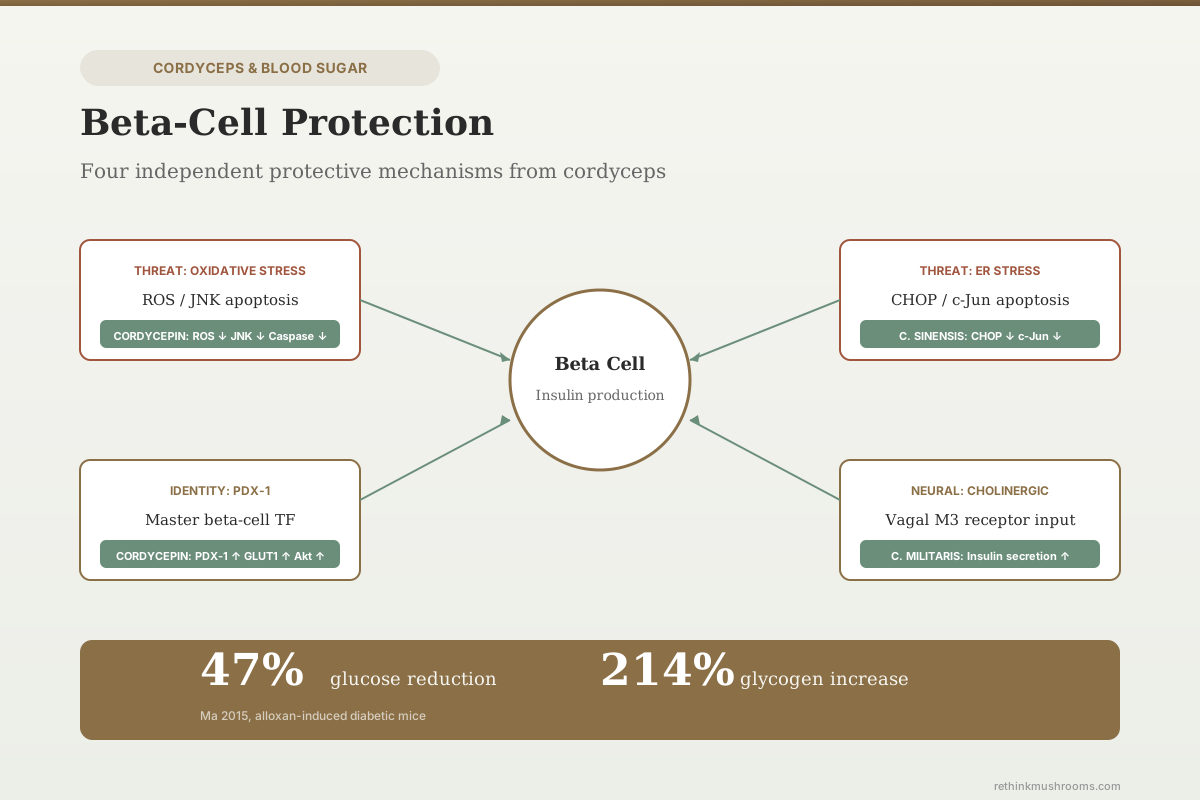
- Cordycepin reduced blood glucose by 47% and increased hepatic glycogen by 214% in alloxan-induced diabetic mice, a model of near-complete beta-cell destruction (Ma et al., Nutrition Research, 2015)
- C. militaris aqueous extracts lowered blood glucose in both normal and STZ-diabetic rats by stimulating cholinergic activation and enhancing insulin secretion from remaining beta cells (Cheng et al., Phytotherapy Research, 2012; International Journal of Medicinal Mushrooms, 2013)
- C. militaris polysaccharides reduced blood glucose and shifted metabolomic profiles in STZ-induced diabetic mice, with effects on multiple metabolic pathways (Shang et al., Natural Product Research, 2020)
Why Beta Cells Die in Diabetes
Beta-cell failure follows a predictable cascade. Chronic hyperglycemia increases glucose flux through beta cells, generating excess reactive oxygen species (ROS) from mitochondrial electron transport. Beta cells have inherently low antioxidant defenses (low catalase, low SOD relative to other cell types), making them disproportionately vulnerable to oxidative damage. Simultaneously, elevated free fatty acids cause lipotoxic stress through ceramide accumulation and ER membrane disruption. The ER stress response (unfolded protein response) initially protects by reducing protein synthesis load, but sustained ER stress activates pro-apoptotic CHOP and JNK signaling, pushing beta cells toward programmed death.
This vulnerability means a compound that reduces ROS, protects mitochondrial function, blocks JNK-mediated apoptosis, or attenuates ER stress could theoretically preserve beta-cell mass longer during the progression from insulin resistance to diabetes. The studies below test whether cordyceps compounds do any of these things.
Yan 2023: Cordycepin vs. Glucotoxicity and Lipotoxicity
Yan and colleagues exposed INS-1 beta cells to high glucose and high lipid conditions to model the dual metabolic stress that beta cells face in type 2 diabetes.[1] Cordycepin treatment produced:
- Improved cell viability under both glucotoxic and lipotoxic conditions
- Reduced intracellular ROS levels
- Increased cellular ATP content
- Induced membrane depolarization and stabilized intracellular calcium concentration
- Enhanced insulin synthesis and secretion
- Upregulated PDX-1 protein expression
- Downregulated phosphorylated JNK, cytochrome c release, and cleaved caspase-3
The PDX-1 finding is mechanistically central. PDX-1 (pancreatic and duodenal homeobox 1) is the master transcription factor for beta-cell identity. It drives insulin gene transcription, maintains beta-cell differentiation, and is required for beta-cell survival. Glucotoxicity and oxidative stress downregulate PDX-1, and its loss is an early marker of beta-cell dedifferentiation in type 2 diabetes. Cordycepin restoring PDX-1 expression under metabolic stress suggests it may help maintain beta-cell identity rather than just preventing cell death.
The JNK pathway is the apoptotic executor. High glucose and ROS activate JNK through ASK1 (apoptosis signal-regulating kinase 1), which phosphorylates downstream targets that trigger mitochondrial outer membrane permeabilization, cytochrome c release, and caspase activation. Cordycepin interrupting this cascade at the JNK level is consistent with its known AMPK activation (AMPK can inhibit ASK1-JNK signaling). For how cordycepin activates AMPK through its conversion to cordycepin monophosphate, see the cordyceps and insulin sensitivity article.
Sun 2022: Insulin Synthesis Enhancement in Normal and Damaged Cells
Sun and colleagues published complementary work testing cordycepin in both normal and oxidatively damaged INS-1 cells.[7] The study established dose-response boundaries: cordycepin at 0.1-20 uM showed no toxicity, while concentrations above 20 uM caused cell death.
In normal INS-1 cells, cordycepin:
- Enhanced insulin synthesis and secretion
- Increased ATP content, triggering membrane depolarization
- Raised intracellular calcium concentration (the direct trigger for insulin granule exocytosis)
- Upregulated mRNA and protein levels of insulin, PDX-1, and GLUT1
- Increased phosphorylated Akt (pAkt)
In oxidatively damaged cells (H2O2-treated), cordycepin additionally:
- Reduced ROS levels
- Restored mitochondrial membrane potential
- Increased SOD activity and catalase expression
The dual effect (enhancing normal function and rescuing damaged function) is relevant because beta cells in the prediabetic state exist along a spectrum from healthy to severely stressed. A compound that both boosts insulin output from functional cells and protects dying cells from further damage addresses both ends of the disease progression. The Akt upregulation connects to insulin signaling: Akt phosphorylation in beta cells promotes cell survival through Bad phosphorylation and mTORC1 activation.
Liu 2016: Protection from ER Stress
Liu and colleagues tested Cordyceps sinensis in rats with STZ-induced type 2 diabetes (combined with high-energy fat diet) for 4 weeks.[2]
The treatment:
- Reduced the number of apoptotic beta cells in pancreatic islets
- Decreased protein expression of CHOP (C/EBP homologous protein)
- Decreased c-Jun expression
- Improved fasting glucose levels
- Restored insulin sensitivity indices
CHOP is the terminal effector of the unfolded protein response (UPR) gone wrong. Under mild ER stress, the UPR activates adaptive pathways (ATF6, IRE1, PERK) that reduce protein synthesis and increase chaperone production, giving the ER time to recover. Under sustained stress, PERK phosphorylates eIF2-alpha, which induces ATF4, which induces CHOP. CHOP downregulates the anti-apoptotic protein Bcl-2 and upregulates pro-apoptotic BIM and death receptor 5, committing the cell to apoptosis.
Cordyceps sinensis reducing CHOP expression in vivo means it either reduced the ER stress itself (by reducing the protein folding burden) or blocked the signaling cascade upstream of CHOP induction (at the PERK-eIF2alpha-ATF4 axis). The study did not distinguish between these possibilities. Either mechanism would preserve beta-cell mass in the context of chronic metabolic stress.
Ma 2015: Cordycepin in Near-Complete Beta-Cell Destruction
Ma and colleagues tested purified cordycepin in alloxan-induced diabetic mice.[3] Alloxan selectively destroys beta cells through ROS generation and NAD+ depletion, creating a model of severe insulin deficiency (type 1-like diabetes).
Results:
- 47% reduction in blood glucose
- 214% increase in hepatic glycogen content
- Improved glucose tolerance
In a model where most beta cells are already destroyed, blood glucose reduction must come from enhanced function of surviving beta cells, improved peripheral insulin sensitivity, or both. The hepatic glycogen increase (requiring insulin-dependent glycogen synthase activation) suggests that cordycepin improved the efficiency of whatever insulin remained. Whether cordycepin also protected a subset of beta cells from alloxan-induced destruction (as it protected INS-1 cells from oxidative damage in the Sun 2022 study) was not directly measured. For how cordyceps affects hepatic glycogen synthesis through the IRS-1/Akt/GLUT4 pathway, see the cordyceps and insulin sensitivity article.
Cheng 2012/2013: Cholinergic Enhancement of Insulin Secretion
Cheng and colleagues published two studies testing C. militaris aqueous extracts in normal (2012) and STZ-diabetic (2013) rats.[4][5]
The mechanism identified was cholinergic activation leading to enhanced insulin secretion. Parasympathetic (vagal) innervation of pancreatic islets amplifies glucose-stimulated insulin secretion through muscarinic M3 receptors on beta cells. M3 receptor activation increases phospholipase C signaling, which raises intracellular calcium and DAG, potentiating insulin granule fusion with the plasma membrane.
In STZ-diabetic rats with reduced beta-cell mass, C. militaris extracts still lowered blood glucose through this cholinergic mechanism. This suggests the extract enhanced secretory function of remaining beta cells rather than regenerating lost ones. The cholinergic pathway is distinct from the ROS/JNK mechanism (Yan 2023) and the ER stress pathway (Liu 2016), representing a third independent route by which cordyceps compounds could support beta-cell function. For how other mushroom species address diabetes through different mechanisms, see comparing mushroom species for diabetes management.
Shang 2020: Polysaccharide Effects on Diabetic Metabolism
Shang and colleagues used 1H NMR-based metabolomics to profile the hypoglycemic effect of Cordyceps militaris polysaccharides in STZ-induced diabetic mice.[6] The polysaccharide fraction reduced blood glucose and shifted metabolite profiles toward normal patterns.
Metabolomic profiling captures the sum of all metabolic changes, including those in beta-cell function, hepatic glucose output, peripheral uptake, and lipid metabolism. The study did not isolate beta-cell-specific effects, but the STZ model's primary pathology is beta-cell destruction, so glucose improvement in this model requires either enhanced function of surviving beta cells or improved peripheral insulin sensitivity.
The Convergent Picture
Five independent research groups using three different experimental systems (INS-1 cells, STZ rats, alloxan mice) identified four distinct beta-cell protective mechanisms:
- ROS reduction and JNK inhibition (Yan 2023, Sun 2022): blocking the oxidative stress-apoptosis cascade at the mitochondrial and kinase levels
- ER stress attenuation (Liu 2016): reducing CHOP-mediated apoptosis from the unfolded protein response
- PDX-1 upregulation (Yan 2023, Sun 2022): maintaining the master transcription factor for beta-cell identity and insulin gene expression
- Cholinergic potentiation (Cheng 2012, 2013): enhancing neural input that amplifies glucose-stimulated insulin secretion
Convergent Evidence
Four Independent Pathways to the Same Beta Cell
Five research groups using three experimental systems identified four mechanistically distinct ways cordyceps compounds protect or support pancreatic beta cells. The pathways do not overlap — mitochondrial, ER-stress, nuclear, and neural — which is why a single compound touching all four is notable. All evidence is preclinical.
ROS / JNK inhibition[1][7]
INS-1 cellsActs at
Mitochondria + kinase cascade
What was shown
Cordycepin lowered intracellular ROS and blocked JNK phosphorylation, cytochrome c release, and cleaved caspase-3 in INS-1 beta cells under glucotoxic/lipotoxic stress.
ER-stress attenuation[2]
STZ ratsActs at
PERK–CHOP pathway
What was shown
C. sinensis reduced CHOP and c-Jun expression and cut beta-cell apoptosis in STZ-induced diabetic rat islets over 4 weeks.
PDX-1 upregulation[1][7]
INS-1 cellsActs at
Nuclear transcription factor
What was shown
Cordycepin restored PDX-1 — the master transcription factor for beta-cell identity and insulin gene expression — under metabolic stress, alongside GLUT1 and phosphorylated Akt.
Cholinergic potentiation[4][5]
Rats (normal + STZ)Acts at
Vagal / muscarinic M3 input
What was shown
C. militaris aqueous extracts enhanced glucose-stimulated insulin secretion from remaining beta cells via cholinergic activation in both normal and STZ-diabetic rats.
The shared limitation cuts across all four cards: INS-1 cells are an immortalized rat insulinoma line, and STZ and alloxan destroy beta cells through mechanisms that do not replicate the gradual metabolic stress of human type 2 diabetes. No human study has measured cordyceps effects on beta-cell function.
Source: Yan 2023; Sun 2022; Liu 2016; Cheng 2012/2013 (see references)
These pathways are mechanistically independent. ROS/JNK operates through the mitochondrial apoptosis pathway. ER stress operates through the PERK-CHOP pathway. PDX-1 is a nuclear transcription factor. Cholinergic signaling is neural. A compound that touches all four pathways simultaneously would be broadly cytoprotective for beta cells, addressing different causes of beta-cell death in the same disease.
The shared limitation: INS-1 cells are an immortalized rat insulinoma line, not primary human beta cells. STZ and alloxan destroy beta cells through mechanisms (selective ROS generation, DNA alkylation) that do not replicate the gradual metabolic stress of human type 2 diabetes. The gap between demonstrating protection in these models and slowing beta-cell loss in a human patient with prediabetes or early diabetes is large. For how cordyceps connects to clinical diabetes data, see cordyceps for type 2 diabetes.
The Bottom Line
Cordycepin and Cordyceps sinensis protect pancreatic beta cells through four independent mechanisms: ROS/JNK-mediated apoptosis inhibition (Yan 2023), ER stress reduction via CHOP downregulation (Liu 2016), PDX-1 upregulation maintaining beta-cell identity (Yan 2023, Sun 2022), and cholinergic enhancement of insulin secretion (Cheng 2012, 2013). In INS-1 cells, cordycepin at 0.1-20 uM enhanced insulin synthesis and secretion while protecting against glucotoxic and lipotoxic damage. In STZ-diabetic rats, Cordyceps sinensis reduced beta-cell apoptosis over 4 weeks. All evidence is from cell lines and animal models. No human study has measured cordyceps effects on beta-cell function.
Frequently Asked Questions
Sources & References
- 1RTHM-00857·Yan, Baiyi et al. (2023). “Cordycepin protects islet β-cells against glucotoxicity and lipotoxicity via modulating related proteins of ROS/JNK signaling pathway..” Biomedicine & pharmacotherapy = Biomedecine & pharmacotherapie.Study breakdown →PubMed →↩
- 2RTHM-00435·Liu, Hong et al. (2016). “The Herbal Medicine Cordyceps sinensis Protects Pancreatic Beta Cells from Streptozotocin-Induced Endoplasmic Reticulum Stress..” Canadian journal of diabetes.Study breakdown →PubMed →↩
- 3RTHM-00396·Ma, Li et al. (2015). “Cordycepin from Cordyceps militaris prevents hyperglycemia in alloxan-induced diabetic mice..” Nutrition research (New York.Study breakdown →PubMed →↩
- 4RTHM-00246·Cheng, Yu-Wen et al. (2012). “Extracts of Cordyceps militaris lower blood glucose via the stimulation of cholinergic activation and insulin secretion in normal rats..” Phytotherapy research : PTR.Study breakdown →PubMed →↩
- 5RTHM-00281·Cheng, Yu-Wen et al. (2013). “Aqueous extracts of Cordyceps militaris (Ascomycetes) lower the levels of plasma glucose by activating the cholinergic nerve in streptozotocin-induced diabetic rats..” International journal of medicinal mushrooms.Study breakdown →PubMed →↩
- 6RTHM-00653·Shang, Xiao-Lan et al. (2020). “1H NMR-based metabonomics of the hypoglycemic effect of polysaccharides from Cordyceps militaris on streptozotocin-induced diabetes in mice..” Natural product research.Study breakdown →PubMed →↩
- 7RTHM-02265·Sun, Huizhen et al. (2022). “Improving effect of cordycepin on insulin synthesis and secretion in normal and oxidative-damaged INS-1 cells..” European journal of pharmacology.Study breakdown →PubMed →↩