Reishi — Ganoderic Acids & Triterpenes
16 studies analyzed
A 2024 study found that ganoderic acid T enhances checkpoint immunotherapy by suppressing a tumor immune-evasion protein, a finding that illustrates why individual ganoderic acids are studied as distinct pharmaceutical leads.
Chen et al., Toxicol Appl Pharmacol, 2024
7
most-studied individual ganoderic acids, each with distinct protein targets
14
confirmed biological activities catalogued in the 2024 Wang review
GA-A
longest plasma half-life of the 7, and the most bioavailable after oral intake
Chen et al., Toxicol Appl Pharmacol, 2024
 View as image ↗
In 2024, a research team combined ganoderic acid T with checkpoint immunotherapy drugs and found that the combination suppressed tumors more effectively than either treatment alone. The mechanism was specific: GA-T was downregulating galectin-1, an immune-suppressive protein that tumors use to hide from immune cells, making the checkpoint blockade more effective.[13] That finding represents one piece of a larger research program: the systematic study of individual ganoderic acids as distinct molecular entities, each acting through its own pathway.
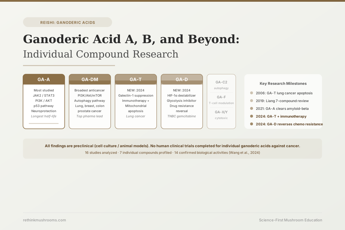
A 2024 systematic review by Wang and colleagues catalogued 14 confirmed biological activities of ganoderic acids as a class, from anti-cancer and anti-inflammatory to hepatoprotection, neuroprotection, and anti-radiation effects.[1] But the most important thing the research has established is that these activities are not uniformly distributed across all ganoderic acids. GA-A, GA-DM, GA-T, and GA-D work through different mechanisms, target different proteins, and show activity against different cancer types. Understanding them individually is not an academic exercise: it changes what researchers look for in a Reishi extract and what pharmaceutical developers are trying to synthesize.
This article examines what the research has found about the 7 most-studied ganoderic acids, compound by compound. For an overview of the full Reishi bioactive landscape, including how polysaccharides and triterpenes complement each other, see What Is Reishi Mushroom: The Science Behind the Mushroom of Immortality.
Key Takeaways
- Reishi contains at least 150 distinct triterpene compounds; the 7 most-studied ganoderic acids (A, C2, D, DM, F, T, X/Y) work through mechanistically distinct pathways and cannot be treated as interchangeable
- GA-A is the most researched and most bioavailable compound, with confirmed activity against breast, liver, brain, and cervical cancers via JAK2/STAT3, PI3K/AKT, and p53 pathways
- GA-DM is the leading anticancer candidate: a 2017 review identified activity across lung, prostate, colon, and breast cancer models via both apoptosis and autophagy, and it remains the strongest pharmaceutical development candidate
- GA-T, known since 2006 for lung cancer activity, gained new relevance in 2024 when researchers showed it enhances checkpoint immunotherapy efficacy by downregulating galectin-1, an immune-evasion protein
- GA-D reversed gemcitabine resistance in triple-negative breast cancer cells in a 2024 study by destabilizing HIF-1α, the protein that drives cancer cell glycolysis under low oxygen
- Pharmacokinetic data from rat studies shows GA-A has the longest plasma half-life of the 7 tested compounds, with highest tissue concentrations in the liver and kidney, which aligns with GA-A's well-documented hepatoprotective effects
The 7 Most-Studied GAs: Why They Are Not Interchangeable
The first thing to understand about ganoderic acid research is why individual compounds matter. A 2019 review by Liang and colleagues in the European Journal of Medicinal Chemistry analyzed seven specific ganoderic acids (A, C2, D, DM, F, X, and Y) and found that each one acts through a distinct combination of signaling pathways.[2]
GA-A primarily targets JAK2/STAT3 and PI3K/AKT signaling, which are cancer cell survival pathways active in breast, liver, and brain tumors. GA-C2 works through autophagy, a cellular recycling process that can be weaponized to destroy cancer cells. GA-DM targets the same PI3K/Akt/mTOR axis as GA-A but relies on autophagy as its primary killing mechanism rather than apoptosis. GA-F modulates T-cell function rather than directly killing cancer cells. GA-X and GA-Y are primarily cytotoxic through less-defined mechanisms. GA-D, covered later in this article, turns out to operate through a completely different metabolic pathway.
This diversity is not incidental. It reflects how Reishi synthesizes these compounds through overlapping but branching biosynthetic pathways, and it explains why a high-triterpene extract containing a full spectrum of ganoderic acids exposes cancer cells to multiple simultaneous stresses, rather than just one. For the broader context of how Reishi's bioactive classes work together, see Reishi Bioactive Compounds: Triterpenes, Polysaccharides, and Sterols.
GA-A: The Most Studied Compound, and Why That Matters
GA-A is the most abundant ganoderic acid in Reishi and the most extensively characterized. A 2025 in-depth review by Sui and colleagues in the Journal of Ethnopharmacology identified GA-A's primary molecular targets using both experimental data and computational docking: PI3K, JAK2, p53, and HMG-CoA reductase.[3]
Each of those targets has a different health implication. PI3K and JAK2 are survival and proliferation switches that cancer cells exploit. p53 is the "guardian of the genome," the tumor suppressor protein that mutant cancers disable. HMG-CoA reductase is the same enzyme that statins block to lower cholesterol. GA-A's ability to dock with all four has made it a central figure in Reishi pharmacology research.
The cancer cell evidence is specific. In triple-negative breast cancer cells, Yang and colleagues found in 2018 that GA-A directly inhibited JAK2 phosphorylation, preventing downstream STAT3 activation; it also reduced MMP-9, a matrix metalloproteinase that cancer cells use to invade surrounding tissue.[4]
In liver cancer cells, Wang and colleagues showed in 2017 that GA-A inhibited proliferation and invasion in a dose-dependent manner, triggering apoptosis via increased Bax and decreased Bcl-2 expression, the classic ratio-shift that tilts a cell from survival toward programmed death.[5]
In glioblastoma (brain cancer) cells, Cheng and Xie found in 2019 that GA-A induced both apoptosis and autophagy simultaneously, inactivating PI3K/AKT while upregulating Bax, caspase-3, and caspase-9.[6]
Liu and colleagues demonstrated in 2015 that structurally related compounds GA-I and GA-J, which share GA-A's core triterpene scaffold, induced apoptosis in cervical cancer cells through reactive oxygen species (ROS) generation and suppression of the cell's own antioxidant defenses.[7]
All of this is preclinical: cell culture and animal model data. No human trials have tested GA-A against any cancer type. The evidence base establishes mechanism and target; it does not establish clinical efficacy.
GA-A Beyond Cancer: The Pharmaceutical Development Case
Two additional lines of GA-A research point in directions that go beyond tumor biology.
In 2023, Jia and colleagues synthesized a series of amide derivatives of GA-A and tested them against the p53-MDM2 protein interaction, a pathway that many cancers hijack by causing MDM2 to mark p53 for destruction.[8] The modified GA-A derivatives showed improved potency over the natural compound. This is pharmaceutical chemistry using GA-A as a scaffold rather than as a drug candidate itself. The significance: natural GA-A already engages the p53 pathway well enough that medicinal chemists see it as a starting point for synthetic optimization.
In 2021, Qi and colleagues went in a different direction entirely, testing GA-A against Alzheimer's disease pathology in mouse models.[9] They found that GA-A induced amyloid-beta clearance through autophagy activation via the Axl receptor, and cognitive deficits improved in the treated animals. This was the first evidence for GA-A's neuroprotective mechanism beyond its known anti-inflammatory effects. The result implies that GA-A can cross the blood-brain barrier in sufficient quantities to produce measurable effects in neurons, a pharmacokinetically non-trivial finding, since most large hydrophobic compounds struggle to enter the CNS.
GA-DM: The Strongest Anticancer Candidate Across Tumor Types
If GA-A is the most studied individual compound, GA-DM is the one with the strongest anticancer profile across multiple cancer types. Bryant and colleagues' 2017 review in the Journal of Clinical and Cellular Immunology, dedicated specifically to GA-DM anticancer activity, found evidence of anti-proliferative effects in lung, prostate, colon, and breast cancer models, noting GA-DM as the strongest pharmaceutical development candidate among ganoderic acids at that time.[10]
The mechanism that distinguishes GA-DM from GA-A is its reliance on autophagy as the primary killing mechanism rather than apoptosis. In 2020, Xia and colleagues demonstrated this specifically in non-small cell lung cancer (NSCLC) cells: GA-DM inhibited PI3K/Akt/mTOR signaling, inducing autophagic flux.[11] Crucially, when they blocked autophagy pharmacologically, the cytotoxic effect of GA-DM was significantly reduced, confirming that autophagy is not a secondary pathway but the primary mechanism. This matters because some cancer cells develop resistance to apoptosis-inducing drugs by upregulating anti-apoptotic proteins. Those same cells may remain susceptible to autophagy-mediated death, which GA-DM activates through a different entry point.
The practical implication of the GA-A vs. GA-DM mechanism difference is that a cancer cell resistant to one may be vulnerable to the other. Combination approaches using multiple ganoderic acids are an active research direction, though not yet clinically tested.
GA-T: From a 2006 Lung Cancer Study to a 2024 Immunotherapy Discovery
GA-T entered the scientific literature in 2006, when Tang and colleagues showed it induced apoptosis in lung cancer cells via the intrinsic mitochondrial pathway: cytochrome-c release from mitochondria, activation of caspase-3 but not caspase-8, and eventual cell death through the inside-out cellular machinery rather than through external death-receptor signaling.[12] That finding was well-characterized and the paper is frequently cited, but GA-T remained a less prominent compound than GA-A or GA-DM in terms of research attention.
The 2024 Chen study changed that. Chen and colleagues found that GA-T modulates the tumor microenvironment by downregulating galectin-1, a protein that tumors secrete to suppress immune cell attack.[13] Galectin-1 is one of the mechanisms by which tumors create an immunosuppressive environment that blocks T-cell infiltration and reduces the efficacy of checkpoint inhibitors. By reducing galectin-1 levels, GA-T rendered the tumor microenvironment more permissive to immune cell attack. When combined with paclitaxel chemotherapy, GA-T significantly enhanced efficacy. When combined with immune checkpoint therapy, the combined tumor suppression was greater than either alone.
This is a distinct mechanism from GA-T's direct apoptosis induction in 2006. The same compound appears to both kill cancer cells directly (via mitochondrial apoptosis) and change the tumor environment to make immunotherapy more effective. Whether this dual activity is present in all tumor types or specific to certain contexts is not yet established.
For more on how Reishi compounds affect the tumor microenvironment in the broader sense, see Ganoderic Acids and Cancer: Specific Anti-Tumor Research.
GA-D: A 2024 Discovery About Drug-Resistant Cancers
GA-D has an unusual research profile. It was first studied for its ability to induce autophagy in esophageal squamous cell carcinoma (ESCC) cells: Shao and colleagues found in 2020 that GA-D triggered autophagic cell death rather than classic apoptosis, which matters because ESCC cells can be resistant to apoptosis-inducing agents.[14]
The 2024 Luo paper gave GA-D a new research identity. In triple-negative breast cancer cells that had developed resistance to gemcitabine (a common chemotherapy), Luo and colleagues found that GA-D restored drug sensitivity by inhibiting the cells' use of glycolysis, the metabolic process that cancer cells rely on even when oxygen is available, known as the Warburg effect.[15]
The specific target was HIF-1α, a protein that stabilizes in low-oxygen environments (common in tumors) and activates genes driving glycolysis and drug resistance. GA-D destabilized HIF-1α, which disrupted the metabolic adaptations that had made these cancer cells resistant to gemcitabine. This metabolic entry point is entirely distinct from the mechanisms of GA-A (JAK2/PI3K), GA-DM (PI3K/Akt autophagy), or GA-T (mitochondrial apoptosis, galectin-1). Drug resistance in triple-negative breast cancer is a major clinical problem with few effective solutions; the GA-D finding positions it as a research priority for further investigation.
GA-C2, GA-F, GA-X, and GA-Y: The Remaining Four
Four additional ganoderic acids are covered in the Liang 2019 review framework, each with a preliminary research profile but less experimental depth than the four discussed above.[2]
GA-C2 induces autophagy as its primary mechanism. In the cancer cell context, autophagy can be either protective (recycling damaged components to keep a cell alive) or lethal (when autophagy degrades cell contents past a point of no return). GA-C2 appears to push the latter pathway.
GA-F modulates T-cell function rather than directly killing cancer cells. It represents the immune-modulatory arm of ganoderic acid pharmacology, a category distinct from direct cytotoxicity.
GA-X and GA-Y show anti-tumor cytotoxicity through less-defined pathways. Both were included in the Liang 2019 review for their demonstrated anticancer activity, but neither has yet been the subject of a focused mechanistic study comparable to GA-A or GA-DM.
This unevenness in the research record is worth acknowledging. The seven compounds have been studied at very different intensities, and the apparent strength of GA-A and GA-DM's evidence base partly reflects how much more research attention they have received rather than an inherent superiority.
For the context of how extraction method determines which of these compounds reach the final product, see Ganoderic Acid Extraction Methods: How Processing Affects Potency.
Pharmacokinetics: How the Body Handles Individual Ganoderic Acids
All of the above research is mechanistic: what each compound does to cancer cells in laboratory conditions. A separate question is what happens after oral ingestion in a living organism.
Cheng and colleagues published pharmacokinetic data for seven ganoderic acids in rats using LC-MS/MS analysis in 2016.[16] All seven were absorbed through the small intestine. Plasma half-lives varied across the compounds, with GA-A showing the longest half-life of the seven tested, meaning it persists in the bloodstream longer than the others after the same dose. Tissue distribution analysis showed highest concentrations in the liver and kidney across all compounds.
The liver concentration has direct mechanistic relevance: GA-A's hepatoprotective effects (protecting liver cells from toxicity and injury, documented separately in the Sui 2025 review) align with where the compound actually accumulates. The compound travels to the organ it protects.
The Alzheimer's model result from Qi 2021 implies CNS penetration adequate to produce measurable effects in brain tissue, which is not obvious from the pharmacokinetics data alone. How much GA-A actually crosses the blood-brain barrier in humans, and what dose would be needed for meaningful CNS concentrations, remains an open question.
These pharmacokinetic studies were done in rats, not humans. Interspecies differences in absorption, metabolite formation, and tissue distribution can be substantial. The data provides a framework for understanding where these compounds go, but human pharmacokinetic studies have not yet been published.
For the broader question of how all Reishi compounds work together at the systems level, see How Reishi Triterpenes Differ from Polysaccharides in Mechanism and Mushroom Consumption and Cancer Risk Reduction: Meta-Analysis of Population Studies.
The Bottom Line
The 7 most-studied ganoderic acids in Reishi are mechanistically distinct compounds, not variations on a single theme. GA-A is the most abundant and most bioavailable, with confirmed activity across breast, liver, and brain cancer models via JAK2/STAT3 and PI3K/AKT pathways, plus neuroprotective effects in Alzheimer's disease models. GA-DM shows the broadest anticancer profile across tumor types, working through autophagy rather than apoptosis. GA-T gained a new identity in 2024 as an immunotherapy sensitizer through galectin-1 suppression, in addition to its established lung cancer apoptosis activity. GA-D reversed chemotherapy resistance in triple-negative breast cancer cells in 2024 via HIF-1α destabilization and glycolysis inhibition. All findings to date are preclinical; no human clinical trial has been completed for any individual ganoderic acid against cancer.
Frequently Asked Questions
Sources & References
- 1RTHM-02014·Wang, Siyi et al. (2024). “Research Progress on the Biological Activity of Ganoderic Acids in Ganoderma lucidum over the Last Five Years..” Life (Basel.Study breakdown →PubMed →↩
- 2RTHM-01092·Liang, Chengyuan et al. (2019). “Review of the molecular mechanisms of Ganoderma lucidum triterpenoids: Ganoderic acids A, C2, D, F, DM, X and Y..” European journal of medicinal chemistry.Study breakdown →PubMed →↩
- 3RTHM-02188·Sui, Qi et al. (2025). “Ganoderic acid A: an in-depth review of pharmacological effects and molecular docking analysis..” Journal of ethnopharmacology.Study breakdown →PubMed →↩
- 4RTHM-01021·Yang, Yuguang et al. (2018). “Ganoderic acid A exerts antitumor activity against MDA-MB-231 human breast cancer cells by inhibiting the Janus kinase 2/signal transducer and activator of transcription 3 signaling pathway..” Oncology letters.Study breakdown →PubMed →↩
- 5RTHM-00927·Wang, Xu et al. (2017). “Ganoderic acid A inhibits proliferation and invasion, and promotes apoptosis in human hepatocellular carcinoma cells..” Molecular medicine reports.Study breakdown →PubMed →↩
- 6RTHM-01043·Cheng, Yong et al. (2019). “Ganoderic acid A holds promising cytotoxicity on human glioblastoma mediated by incurring apoptosis and autophagy and inactivating PI3K/AKT signaling pathway..” Journal of biochemical and molecular toxicology.Study breakdown →PubMed →↩
- 7RTHM-00721·Liu, Ru-Ming et al. (2015). “Structurally related ganoderic acids induce apoptosis in human cervical cancer HeLa cells: Involvement of oxidative stress and antioxidant protective system..” Chemico-biological interactions.Study breakdown →PubMed →↩
- 8RTHM-01712·Jia, Yi et al. (2023). “Ganoderic Acid A and Its Amide Derivatives as Potential Anti-Cancer Agents by Regulating the p53-MDM2 Pathway: Synthesis and Biological Evaluation..” Molecules (Basel.Study breakdown →PubMed →↩
- 9RTHM-00712·Qi, Li-Feng-Rong et al. (2021). “Ganoderic Acid A Promotes Amyloid-β Clearance (In Vitro) and Ameliorates Cognitive Deficiency in Alzheimer's Disease (Mouse Model) through Autophagy Induced by Activating Axl..” International journal of molecular sciences.Study breakdown →PubMed →↩
- 10RTHM-00852·Bryant, John Matthew et al. (2017). “Anticancer Activity of Ganoderic Acid DM: Current Status and Future Perspective..” Journal of clinical & cellular immunology.Study breakdown →PubMed →↩
- 11RTHM-01303·Xia, Junbo et al. (2020). “Ganoderic acid DM induces autophagic apoptosis in non-small cell lung cancer cells by inhibiting the PI3K/Akt/mTOR activity..” Chemico-biological interactions.Study breakdown →PubMed →↩
- 12RTHM-00201·Tang, Wen et al. (2006). “Ganoderic acid T from Ganoderma lucidum mycelia induces mitochondria mediated apoptosis in lung cancer cells..” Life sciences.Study breakdown →PubMed →↩
- 13RTHM-01869·Chen, Suyu et al. (2024). “Ganoderic acid T, a Ganoderma triterpenoid, modulates the tumor microenvironment and enhances the chemotherapy and immunotherapy efficacy through downregulating galectin-1 levels..” Toxicology and applied pharmacology.Study breakdown →PubMed →↩
- 14RTHM-01285·Shao, Chang-Sheng et al. (2020). “Ganoderic acid D induces synergistic autophagic cell death except for apoptosis in ESCC cells..” Journal of ethnopharmacology.Study breakdown →PubMed →↩
- 15RTHM-01940·Luo, Binbin et al. (2024). “Ganoderic acid D attenuates gemcitabine resistance of triple-negative breast cancer cells by inhibiting glycolysis via HIF-1α destabilization..” Phytomedicine : international journal of phytotherapy and phytopharmacology.Study breakdown →PubMed →↩
- 16RTHM-00774·Cheng, Chun-Ru et al. (2016). “Pharmacokinetic Studies of Ganoderic Acids from the Lingzhi or Reishi Medicinal Mushroom, Ganoderma lucidum (Agaricomycetes), by LC-MS/MS..” International journal of medicinal mushrooms.Study breakdown →PubMed →↩