Reishi for Liver Health
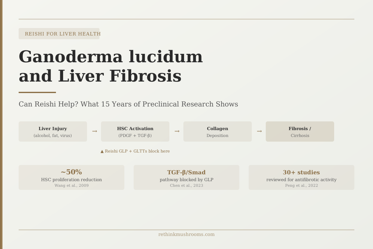
50% HSC proliferation reduction
Ganoderma lucidum extract reduced hepatic stellate cell proliferation by approximately 50% in culture by blocking the PDGF-BB receptor, establishing the core anti-fibrotic mechanism studied in subsequent research.
Wang et al., World Journal of Gastroenterology, 2009
TGF-β/Smad
signaling pathway blocked by GL polysaccharides to halt fibrosis progression (Chen 2023)
Dual Mechanism
both GLP polysaccharides and GLTTs triterpenoids independently inhibit hepatic stellate cell activation
Preclinical Only
no human clinical trials have tested Reishi for liver fibrosis; all evidence is animal or cell culture
Wang et al., World Journal of Gastroenterology, 2009
 View as image ↗
Liver fibrosis is the liver's scar-building response to chronic injury. Whether the injury is from alcohol, hepatitis B, or fat accumulation, the body's response converges on the same cell type: hepatic stellate cells (HSCs), which produce the collagen that progressively hardens liver tissue. Ganoderma lucidum (reishi) has been studied specifically at this step; researchers have been asking whether its compounds can interfere with the stellate cell activation process that drives scarring.
The answer, from 15 years of preclinical research, is that two separate classes of reishi bioactives both reach the same target through different molecular routes. What that means for ganoderma lucidum liver fibrosis research, and where the evidence currently sits, is the subject of this article. For the broader picture of reishi's hepatoprotective effects across multiple conditions, see What Is Reishi Mushroom? The Science Behind the Mushroom of Immortality.
Key Takeaways
- Liver fibrosis is driven by hepatic stellate cells (HSCs), which become activated by liver injury and produce collagen; reishi research has focused specifically on blocking HSC activation
- A 2009 study found that Ganoderma lucidum extract reduced HSC proliferation by approximately 50% by blocking the PDGF-BB receptor and reducing collagen I and III expression (Wang et al.)
- A 2023 study identified TGF-β/Smad signaling as the pathway through which Ganoderma polysaccharides (GLP) block HSC activation, induce HSC apoptosis via cell cycle arrest, and reduce extracellular matrix deposition (Chen et al.)
- A comprehensive 2022 review of 30+ studies found both GL triterpenoids (GLTTs) and GL polysaccharides (GLP) have antifibrotic activity, acting through TGF-β1/Smad, NF-κB, and PDGF pathways simultaneously (Peng et al.)
- No human clinical trials have tested reishi for liver fibrosis; all fibrosis-specific evidence is from animal models or cell culture systems
- The antifibrotic mechanisms identified in preclinical research are mechanistically plausible and well-characterized, but clinical translation to human fibrosis remains unconfirmed
The Stellate Cell Problem: What Actually Causes Liver Fibrosis
Liver fibrosis is not a disease in itself; it is the liver's wound-healing response gone chronic. When the liver is repeatedly injured (by alcohol, viral infection, excess fat, or toxins), it repairs itself by depositing collagen at the site of damage. This is normal after a single injury. When the injury is continuous, collagen deposition becomes continuous too, and the resulting scar tissue progressively replaces functional liver cells.
The cells responsible for producing this scar tissue are hepatic stellate cells (HSCs). In a healthy liver, HSCs are quiescent; they sit in a dormant, fat-storing state. When the liver is damaged, chemical signals (particularly TGF-β1 and PDGF) activate HSCs. An activated HSC transforms from a dormant cell into a proliferating, collagen-secreting machine. If nothing stops this activation process, collagen accumulates, liver architecture distorts, and the condition progresses from fibrosis toward cirrhosis.
This is the specific problem reishi research has targeted: interfering with the molecular signals that convert dormant HSCs into scar-tissue producers. Two types of reishi compounds have been studied for this purpose, and they reach the HSC through different molecular entry points.
Two Compounds, Two Routes: How GLP and Triterpenoids Both Target Fibrosis
Ganoderma lucidum produces two main classes of bioactive compounds relevant to liver biology: polysaccharides (abbreviated GLP, for Ganoderma lucidum polysaccharides) and triterpenoids (abbreviated GLTTs, for Ganoderma lucidum triterpenoids, primarily ganoderic acids).
A 2022 review by Peng et al. in Frontiers in Pharmacology examined over 30 studies on GL and liver fibrosis.[2] Both compound classes showed antifibrotic activity, but through distinct mechanisms: GLTTs (triterpenoids) primarily act through NF-κB inhibition, reducing the inflammatory signaling that activates HSCs in the first place. GLP (polysaccharides) primarily acts directly on HSCs through TGF-β1/Smad and PDGF receptor pathways, blocking the activation process itself.
The significance of having two independent routes is that they address different stages and triggers of fibrosis. The triterpenoid route reduces the inflammatory signals that tell HSCs to activate. The polysaccharide route blocks the activation machinery within HSCs directly. Both approaches converge on the same outcome: less collagen, less scarring.
For context on how ganoderic acids work across multiple liver conditions beyond just fibrosis, see Reishi for Liver Health: Hepatoprotective Research.
The Foundational Finding: Blocking the PDGF-BB Receptor by ~50% (Wang 2009)
Wang et al. established the foundational anti-fibrotic mechanism in 2009, using a hepatic stellate cell line called HSC-T6.[1] They found that Ganoderma lucidum extract reduced HSC-T6 proliferation by approximately 50% compared to untreated controls.
The mechanism was at the receptor level: the extract blocked the PDGF-BB receptor. PDGF-BB (platelet-derived growth factor BB) is one of the primary signals that tells dormant HSCs to divide and become active. By blocking the receptor that PDGF-BB binds to, the extract interrupted the activation signal before it could reach the inside of the cell.
The downstream effects followed from this receptor blockade: collagen type I expression fell, collagen type III expression fell, and TGF-β1 signaling was reduced. The study established that reishi extract was not generally toxic to stellate cells; it was specifically interfering with the growth and activation pathways, not killing all cells indiscriminately.
This is the mechanistic anchor for all subsequent reishi fibrosis research: if an extract can reduce HSC proliferation by blocking PDGF-BB, that's a biologically meaningful interaction with the key driver of fibrosis progression.
The 2023 TGF-β Discovery: How GLP Shuts Down the Fibrosis Signaling Chain
The 2023 study by Chen et al. in Phytomedicine is the most mechanistically detailed reishi fibrosis paper available as of 2026.[3] It focused on Ganoderma lucidum polysaccharides (GLP) specifically, rather than whole extract, and examined the TGF-β/Smad signaling axis.
TGF-β (transforming growth factor beta) is often described as the master regulator of fibrosis. It is the chemical signal most responsible for converting dormant HSCs into collagen-secreting activated cells. TGF-β signals through a set of intracellular proteins called Smad proteins. When TGF-β binds its receptor, it phosphorylates Smad2 and Smad3, which then enter the cell nucleus and activate fibrosis-related genes.
Chen et al. found that GLP inhibits this TGF-β/Smad axis. The polysaccharide blocked HSC activation by reducing Smad signaling, which in turn reduced collagen type I and type III deposition. But the 2023 study also identified something the 2009 Wang study hadn't captured: GLP was inducing HSC apoptosis (programmed cell death) through cell cycle arrest. It was not only preventing new HSC activation but also eliminating already-activated HSCs.
The study also found GLP modulated ECM-receptor interaction: the way activated HSCs anchor themselves to the extracellular matrix (the protein scaffold surrounding cells). Disrupting this anchoring reduces the structural reinforcement that allows scar tissue to persist even when HSC activity decreases.
Three converging effects from a single compound: blocking activation signals, inducing death of already-activated cells, and weakening the structural infrastructure of established scar tissue.
The Triterpenoid Angle: Ganoderic Acids Approach Fibrosis from the Inflammatory Side
While polysaccharides act at the TGF-β/PDGF level to block HSC activation directly, triterpenoids (ganoderic acids) approach the fibrosis problem one step earlier in the causal chain: they reduce the inflammation that triggers HSC activation in the first place.
Zhou et al.'s 2019 review of molecular mechanisms of Ganoderma triterpenoids identified the NF-κB pathway as a primary target.[4] NF-κB is the master transcription factor for inflammatory gene expression. When the liver is injured, NF-κB drives the production of inflammatory cytokines including TNF-α and IL-1β, and these cytokines are among the signals that activate HSCs. Ganoderic acids suppress NF-κB, which reduces the downstream inflammatory cascade that HSC activation depends on.
The ganoderic acid A is particularly studied in the anti-fibrotic context. It is also found to directly inhibit TGF-β1 signaling in some models, creating an overlap with the polysaccharide mechanism at the TGF-β level. In this sense, the two compound classes are not strictly independent in their anti-fibrotic effects; they share the TGF-β target while approaching it through different entry points (ganoderic acids via NF-κB-driven cytokine suppression, GLP via direct Smad pathway inhibition).
The practical implication is that extracts containing both polysaccharides and triterpenoids may have complementary anti-fibrotic activity. Most commercial reishi extracts contain both compound classes, though the ratio varies depending on whether extraction was done with hot water (favoring polysaccharides) or alcohol (favoring triterpenoids).
Where Reishi's Fibrosis Evidence Sits Among Its Other Liver Research
Ahmad et al.'s 2023 review in Nutrients cataloged six distinct hepatoprotective mechanisms for Ganoderma lucidum: modulation of Phase I/II liver enzymes, beta-glucuronidase suppression, antifibrotic effects, antiviral activity (HBV), nitric oxide regulation, and hepatocellular calcium homeostasis.[5]
Within this picture, the antifibrotic mechanism is one of the better-characterized ones. The PDGF-BB, TGF-β/Smad, and NF-κB data collectively provide three independent lines of mechanistic evidence. Compared to the antiviral HBV evidence (which relies more heavily on a single class of in vitro studies) or the NAFLD evidence (strong for polysaccharides, weaker for triterpenoids), the fibrosis picture has both compound classes represented and a progression from 2009 (PDGF mechanism) to 2022 (comprehensive review) to 2023 (TGF-β/Smad precision) that shows active research accumulation.
Where reishi's fibrosis evidence is weaker than its general hepatoprotective data is on the human side. The general hepatoprotective literature includes a small human trial on HBV and case reports (both protective and cautionary) that provide some human biology context. The fibrosis literature has no human data at all.
For the NAFLD angle, where polysaccharide data is also strong, see Reishi and Non-Alcoholic Fatty Liver Disease (NAFLD): Emerging Evidence. Other mushroom compounds also show hepatoprotective activity through independent mechanisms, including Ergothioneine and Liver Health: Hepatoprotective Research.
The Evidence Gap: No Human Fibrosis Trials, and What That Means
Every study cited in this article is preclinical. Wang 2009 used a stellate cell line. Chen 2023 used animal models and cell culture. Peng 2022's review synthesized animal and in vitro studies. There are no published randomized controlled trials, and no completed human cohort studies, testing reishi against liver fibrosis as a primary endpoint in humans.
This gap is significant because the biology of liver fibrosis in humans is more complex than animal models capture. Humans develop fibrosis over years or decades, not over weeks as in the DSS and CCl4 mouse models used in many of these studies. The dose-response relationship in humans is unknown. Whether the oral bioavailability of ganoderic acids and GLP polysaccharides in a commercially available supplement is sufficient to reach the HSC concentrations used in the preclinical studies has not been established.
What the preclinical evidence provides is mechanistic plausibility. The biological pathway is real: TGF-β/Smad drives HSC activation in humans, and blocking this pathway reduces fibrosis in animal models. Reishi compounds demonstrably reach this pathway in cell and animal systems. That is a meaningful finding; it explains why reishi's anti-fibrotic potential warrants clinical investigation. It does not confirm clinical benefit in human fibrosis.
The honest position is: reishi has among the better-characterized preclinical antifibrotic mechanisms of any dietary supplement studied for liver conditions, and no human clinical evidence that this mechanism translates to benefit in human liver fibrosis.
For context on how reishi's alcohol-related liver effects (a related condition) are also all preclinical, see Reishi and Alcohol-Related Liver Damage: Preclinical Evidence. For antiviral effects on hepatitis B, see Reishi and Hepatitis B: Antiviral Research.
The Bottom Line
Ganoderma lucidum contains two classes of compounds, polysaccharides (GLP) and triterpenoids (GLTTs), that independently inhibit hepatic stellate cell activation through complementary pathways: PDGF-BB receptor blockade (Wang 2009), TGF-β/Smad inhibition (Chen 2023), and NF-κB-mediated inflammatory suppression (Zhou 2019). A 2022 review synthesized 30+ studies confirming antifibrotic activity across both compound classes. No human clinical trials have tested reishi for liver fibrosis; all evidence is preclinical.
Frequently Asked Questions
Sources & References
- 1. Source details for this reference are not yet available.
- 2. Source details for this reference are not yet available.
- 3. Source details for this reference are not yet available.
- 4. Source details for this reference are not yet available.
- 5. Source details for this reference are not yet available.