Cordyceps & Cellular Energy
2 distinct signaling pathways
A 2020 Cell Chemical Biology study showed that cordycepin activates AMPK not by binding adenosine receptors but through a completely separate phosphorylation pathway, overturning a decade of assumptions about how this compound works.
Hawley et al., Cell Chemical Biology, 2020
AMPK
the master cellular energy sensor activated via cordycepin monophosphate phosphorylation
A1 receptor
adenosine receptor through which cordycepin increases NREM sleep in animal studies
PGC-1α
the mitochondrial biogenesis regulator activated downstream of cordycepin's AMPK pathway
Hawley et al., Cell Chemical Biology, 2020
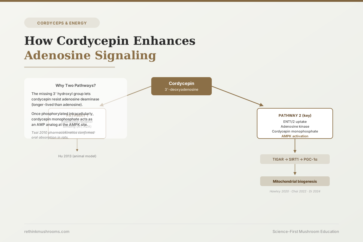 View as image ↗
For years, the conventional explanation for cordycepin's effects on energy and metabolism went roughly like this: cordycepin looks like adenosine, so it probably works like adenosine, activating the same receptors and producing the same downstream signals. That explanation is not wrong, but a 2020 paper in Cell Chemical Biology showed it is incomplete in a way that matters. The more important pathway for energy metabolism operates through a completely different mechanism, does not involve adenosine receptor binding at all, and explains effects that the receptor pathway alone cannot account for.[1]
Cordycepin has two parallel signaling pathways that operate simultaneously. Understanding both is necessary for understanding why it does what it does. This article traces each pathway from the molecule's entry into the cell to its downstream effects on energy metabolism and mitochondrial function. For the broader context of what Cordyceps is and why cordycepin is its most studied active compound, see Cordyceps: The Science Behind the Energy Mushroom.
Key Takeaways
- Cordycepin (3'-deoxyadenosine) is a structural analog of adenosine that differs by a single missing hydroxyl group at the 3' carbon position; this small difference gives it distinct pharmacology from adenosine
- Pathway 1: cordycepin binds adenosine receptors (primarily A1 and A2A) and produces downstream effects including cAMP elevation and, in animal studies, increased NREM sleep duration
- Pathway 2 (the more consequential for energy): cordycepin is transported into cells via ENT1/ENT2 transporters, phosphorylated by adenosine kinase to cordycepin monophosphate, and then activates AMPK as an AMP analog, entirely independent of adenosine receptor binding (Hawley et al., Cell Chemical Biology, 2020)
- AMPK activation downstream triggers a cascade: TIGAR activation, SIRT1 upregulation, and PGC-1α stimulation, which drives mitochondrial biogenesis and increased cellular energy capacity
- Rat pharmacokinetic data confirms cordycepin is absorbed orally and rapidly phosphorylated to active forms; the mechanism is not theoretical but confirmed to occur after oral ingestion
- The two pathways operate at different effective concentrations and may be relevant to different physiological outcomes; this distinction is largely absent from consumer-facing content
Cordycepin vs. Adenosine: One Missing Atom, Two Different Profiles
Adenosine is one of the most important signaling molecules in the body. It is a component of ATP (the cell's primary energy currency), a neuromodulator that promotes sleep when it accumulates in the brain, and a regulator of inflammation and immune responses. Understanding adenosine's role makes it clear why a compound that mimics adenosine's structure would attract research attention.
Cordycepin (chemically called 3'-deoxyadenosine) differs from adenosine at one position: the hydroxyl group at the 3' carbon of the ribose sugar is replaced by a hydrogen atom. This is a minimal structural change, but it has two important consequences. First, it allows cordycepin to partially mimic adenosine's receptor-binding activity, binding to some of the same cell-surface proteins. Second, it causes cordycepin to behave differently once inside the cell, because that missing hydroxyl group changes how cordycepin is processed by intracellular enzymes.
Tsai and colleagues characterized cordycepin's pharmacokinetics in rats in 2010, establishing that oral cordycepin is absorbed from the gut and rapidly phosphorylated inside cells to form active phosphorylated metabolites.[3] The pharmacokinetics differ measurably from adenosine itself. This matters because it means the active compounds reaching cells after oral cordycepin supplementation are not identical to cordycepin as ingested: they are phosphorylated derivatives, and these derivatives have their own distinct biological activity.
Pathway 1: How Cordycepin Binds Adenosine Receptors
The first pathway is the more intuitive one. Cordycepin's structural similarity to adenosine allows it to bind to cell-surface adenosine receptors, a family of four G-protein-coupled receptors (A1, A2A, A2B, and A3) that regulate a broad range of physiological processes.
When adenosine binds A1 receptors, it decreases cAMP levels in the cell, slowing metabolic activity, which is part of how adenosine promotes sleep when it accumulates in the brain during waking hours. When adenosine binds A2A receptors, it increases cAMP, producing vasodilatory and anti-inflammatory effects.
Cordycepin can engage this system. Hu and colleagues demonstrated in 2013 using rat models that cordycepin significantly increased NREM (non-rapid eye movement) sleep duration, and that this effect was blocked when A1 receptors were pharmacologically antagonized.[2] The A1 receptor blockade abolishing the sleep effect is direct mechanistic evidence for functional A1 receptor agonism by cordycepin.
This pathway is real, demonstrated in vivo, and explains some of cordycepin's effects. But it is not the pathway responsible for its energy metabolism and anti-fatigue effects. For that, a different mechanism operates.
Pathway 2: The AMPK Mechanism Hawley 2020 Discovered
The 2020 Hawley et al. paper in Cell Chemical Biology resolved a long-standing question about cordycepin's energy effects by tracing the mechanism step by step, then testing each step with specific pharmacological inhibitors.[1]
AMPK (AMP-activated protein kinase) is sometimes called the cell's master energy sensor. When the ratio of AMP to ATP rises, meaning the cell is running low on energy, AMPK activates. It then switches off energy-consuming processes and switches on energy-generating processes, including fatty acid oxidation, glucose uptake, and ultimately mitochondrial biogenesis. Activating AMPK is one of the mechanisms by which exercise produces long-term metabolic adaptations. It is also the target of metformin, a widely used diabetes drug.
Hawley showed that cordycepin activates AMPK, but not in the way anyone had assumed. The route is:
- Cordycepin enters the cell via equilibrative nucleoside transporters ENT1 and ENT2 (specialized proteins on cell membranes that transport nucleoside compounds into cells)
- Inside the cell, the enzyme adenosine kinase phosphorylates cordycepin, attaching a phosphate group to create cordycepin monophosphate
- Cordycepin monophosphate structurally mimics AMP (adenosine monophosphate) because the missing 3' hydroxyl group is not critical for the AMP-binding site on AMPK
- Cordycepin monophosphate binds the AMP-binding site on AMPK and activates it, signaling to the cell that energy is low
The critical experimental test: when ENT1/ENT2 transporters were pharmacologically blocked, cordycepin could no longer enter cells efficiently, cordycepin monophosphate could not be formed, and AMPK activation was abolished. This definitively rules out adenosine receptor binding as the mechanism for AMPK activation: the effect requires intracellular processing, not cell-surface receptor engagement.
This pathway is entirely distinct from Pathway 1. Two separate mechanisms, operating in parallel, activated by the same molecule entering the body.
The Downstream Cascade: From AMPK to Mitochondrial Biogenesis
AMPK activation is not itself the endpoint. It triggers a downstream cascade with consequences for mitochondrial function and long-term cellular energy capacity.
Chai and colleagues traced this cascade in 2022, showing that cordycepin-induced AMPK activation proceeded through the following sequence.[4] AMPK activates TIGAR (TP53-induced glycolysis and apoptosis regulator), a metabolic regulator that reduces reliance on glycolysis and pushes cells toward more efficient oxidative phosphorylation. TIGAR activation leads to SIRT1 upregulation. SIRT1 is a sirtuin, a class of proteins involved in NAD-dependent metabolic regulation. Activated SIRT1 then stimulates PGC-1α (peroxisome proliferator-activated receptor-gamma coactivator 1-alpha), the master regulator of mitochondrial biogenesis: the process by which cells generate more mitochondria and increase their capacity for sustained energy production.
In the Chai 2022 models, cordycepin reduced fatigue biomarkers including lactic acid and creatine kinase, consistent with improved metabolic efficiency. The paper provided the first explicit mechanistic chain connecting the AMPK pathway to the anti-fatigue effects documented in animal models.
Di Lorenzo and colleagues found related evidence in 2024, showing that Cordyceps sinensis extract elevated intracellular NAD+ levels and activated SIRT1 and SIRT3 sirtuins, enhancing mitochondrial energy metabolism markers in cell culture.[5] The NAD+/sirtuin connection links cordycepin's effects to the broader longevity and metabolic regulation pathways that NAD+ precursors like NMN and NR also target.
Choi and colleagues provided the most direct endpoint measurement in 2020, showing that C. militaris extracts increased cellular ATP content and mitochondrial membrane potential (a measure of how actively mitochondria are producing energy) in cultured cells, with simultaneous reductions in reactive oxygen species.[6] These measurements represent the output of the cascade: more ATP, healthier mitochondria, less oxidative stress.
For a deeper look at how these effects translate to mitochondrial function specifically, see Cordyceps and Mitochondrial Function: Research on Energy Metabolism.
Why Oral Cordycepin Reaches These Pathways: Pharmacokinetics
A compound can have the most elegant cellular mechanism and still fail to do anything clinically useful if it cannot survive digestion and reach tissues in active form. Cordycepin's pharmacokinetics address this concern.
Tsai and colleagues' 2010 rat study established several relevant facts.[3] Cordycepin is absorbed through the gut wall. Once in circulation, it distributes to tissues and is phosphorylated to active forms including cordycepin monophosphate. The pharmacokinetic profile differs from adenosine, which has a very short half-life in plasma due to rapid enzymatic degradation. Cordycepin's missing 3' hydroxyl group makes it resistant to adenosine deaminase, one of the enzymes that would otherwise break it down quickly, and this contributes to its different pharmacokinetic behavior.
This has a practical implication for interpreting the Hawley mechanism: the ENT1/ENT2 uptake and adenosine kinase phosphorylation steps that Hawley described are downstream of absorption. Oral cordycepin is absorbed, reaches cells throughout the body, enters those cells via ENT transporters, and is phosphorylated to cordycepin monophosphate. The mechanism is not theoretical: the pharmacokinetic data confirms each step occurs in living systems after oral dosing.
The data is from rats, not humans. Whether the phosphorylation kinetics and tissue distribution are sufficiently similar in humans to produce equivalent AMPK activation at typical supplement doses is not yet established by human pharmacokinetic studies.
Two Pathways, One Molecule: What This Means for Research
The existence of two parallel pathways has practical implications for how cordycepin research should be designed and interpreted.
First, the pathways likely have different effective concentration thresholds. Adenosine receptor binding typically occurs at nanomolar concentrations (very low amounts). AMPK activation via cordycepin monophosphate is an intracellular event that requires uptake and phosphorylation. Whether typical oral doses of Cordyceps mushrooms or extracts produce sufficient intracellular cordycepin monophosphate to meaningfully activate AMPK is a question the current literature cannot fully answer, because the precise dose-response relationship in human tissues has not been characterized.
Second, the two pathways could produce opposing effects in some contexts. Adenosine receptor activation (especially A1) is associated with reduced metabolic activity and sleep promotion. AMPK activation is associated with increased metabolic activity. Whether these effects cancel each other, operate in different tissues at the same dose, or are concentration-dependent is not yet clear.
Third, much of the mechanistic work (Hawley 2020, Chai 2022) was done in cell culture or animal models. The specific pathway activations confirmed in those systems may not translate directly to human doses available from Cordyceps supplementation.
For the clinical evidence on what cordycepin-containing Cordyceps supplements actually do in humans for fatigue and energy, see Cordyceps for Fatigue: Clinical Evidence on Energy Levels and Cordyceps and ATP: How It Boosts Cellular Energy.
The Bottom Line
Cordycepin operates through two parallel signaling pathways that are mechanistically independent. The first pathway involves direct binding to adenosine receptors (primarily A1), demonstrated in vivo by Hu 2013's sleep study. The second and more consequential pathway for energy metabolism was clarified by Hawley 2020 in Cell Chemical Biology: cordycepin enters cells via ENT1/ENT2 transporters, is phosphorylated to cordycepin monophosphate by adenosine kinase, and activates AMPK as an AMP analog, not via adenosine receptor binding. Downstream, AMPK activation proceeds through TIGAR, SIRT1, and PGC-1α to drive mitochondrial biogenesis (Chai 2022). Pharmacokinetic data from rat studies (Tsai 2010) confirms this intracellular processing occurs after oral ingestion. Whether the AMPK pathway is activated at doses achievable from dietary Cordyceps supplementation in humans remains an open question.
Frequently Asked Questions
Sources & References
- 1RTHM-00618·Hawley, Simon A et al. (2020). “Mechanism of Activation of AMPK by Cordycepin..” Cell chemical biology.Study breakdown →PubMed →↩
- 2RTHM-00288·Hu, Zhenzhen et al. (2013). “Cordycepin Increases Nonrapid Eye Movement Sleep via Adenosine Receptors in Rats..” Evidence-based complementary and alternative medicine : eCAM.Study breakdown →PubMed →↩
- 3RTHM-00199·Tsai, Yung-Jen et al. (2010). “Pharmacokinetics of adenosine and cordycepin, a bioactive constituent of Cordyceps sinensis in rat..” Journal of agricultural and food chemistry.Study breakdown →PubMed →↩
- 4RTHM-00749·Chai, Xiaoming et al. (2022). “Cordycepin exhibits anti-fatigue effect via activating TIGAR/SIRT1/PGC-1α signaling pathway..” Biochemical and biophysical research communications.Study breakdown →PubMed →↩
- 5RTHM-00873·Di Lorenzo, Ritamaria et al. (2024). “NAD-Driven Sirtuin Activation by Cordyceps sinensis Extract: Exploring the Adaptogenic Potential to Promote Skin Longevity..” International journal of molecular sciences.Study breakdown →PubMed →↩
- 6RTHM-00613·Choi, Eunhyun et al. (2020). “Beneficial Effect of Cordyceps militaris on Exercise Performance via Promoting Cellular Energy Production..” Mycobiology.Study breakdown →PubMed →↩