Mushrooms & Neuroinflammation
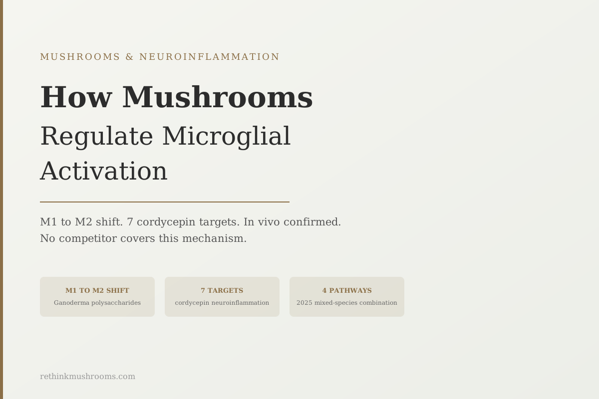
7 molecular targets inhibited by cordycepin
Cordycepin from Cordyceps mushrooms inhibits neuroinflammation through at least seven distinct molecular targets simultaneously: adenosine A2 receptor, NF-κB, NLRP3 inflammasome, IL-1β, iNOS, COX-2, and HMGB1. Ganoderma polysaccharides and lion's mane extract target complementary pathways in a different brain immune cell mechanism.
Govindula et al., Eur J Pharmacol, 2021; Cai et al., J Neuroinflammation, 2017
M1 to M2
Ganoderma polysaccharides shift microglia from inflammatory to protective
4 pathways
JNK, ERK, p38, and IκB all suppressed by mixed mushroom extract in 2025 data
In vivo confirmed
Ganoderma microglial suppression confirmed in Parkinson's mouse brain tissue
Govindula et al., Eur J Pharmacol, 2021; Cai et al., J Neuroinflammation, 2017
 View as image ↗
Chronic neuroinflammation is one of the defining features of Alzheimer's disease, Parkinson's disease, and most other neurodegenerative conditions. Its primary driver is not neurons themselves but a different brain cell type called microglia, which function as the brain's resident immune cells. When microglia are chronically overactivated, they shift from a state that supports neural repair to one that damages and kills neurons. This shift is the mechanistic heart of neuroinflammatory disease.
Several mushroom compounds have been shown to reverse this shift. Ganoderma polysaccharides, cordycepin from Cordyceps, and lion's mane extract each act on different molecular pathways to suppress the damaging microglial state and restore the protective one. Understanding how they do this requires understanding what microglia are and what the M1/M2 distinction actually means in brain tissue.
For the broader inflammation context, see How Mushrooms Fight Inflammation: NF-κB, COX-2, and Beyond and Mushroom Anti-Inflammatory Mechanisms: A Comprehensive Guide.
Key Takeaways
- Microglia are the brain's immune cells; when chronically activated in a pro-inflammatory (M1) state, they produce cytokines and reactive oxygen species that damage neurons; mushroom compounds shift them toward the protective (M2) state
- Ganoderma lucidum polysaccharides suppressed IL-1β, IL-6, and iNOS in LPS-activated BV2 microglia while upregulating the protective cytokine TGF-β, and also modulated microglial phagocytosis and migration behavior (Cai et al., J Neuroinflammation, 2017)
- Cordycepin from Cordyceps inhibits neuroinflammation via seven distinct molecular targets: adenosine A2 receptor, NF-κB, NLRP3 inflammasome, IL-1β, iNOS, COX-2, and HMGB1, the most comprehensively mapped mushroom neuroinflammatory mechanism
- Lion's mane ethanolic extract directly reduced nitric oxide production in LPS-activated BV2 microglia while simultaneously protecting hippocampal neurons from oxidative damage (Kushairi et al., Antioxidants, 2019)
- A 2025 study found a mixed mushroom mycelia combination suppressed BV2 neuroinflammation through four simultaneous MAPK/NF-κB pathways: JNK, ERK, p38, and IκB phosphorylation
- The Ganoderma microglial suppression data was confirmed in living animal brain tissue in the nigrostriatal region of Parkinson's disease mice, validating the cell culture data in a disease-relevant in vivo context
What Microglia Are and Why M1/M2 Polarization Matters
Microglia are the resident immune cells of the central nervous system. They make up roughly 10-15% of all brain cells. In a healthy brain, they perform surveillance functions: scanning for damaged neurons, clearing cellular debris, modulating synaptic connections, and responding to pathogens. This protective, alert state is sometimes called M2 or ramified microglial activity.
When microglia detect damage signals (pro-inflammatory cytokines, amyloid beta aggregates, LPS from gut bacteria that cross a compromised blood-brain barrier, or other inflammatory triggers), they activate into a reactive state. This M1 activation involves: secreting IL-1beta, IL-6, TNF-alpha, and IL-18 (pro-inflammatory cytokines that recruit more immune cells), releasing nitric oxide via iNOS and prostaglandins via COX-2 (reactive molecules that directly damage nearby neurons), and increasing phagocytic activity (which, when excessive, can engulf healthy synapses).
In acute injury, M1 activation is appropriate and necessary. In chronic neurodegeneration, it becomes self-sustaining and destructive. Damaged neurons release signals that keep microglia activated; activated microglia damage more neurons; more damaged neurons release more activating signals. This cycle is called "reactive microgliosis," and it is a core mechanism in Alzheimer's and Parkinson's disease progression.
The M1/M2 framework is a simplification of a spectrum of microglial states, but it is the most commonly used shorthand in the literature and captures the clinically relevant distinction: too much M1 activity damages the brain, and compounds that shift toward M2 activity tend to be neuroprotective. The question for mushroom research is which compounds produce this shift, at what concentrations, and through which molecular routes.
Ganoderma Polysaccharides and the Definitive BV2 Study
The most rigorously published study on mushrooms and microglial activation was conducted by Cai and colleagues in 2017 and published in the Journal of Neuroinflammation, the journal specifically dedicated to this research field.[1]
The study used BV2 microglia, the standard laboratory cell line for microglial research, activated with either LPS (the bacterial endotoxin used to induce M1 microglial activation in experiments) or amyloid beta. Ganoderma lucidum polysaccharides (GLP) were added at various concentrations.
The results established three distinct types of microglial modulation:
Pro-inflammatory cytokine suppression. GLP significantly inhibited LPS-induced IL-1beta, IL-6, and iNOS production in activated BV2 microglia, reducing the pro-inflammatory output that damages surrounding neurons. The suppression was dose-dependent.
Anti-inflammatory cytokine upregulation. GLP increased TGF-beta expression in the same cells, shifting the cytokine balance from M1 toward M2. TGF-beta is a critical anti-inflammatory signal that promotes tissue repair and suppresses excessive immune responses.
Phagocytosis and migration modulation. GLP also modified microglial behavior beyond cytokine output, altering phagocytic capacity and migration patterns. This is significant because it suggests Ganoderma polysaccharides don't simply suppress microglial activity; they reshape what the cells do.[1]
The amyloid beta component of the experiment is particularly relevant: GLP suppressed microglia even when activated by the same protein aggregates found in Alzheimer's disease brain tissue, not just the synthetic LPS stimulus. This improves the translational relevance from laboratory model to disease context.
Cordycepin: Seven Molecular Targets, One Compound
Cordycepin (3'-deoxyadenosine), the primary bioactive compound in Cordyceps militaris, has been studied against neuroinflammation more systematically than almost any other mushroom compound.
Govindula and colleagues published a comprehensive mechanistic mapping in the European Journal of Pharmacology in 2021, identifying at minimum seven distinct molecular targets through which cordycepin inhibits microglial activation and neuroinflammation:[2]
- Adenosine A2 receptor downregulation. Microglial activation is partly driven by adenosine receptor signaling; cordycepin's structural similarity to adenosine allows it to interfere with this pathway.
- NF-κB inhibition. The master transcription factor for inflammatory gene expression, suppressed downstream of the adenosine receptor interaction.
- NLRP3 inflammasome inhibition. The same cellular alarm system that triggers pyroptotic inflammatory cell death in liver macrophages and in brain microglia, where it amplifies neuroinflammation.
- IL-1beta suppression. The inflammasome's primary output and a potent pro-inflammatory cytokine.
- iNOS suppression. Reduces nitric oxide production, one of the most directly neurotoxic reactive species from activated microglia.
- COX-2 suppression. Reduces prostaglandin production and the pain/inflammation cascade.
- HMGB1 suppression. High mobility group box 1 protein is a late-stage inflammatory signal that sustains chronic inflammation after the initial trigger has resolved.
Seven simultaneous molecular targets in a single compound is unusual. Most pharmaceutical anti-inflammatory agents target one pathway specifically (aspirin inhibits COX; NSAIDs inhibit COX-1/2; TNF inhibitors block TNF). Cordycepin appears to engage multiple routes at once, which may explain why it shows broad neuroinflammatory suppression across multiple experimental models of brain disease.
Lion's Mane Extract in BV2 Microglia: The Direct Cellular Evidence
While most Lion's Mane neuroinflammation research focuses on erinacines and their effects in Alzheimer's models, Kushairi and colleagues provided direct BV2 microglial activation data in 2019, published in Antioxidants.[3]
Their experiment added Lion's Mane ethanolic extract to BV2 microglia activated with LPS. The extract significantly reduced nitric oxide production, one of the primary outputs of M1-activated microglia and one of the most directly damaging molecules microglia produce.
The study used two cell types simultaneously: BV2 microglia and HT22 hippocampal neurons. The same Lion's Mane extract that suppressed microglial nitric oxide also protected HT22 neurons from oxidative damage induced by hydrogen peroxide. Running both experiments in the same study tested whether the extract could address both sides of the neuroinflammation problem: the immune cells causing damage and the neurons being damaged.[3]
It did. Both effects were significant, suggesting Lion's Mane compounds act on both the inflammatory cell population and the target cell population. This bidirectional activity makes biological sense given that erinacines and hericenones promote NGF (which supports neuron survival) while also suppressing the inflammatory signals that harm neurons.
For the full picture of Lion's Mane's anti-inflammatory action including its effects on cytokines in metabolic inflammation contexts, see Lion's Mane and Anti-Inflammatory Cytokines: The Mood-Inflammation Link.
Multi-Species Combinations: The 2025 Four-Pathway Data
Individual species data establishes that mushroom compounds affect microglia. The question of whether combinations of mushroom species produce broader or deeper effects was addressed directly in a 2025 study.
Lee and colleagues tested a mixed mushroom mycelia extract (designated GMK, a combination of multiple species) against LPS-induced neuroinflammation in BV2 microglia and neurotoxicity in PC12 neurons, published in Cells in 2025.[4]
The mechanistic profile was striking. The GMK extract suppressed BV2 microglial activation by inhibiting four distinct MAPK and NF-κB signaling pathways simultaneously:
- JNK (c-Jun N-terminal kinase) phosphorylation
- ERK (extracellular signal-regulated kinase) phosphorylation
- p38 MAPK phosphorylation
- IκB phosphorylation (the gateway to NF-κB nuclear translocation)
Together these pathways account for a large fraction of the known microglial activation signaling network. By targeting all four simultaneously, the combination extract suppressed NO, TNF-alpha, and IL-6 production while protecting PC12 neurons from neurotoxic damage.[4]
The four-pathway inhibition profile is more comprehensive than any single-species study reviewed here, which suggests that species combinations may produce mechanistic breadth that single species cannot. This is relevant for supplement formulations: products that combine multiple mushroom species may have a different microglial modulation profile than products using a single species, though this remains an early observation from one study.
From BV2 Cells to Animal Brain Tissue: Ganoderma in Parkinson's Disease
Cell culture data establishes that mushroom compounds can suppress microglial activation in laboratory conditions. Animal model data tests whether those effects translate to living brain tissue in the context of actual neurological disease.
Ren and colleagues published a Parkinson's disease model study in Nutrients in 2022 that provided exactly this validation for Ganoderma.[5] The MPTP model (1-methyl-4-phenyl-1,2,3,6-tetrahydropyridine injection) is the standard laboratory model for Parkinson's disease: MPTP selectively destroys dopaminergic neurons in the nigrostriatal pathway, producing the motor and neurochemical features of Parkinson's.
In this model, Ganoderma lucidum extract:
- Repressed MPTP-induced microglial activation specifically in the nigrostriatal region, the brain area where dopaminergic neurons are lost in Parkinson's disease
- Modulated the abnormal cytokine expression profile that accompanies MPTP-induced neurodegeneration
- Reduced iNOS and NLRP3 inflammasome activity in the same region
- Also regulated microglial autophagy, the cellular process by which microglia clear damaged proteins
This is in vivo confirmation that Ganoderma's microglial suppressive effects, first observed in BV2 cell culture by Cai 2017, operate in real brain tissue in the context of a validated Parkinson's disease model.[5] The iNOS/NLRP3 pathway overlap between the 2017 BV2 data and the 2022 animal data strengthens the mechanistic consistency across these experimental systems.
For the clinical evidence on mushrooms in Alzheimer's and Parkinson's, see Mushrooms for Neuroinflammatory Conditions: Research Guide, which covers the Li 2020 49-week double-blind Alzheimer's trial, the strongest clinical evidence in this research area.
The Honest Assessment: What This Evidence Establishes
The five studies reviewed here establish a consistent pattern: multiple mushroom compounds, from multiple species, using multiple molecular mechanisms, suppress M1-type microglial activation in laboratory cell models and at least one animal disease model. The mechanistic breadth is genuine and would be difficult to dismiss as coincidence or artifact.
What the evidence does not establish: that consuming mushroom supplements reduces clinically significant neuroinflammation in humans with neurological disease. That step requires human clinical trials measuring biomarkers of microglial activation or neuroinflammation. Very few such trials exist. The Li 2020 Alzheimer's RCT (erinacine A-enriched lion's mane mycelium, 49 weeks, significant MMSE improvement) is the most compelling clinical signal in the broader literature, but it measures cognitive outcomes rather than microglial biology directly.
The M1/M2 mechanism described in this article is the most plausible pathway connecting the preclinical mushroom data to the cognitive and neuroprotective outcomes observed in animal models and the one human RCT. It is not yet directly confirmed in human brain tissue.
For a broader comparison of mushroom species and their neuroprotective evidence, see Mushrooms and Brain Inflammation: Neuroprotective Effects.
The Bottom Line
Microglia shift between a pro-inflammatory (M1) state that damages neurons and a protective (M2) state that supports repair. Multiple mushroom compounds have been shown to drive this shift in laboratory models. Ganoderma lucidum polysaccharides suppressed IL-1beta, IL-6, and iNOS in LPS-activated BV2 microglia while upregulating TGF-beta and modulating phagocytosis behavior, as published in the Journal of Neuroinflammation. Cordycepin from Cordyceps inhibits neuroinflammation via seven distinct molecular targets. Lion's mane ethanolic extract reduced nitric oxide in BV2 microglia while protecting hippocampal neurons simultaneously. A 2025 multi-species combination suppressed four MAPK/NF-κB pathways at once. Ganoderma's microglial suppression was confirmed in living Parkinson's disease mouse brain tissue, validating the cell culture data in an in vivo disease model. Clinical translation of these preclinical findings remains limited.
Frequently Asked Questions
Sources & References
- 1RTHM-00462·Cai, Qing et al. (2017). “Polysaccharides from Ganoderma lucidum attenuate microglia-mediated neuroinflammation and modulate microglial phagocytosis and behavioural response..” Journal of neuroinflammation.Study breakdown →PubMed →↩
- 2RTHM-01359·Govindula, Anusha et al. (2021). “Molecular mechanisms of cordycepin emphasizing its potential against neuroinflammation: An update..” European journal of pharmacology.Study breakdown →PubMed →↩
- 3RTHM-01078·Kushairi, Naufal et al. (2019). “Lion's Mane Mushroom, Hericium erinaceus (Bull.: Fr.) Pers. Suppresses H2O2-Induced Oxidative Damage and LPS-Induced Inflammation in HT22 Hippocampal Neurons and BV2 Microglia..” Antioxidants (Basel.Study breakdown →PubMed →↩
- 4RTHM-02125·Lee, Sang-Seop et al. (2025). “Neuroprotective Effect of Mixed Mushroom Mycelia Extract on Neurotoxicity and Neuroinflammation via Regulation of ROS-Induced Oxidative Stress in PC12 and BV2 Cells..” Cells.Study breakdown →PubMed →↩
- 5RTHM-01609·Ren, Zhili et al. (2022). “Ganoderma lucidum Modulates Inflammatory Responses following 1-Methyl-4-Phenyl-1,2,3,6-Tetrahydropyridine (MPTP) Administration in Mice..” Nutrients.Study breakdown →PubMed →↩