Maitake & Metabolic Health
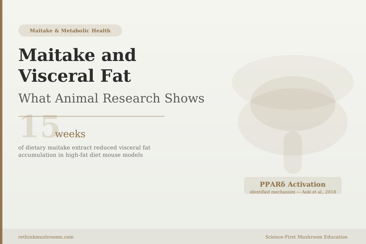
15 weeks
In a 15-week high-fat diet mouse study, maitake extract significantly reduced visceral fat accumulation by activating PPARδ, a regulatory switch inside fat cells that governs fat storage versus fat burning.
Aoki et al., Biosci Biotechnol Biochem, 2018
PPARδ
fat-metabolism regulatory pathway activated by maitake
2 fat cell genes
reduced at the mRNA level in Chen 2021 (leptin, FABP4)
0 human RCTs
all visceral fat evidence for maitake is preclinical
Aoki et al., Biosci Biotechnol Biochem, 2018
 View as image ↗
A 2018 experiment gave obese mice a diet where 0.4% of total food weight was dried maitake extract, and ran it for 15 weeks. By the end, the treated animals had significantly less visceral fat — the fat stored around their internal organs — compared to mice eating the same high-fat diet without any mushroom extract.[1] The researchers then investigated why. They found that maitake had activated a protein called PPARδ, which functions like a regulatory dial inside fat cells, controlling whether the cell burns incoming fatty acids or packages them for storage.
That result from Aoki and colleagues is one of five animal studies now showing that maitake mushroom measurably affects fat accumulation in rodent models. A separate 2021 study went deeper than fat weight measurements, instead examining what was happening inside fat cells at the gene level, and found that maitake extract reduced the production of two key fat-related proteins at the molecular level.[2] This article covers what those studies found, what the mechanistic evidence suggests, and what remains untested in humans.
Key Takeaways
- Aoki et al. (2018) found that 0.4% maitake extract in diet significantly reduced visceral fat accumulation in obese mice over 15 weeks, with PPARδ activation identified as the primary mechanism
- Chen et al. (2021) found maitake extract reduced leptin mRNA and FABP4 mRNA expression in subcutaneous fat cells of obese mice, showing effects at the gene-activity level
- Li et al. (2019) showed maitake polysaccharides reduced body fat, triglycerides, LDL, and liver fat in high-fat-diet rats while simultaneously restoring gut microbiota diversity
- Guo et al. (2020) found maitake polysaccharides lowered blood glucose and blood lipids simultaneously in obese diabetic mice while modulating gut microflora composition
- Huang et al. (2016) identified that maitake lowers liver cholesterol through two pathways: increased LDL receptor expression and increased bile acid synthesis in liver cells
- All visceral fat evidence for maitake is from rodent studies; no peer-reviewed randomized controlled trial on maitake and visceral fat in humans has been published
Why Visceral Fat Is More Dangerous Than the Fat Under Your Skin
Body fat is not uniform. Subcutaneous fat sits just beneath the skin — around the hips, thighs, and abdomen, where you can physically grab it. Visceral fat sits deeper, packed around internal organs: the liver, pancreas, kidneys, and intestines. You cannot see or feel visceral fat from the outside.
The two types behave differently in the body. Subcutaneous fat acts mostly as energy storage, releasing fatty acids gradually when the body needs fuel. Visceral fat is metabolically active in ways that create measurable health problems. It continuously releases inflammatory signaling molecules — including TNF-alpha and interleukin-6 — directly into the bloodstream. It also drains fatty acids into the portal vein, the blood vessel that carries blood directly from the abdominal organs to the liver.
That direct route to the liver matters. The liver receives a continuous supply of excess fatty acids from visceral fat, which drives fat accumulation in liver cells and contributes to insulin resistance. Over time, this cycle reinforces itself: more visceral fat produces more free fatty acids, which worsen liver function, which impairs the signaling systems that regulate fat storage.
This is why visceral fat is a stronger predictor of metabolic disease risk than total body weight or body mass index. People with high visceral fat develop type 2 diabetes and cardiovascular disease at higher rates even when their overall weight falls in a "normal" range.
Reducing visceral fat specifically, rather than just total body weight, is a distinct and meaningful metabolic target. That distinction makes the Aoki 2018 study notable: the researchers measured visceral fat depot weight directly, not just overall body weight change.
15 Weeks, 0.4%: What the Aoki 2018 Experiment Found
Aoki and colleagues put high-fat-diet-induced obese mice on a diet where 0.4% of total food weight was dried Grifola frondosa (maitake) extract, and ran the experiment for 15 weeks.[1] A control group ate the identical high-fat diet without any mushroom extract.
At the end of 15 weeks, the maitake group showed four significant changes compared to controls: lower body weight gain, reduced visceral fat accumulation, improved glucose tolerance, and lower hepatic triglyceride levels (less fat stored in liver cells).
The team then worked backward to find the cause. Testing the extract's molecular effects, they found that maitake activated PPARδ in fat tissue. PPARδ activation was identified as the primary mechanism connecting the dietary intervention to the observed changes in fat accumulation and glucose handling.
One practical note: 0.4% of total diet weight in a mouse is not directly translatable to a human supplement dose. Converting rodent doses to human equivalents requires adjustments for differences in body surface area and metabolic rate, and those conversions carry meaningful uncertainty. The study establishes that a biologically active dose exists in this model; it does not establish what a comparable dose would be in humans.
What PPARδ Actually Does — and Why Activating It Changes Fat Storage
PPARδ stands for peroxisome proliferator-activated receptor delta. The name is technical jargon. What it does is not difficult to understand.
PPARδ is a protein that lives inside cell nuclei and functions as a gene regulator. When it gets activated — by a compound binding to it, or by physical exercise, or by certain foods — it attaches to DNA and switches on a specific set of genes. The genes it activates increase the rate at which cells break down and burn fatty acids. Simultaneously, it reduces the activity of genes that promote fat accumulation.
Think of it as a dial rather than an on/off switch. Low PPARδ activity tilts the cell toward fat storage. High PPARδ activity tilts it toward fat burning. In the Aoki experiment, maitake extract raised that dial in fat tissue, shifting the metabolic balance of fat cells away from storage and toward oxidation.
PPARδ is also active in muscle cells, where it increases fat burning during physical activity. Its activation is one mechanism by which regular exercise reduces visceral fat over time. The significance of the Aoki finding is that a dietary compound appears to engage the same regulatory pathway.
One important caveat: the Aoki study measured PPARδ activation in mouse fat tissue. No maitake study has measured PPARδ activity in human fat tissue. The finding is mechanistically compelling but not yet confirmed in humans.
Inside Fat Cells: What the 2021 Gene Expression Evidence Found
Chen and colleagues published a comprehensive anti-obesity study in 2021 using Grifola frondosa extract in obese mice.[2] The results included reduced body weight, reduced visceral fat weight, and lower liver fat accumulation. But the study's most detailed contribution was at the gene expression level.
The researchers measured mRNA activity inside fat cells from treated and untreated animals. mRNA — messenger RNA — is the intermediate step between a gene's instructions and the protein the gene produces. Measuring mRNA levels tells you how actively a gene is being used at a given moment, not just whether it exists in the cell's DNA.
In the subcutaneous fat cells of maitake-treated mice, two genes were significantly less active: leptin and FABP4.
Leptin is produced primarily by fat cells and signals the brain to reduce appetite and increase energy use. In obesity, fat cells produce large amounts of leptin, but the brain gradually stops responding to it — a state called leptin resistance. The brain keeps receiving the "stop eating" signal but ignores it. Reduced leptin mRNA in fat cells means those cells are producing less leptin at the gene level. Whether this reduction could help break the cycle of leptin resistance is a genuinely open question; the Chen study did not measure leptin receptor sensitivity in brain tissue or appetite behavior.
FABP4 — fatty acid binding protein 4 — is a protein that transports fatty acids within cells. High FABP4 expression in fat tissue is associated with inflammation and metabolic dysfunction. Its reduction in maitake-treated animals suggests a shift toward less inflamed, more metabolically normal fat cell behavior.
These are not just measurements of fat weight. They are evidence that maitake alters the internal molecular activity of fat cells. That level of mechanistic specificity goes further than most plant-based nutrition research, and it distinguishes the Chen study from simple "fat was reduced" observations.
For a detailed look at the leptin mechanism and what it means for appetite, see Maitake and Leptin Signaling: Appetite Regulation Research.
Body Fat and Liver Fat: Two More Lines of Animal Evidence
Li and colleagues (2019) fed high-fat-diet rats maitake polysaccharides for several weeks in a study designed to examine both fat metabolism and gut microbiota.[3] The treated animals showed reductions in body fat percentage, blood triglycerides, LDL cholesterol, and liver fat accumulation. The polysaccharide treatment also restored gut microbiota diversity, which had been disrupted by the high-fat diet.
The gut finding is relevant to fat metabolism because the composition of intestinal bacteria influences how dietary fat is processed and how fat cell metabolism is regulated. A gut microbiome disrupted by a high-fat diet — one skewed toward bacteria that extract more calories from food and promote inflammatory signaling — is associated with increased visceral fat accumulation. Restoring that balance may be part of how maitake polysaccharides produce their metabolic effects.
Guo and colleagues (2020) used a more complex model: mice given both a high-fat diet and a drug to induce type 2 diabetes.[4] In this combined obesity-and-diabetes model, maitake polysaccharides simultaneously lowered blood glucose and blood lipid levels while also altering gut microflora composition. The simultaneous effects across multiple metabolic systems suggest these pathways interact: improving one tends to shift the others.
Neither Li 2019 nor Guo 2020 measured visceral fat depot weight specifically. The fat reductions in these studies reflect overall lipid accumulation across tissue types rather than visceral fat targeted directly. Both used high-fat diet models where visceral fat accumulation is a primary feature, so the lipid reductions are almost certainly relevant to visceral fat — but the studies don't isolate it the way Aoki 2018 did.
For the broader picture of how maitake simultaneously affects blood sugar, blood pressure, and blood lipids, see Maitake and Metabolic Syndrome: Multi-Target Effects. For the insulin sensitivity connection, see Maitake SX-Fraction and Insulin Sensitivity.
The Liver Mechanism: How Maitake Affects Hepatic Fat Accumulation
Visceral fat and liver fat are physiologically connected. Fat cells in visceral depots release free fatty acids into the portal vein, which carries blood directly to the liver. The liver absorbs these fatty acids, and when the supply exceeds the liver's processing capacity, fat accumulates in liver cells — a condition called hepatic steatosis, or fatty liver. Visceral fat accumulation and fatty liver tend to occur together and share the same metabolic drivers.
Huang and colleagues (2016) specifically investigated the molecular mechanisms behind maitake's cholesterol-lowering effects in rat liver tissue.[5] They found two parallel mechanisms operating simultaneously.
First, maitake treatment increased LDL receptor expression in liver cells. LDL receptors are the proteins on the liver cell surface that capture and remove LDL cholesterol from the bloodstream. More receptors on the cell surface means more LDL cholesterol gets pulled out of circulation.
Second, maitake increased the activity of bile acid synthesis pathways in the liver. The liver converts cholesterol into bile acids as part of the digestive process; those bile acids are secreted into the intestine and eventually excreted. Increasing bile acid synthesis is one of the main ways the body disposes of excess cholesterol.
These two mechanisms work in parallel: one removes LDL from blood directly, the other disposes of cholesterol by converting it. Together they explain how maitake reduces circulating cholesterol at the molecular level rather than simply observing that blood cholesterol numbers change.
For the specific angle on fatty liver, see Maitake and Hepatic Lipid Metabolism: Research on Fatty Liver Prevention.
What This Research Can and Cannot Tell Us
Five rodent studies showing fat reduction through multiple mechanisms is a meaningful body of preclinical evidence. But there are specific limits to what this evidence supports.
All five studies used mouse or rat models. Rodents metabolize lipids differently than humans, maintain different gut microbiota compositions, and have different adipose tissue distribution patterns. Results from high-fat diet rodent models are useful for identifying mechanisms and guiding hypothesis formation. They do not predict clinical outcomes in humans with the reliability of randomized controlled trials.
Dose translation is also uncertain. A diet containing 0.4% maitake extract by weight in a mouse — from the Aoki study — requires calculation to estimate a human equivalent, and the standard body surface area conversion method carries meaningful uncertainty. The studies don't include dose-response curves that would clarify what the minimum effective dose is even in animals.
No peer-reviewed randomized controlled trial on maitake and visceral fat in humans has been published. There is one historical observation — a 1992 report describing weight changes in patients taking maitake tablets — but that report was published in a Japanese consumer magazine rather than a peer-reviewed journal, did not use a control group, and does not meet current clinical evidence standards. The visceral fat question specifically has not been tested in any structured human study.
What the animal evidence does provide is a mechanistic foundation: PPARδ activation as a plausible pathway, gene expression changes showing effects at the molecular level in fat cells, and lipid metabolism improvements across multiple rodent models. These findings give researchers a basis for designing human trials, and they represent a more detailed mechanistic picture than most dietary compounds have accumulated. But the clinical question — whether maitake reduces visceral fat in humans and at what dose — remains open.
For the broader weight and fat loss evidence, including the historical human observation and an honest assessment of what it does and doesn't show, see Maitake for Weight Loss: What the Research Shows. For the full metabolic hormone picture, see Maitake and Leptin Signaling: Research on Appetite Regulation and Metabolism.
The Bottom Line
Animal studies consistently show that maitake extract reduces fat accumulation in high-fat diet rodent models, with the most specific evidence coming from a 15-week study demonstrating visceral fat reduction via PPARδ activation. Gene expression work adds molecular depth: maitake alters leptin and FABP4 gene activity inside fat cells, suggesting effects beyond simple fat weight changes. The evidence is entirely preclinical; no randomized controlled trial on maitake and visceral fat in humans has been conducted.
Frequently Asked Questions
Sources & References
- 1. Source details for this reference are not yet available.
- 2. Source details for this reference are not yet available.
- 3. Source details for this reference are not yet available.
- 4. Source details for this reference are not yet available.
- 5. Source details for this reference are not yet available.