Maitake & Liver Health
3 documented mechanisms
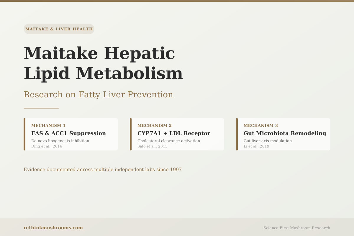
Animal studies have mapped three distinct molecular mechanisms through which maitake polysaccharides reduce liver fat accumulation, with the first controlled evidence appearing nearly 30 years ago.
Ding et al., Frontiers in Microbiology, 2016
FAS & ACC1
fat-synthesis genes suppressed in liver tissue (Ding, 2016)
CYP7A1 + LDL-R
cholesterol clearance genes activated in mouse liver (Sato, 2013)
Since 1997
consistent hepatic lipid inhibition across independent labs
Ding et al., Frontiers in Microbiology, 2016
 View as image ↗
In 1997, Kubo and Nanba fed one group of rats a diet supplemented with maitake fruit body and left another group on standard chow. At the experiment's end, they extracted and weighed the livers and fat deposits. The maitake-fed animals had smaller livers, less accumulated epididymal fat, and lower serum lipid levels.[1] Published in Biological & Pharmaceutical Bulletin, it was the first controlled demonstration that maitake specifically inhibited hepatic lipid accumulation — and it was done nearly 30 years ago.
Since then, four independent research groups have returned to this question and mapped what Kubo's result pointed toward. The picture that has emerged for maitake hepatic lipid metabolism and fatty liver prevention spans three distinct molecular pathways:
- De novo lipogenesis suppression: GF polysaccharides downregulate FAS and ACC1 — the two core fat-synthesis enzymes in the liver — at the level of gene expression.
- Cholesterol clearance activation: Maitake upregulates CYP7A1 (which converts cholesterol to bile acids) and the LDL receptor gene, accelerating cholesterol removal through two independent routes simultaneously.
- Gut microbiota remodeling: GF polysaccharides restore the gut bacterial disruption caused by high-fat feeding, reducing the hepatic lipid load delivered via the gut-liver axis.
The honest framing: all three pathways have been documented in animal studies. No controlled clinical trial has specifically tested maitake for hepatic fat reduction in humans. The preclinical data is unusually detailed for a food ingredient — multiple labs, multiple molecular targets, consistent directional effects — but it remains preclinical.
For a full overview of what maitake is and how its bioactive compounds function, what maitake mushroom is and how it works covers the broader evidence landscape. This article focuses on the liver fat story specifically.
Key Takeaways
- Kubo & Nanba (1997) established the first controlled evidence that maitake reduced liver weight and fat pad size vs. controls in animals, with concurrent serum lipid reduction
- Ding et al. (2016) found maitake significantly reduced liver expression of FAS (fatty acid synthase) and ACC1 (acetyl-CoA carboxylase 1), the two core enzymes that manufacture new fatty acids from carbohydrates in the liver
- Sato et al. (2013) found dietary maitake activated CYP7A1 (bile acid synthesis from cholesterol) and upregulated LDL receptors in mouse liver, while also downregulating HMGCR — the enzyme inhibited by statin drugs
- Li et al. (2019) documented that GF heteropolysaccharide (GFP) reduced ALT, AST, total cholesterol, triglycerides, and LDL-C in NAFLD rats while increasing HDL-C and protective antioxidant enzyme activity
- A separate Li et al. (2019) study showed GF polysaccharides corrected high-fat diet-induced gut microbiota dysbiosis alongside direct hepatic lipid regulation, confirming the gut-liver axis as a distinct second mechanism
- Jiang et al. (2021) identified ceramide pathway modulation as a novel maitake mechanism, connecting its effects to the lipotoxicity pathway that links obesity specifically to hepatic fat accumulation
The 30-Year Evidence Base for Maitake and Hepatic Lipid Accumulation
Kubo and Nanba's 1997 experiment was the foundation.[1] The setup was straightforward: one group of rats received standard feed; the other received feed supplemented with maitake fruit body. At the experiment's end, extracted livers were measurably lighter and epididymal fat deposits were smaller in the maitake group, and serum lipid levels were lower. This was an early study by modern standards, but it established something durable: maitake's effect on liver fat was not a theoretical prediction. It was a physically measurable outcome at the tissue level.
Four years later, Fukushima's team added comparative context by running a head-to-head experiment in cholesterol-fed rats.[2] They tested maitake fiber against shiitake and enokitake fiber. All three mushroom fibers reduced plasma cholesterol, with maitake fiber showing lipid-lowering activity in the direct comparison. Fukushima's study established that dietary fiber from the maitake fruiting body contributes to lipid reduction independent of its polysaccharide fractions — the fiber and polysaccharide components appear to work through different mechanisms and may be additive.
Together, these two studies from 1997 and 2001 established the starting point for a more detailed mechanistic investigation. The question they left unanswered — exactly how maitake was turning down liver fat production — would take another 15 years to answer at the molecular level.
The Fat Factory Shutdown: How Maitake Turns Off Fat-Synthesis Genes
The liver is the body's primary fat-production site. Every day it manufactures fatty acids from carbohydrates and other raw materials through a process called de novo lipogenesis — fat creation from scratch. Two enzymes run the core assembly line. Fatty acid synthase (FAS) assembles long chains of fatty acids from two-carbon building blocks. Acetyl-CoA carboxylase (ACC1) produces the specific raw material that FAS needs to start assembly. Without these two enzymes running at full capacity, the liver's ability to manufacture new fat is significantly reduced.
In 2016, Ding's team in Jiangnan Province set out to identify the molecular mechanism behind maitake's established lipid-reducing effects in hyperlipidemic rats.[3] They found it at the gene level in the liver. Maitake significantly reduced the mRNA expression of both FAS and ACC1 in liver tissue. The genes coding for the fat assembly-line enzymes were being turned down at transcription — not just reduced enzyme activity after the enzymes were built, but reduced instructions for building them in the first place.
This is a more specific finding than simply showing reduced liver fat levels. Gene-level downregulation of FAS and ACC1 identifies where in the biochemical chain maitake is acting and provides a target that researchers can investigate in detail.
Lo's 2021 review of medicinal mushrooms and NAFLD pathogenesis provides useful upstream context for this finding.[4] Maitake's beta-glucans appear to activate AMPK — a molecular energy sensor that cells use to detect low-energy conditions. One of AMPK's downstream jobs is to silence a fat-production coordinator called SREBP-1c, which is the transcription factor that normally promotes FAS and ACC1 gene expression. When SREBP-1c is quieted by active AMPK, the liver receives fewer instructions to run its fat-synthesis machinery. Ding's gene expression data is consistent with exactly this AMPK-SREBP-1c-FAS/ACC1 sequence being active in maitake-treated liver tissue.
For readers who want to understand how maitake's bioactive compounds generate these effects at a broader pharmacological level, the pharmacology of Grifola frondosa covers the beta-glucan and D-fraction mechanisms in detail.
Cholesterol Clearance: The Two Genes Maitake Activates in the Liver
Reducing fat synthesis is one half of the hepatic lipid equation. The other half is accelerating cholesterol removal, and this is where Sato's 2013 mouse study adds a distinct layer of evidence.[5]
Sato's team fed mice a cholesterol-enriched diet with or without dietary maitake mushroom, then extracted the livers and measured gene expression in liver tissue directly. Maitake significantly upregulated two genes involved in cholesterol processing. The first was CYP7A1 — the gene coding for an enzyme that converts excess cholesterol into bile acids inside liver cells. Bile acids are then secreted and eliminated through the digestive tract. When CYP7A1 is more active, the liver disposes of cholesterol faster.
The second gene upregulated was the LDL receptor. LDL receptors are proteins on liver cell surfaces that grab LDL cholesterol particles circulating in the bloodstream and pull them into the liver for processing. More receptors means more LDL removed from circulation per unit of time.
Sato also found that maitake downregulated HMGCR, which codes for HMG-CoA reductase. This is the rate-limiting enzyme in the liver's internal cholesterol production pathway — and it is the exact target that statin drugs inhibit. Maitake appeared to reduce this gene's activity through dietary exposure in mouse liver tissue.
The three-gene pattern in Sato's data is notable: CYP7A1 upregulation, LDL receptor upregulation, and HMGCR downregulation occurring together. The liver was processing cholesterol faster, pulling more out of blood, and producing less on its own — simultaneously.
The Gut-Liver Pipeline: Why Microbiota Remodeling Changes What Ends Up in Your Liver
The liver does not receive its inputs in isolation. The portal vein carries blood directly from the gut to the liver, meaning bacterial metabolites, endotoxins, and short-chain fatty acids produced by gut bacteria arrive at the liver before circulating anywhere else in the body.
When gut bacteria are disrupted by a high-fat diet — a condition called dysbiosis — the composition of bacterial species shifts in ways that increase the hepatic lipid load and promote liver inflammation. Two studies by Li and colleagues in 2019 documented maitake's effect on this gut-liver connection.
The first Li study, published in Food & Function, tested GF polysaccharides in high-fat diet rats.[6] The polysaccharides reduced hepatic lipid accumulation and improved serum lipid profiles. The study also measured gut microbiota composition before and after treatment. Polysaccharide treatment restored the gut bacterial balance that the high-fat diet had disrupted, and this microbiota correction occurred alongside the liver fat reduction — not as a secondary consequence of it.
The second Li study, in the International Journal of Molecular Sciences, used a more specific GF heteropolysaccharide extract (GFP) in a NAFLD rat model.[7] The results included specific liver enzyme measurements: ALT and AST — markers that rise when liver cells are stressed or damaged — both decreased with GFP treatment. Total cholesterol, triglycerides, and LDL-C also decreased. Protective markers moved in the opposite direction: HDL-C increased, as did superoxide dismutase (SOD) and glutathione peroxidase (GSH-Px), two antioxidant enzymes that help liver cells manage oxidative stress.
Two liver-specific genes responded as well. Cyp4a1 — which becomes overactive during liver cell stress and injury — was downregulated in GFP-treated animals. Cyp7a1, which converts cholesterol to bile acids (the same gene Sato documented in mice), was upregulated. The pattern was consistent across both Li 2019 studies: GF polysaccharides reduced markers of liver injury while simultaneously clearing cholesterol and restoring gut microbiota balance.
The Grifola frondosa and liver enzyme regulation article covers the ALT and AST findings in more detail, including what these enzyme markers measure and why their reduction is used to assess liver health status.
Ergothioneine, a compound naturally concentrated in maitake mushrooms, has also been studied separately as an anti-NAFLD agent. The ergothioneine and NAFLD article covers that compound-specific evidence and how it relates to whole-mushroom liver research.
The Ceramide Discovery: A Novel Link Between Obesity and Liver Fat
The mechanisms described above explain how maitake reduces liver fat through direct gene expression changes. Jiang's 2021 study points to something earlier in the causal chain — a mechanism that connects maitake to the specific form of fatty liver disease that develops in people with obesity.[8]
Ceramide is a fat-based signaling molecule. When cells are overwhelmed by excess dietary fat, they produce more of it. High ceramide levels interfere with insulin signaling and push liver cells toward storing fat rather than burning it — contributing to the fat droplet accumulation inside liver cells that defines hepatic steatosis, the earliest stage of fatty liver disease. Ceramide sits at the metabolic intersection of obesity, insulin resistance, and liver fat: it is part of why these three conditions are so tightly linked.
Jiang's team tested Grifola frondosa in high-fat diet mice and tracked the ceramide pathway specifically. The mushroom reduced both obesity and hepatic lipid accumulation, and the mechanism traced through ceramide pathway modulation. This was the first study to identify ceramide as a maitake hepatic target.
The implication matters for people interested in maitake specifically for NAFLD related to obesity. Most of the earlier maitake liver studies used general hyperlipidemia models that don't capture the obesity-specific metabolic disruptions that drive most NAFLD in the population. Ceramide pathway modulation addresses the mechanism that connects excess body fat to liver fat, which is a more relevant model for the largest affected group.
The inflammatory side of the maitake-liver connection covers how maitake's anti-inflammatory effects — including the NF-kappaB pathway suppression and cytokine reduction — run in parallel to this ceramide pathway.
The Evidence Ceiling: What Animal Data Can and Cannot Tell Us
Every study described in this article used animal models — rats or mice on high-fat diets, cholesterol-enriched feed, or chemical liver injury induction. This is the honest limit of what currently exists for maitake and hepatic lipid metabolism specifically.
Lo's 2021 review identifies maitake as one of the most thoroughly studied mushrooms for NAFLD-related liver effects among all medicinal mushrooms reviewed.[4] The mechanistic picture is unusually detailed by food-ingredient standards: multiple labs, multiple molecular pathways, consistent directional effects across different animal models and extract types. But the gap between this preclinical depth and the absence of human liver lipid trials remains real.
The best available human data is indirect. Zhang's 2020 cohort study found adults consuming mushrooms four or more times per week had lower NAFLD prevalence than infrequent mushroom eaters — but that study used mixed mushroom consumption, not maitake specifically, and it was observational. The companion article on maitake for fatty liver disease and NAFLD covers the full picture of NAFLD-specific evidence including that cohort data.
For the broader hepatoprotective evidence base beyond lipid metabolism — including acute liver injury protection and liver enzyme regulation — the maitake and liver health overview covers the complete evidence landscape across all three hepatoprotective mechanisms.
For comparison, reishi mushroom's hepatoprotective research shows a similar pattern: strong preclinical mechanism work, limited human data. This appears to be characteristic of the medicinal mushroom research field broadly.
The evidence hierarchy for maitake hepatic lipid metabolism: multiple animal studies documenting consistent effects through at least three distinct pathways, a 2021 review identifying it among the best-supported mushrooms for this indication, and no dedicated human RCT for the specific endpoint of hepatic fat reduction. The mechanistic pathway from animal evidence to human benefit is plausible and detailed — but not yet directly demonstrated in clinical trials.
The Bottom Line
The animal-based research on maitake and hepatic lipid metabolism spans nearly 30 years across multiple independent labs, documenting consistent effects through at least three distinct molecular pathways: direct suppression of fat-synthesis gene expression, cholesterol clearance activation, and gut microbiota remodeling. The gap between this unusually detailed preclinical picture and the absence of human liver lipid trials is the central limitation readers should weigh.
Frequently Asked Questions
Sources & References
- 1RTHM-00052·Kubo, K et al. (1997). “Anti-hyperliposis effect of maitake fruit body (Grifola frondosa). I..” Biological & pharmaceutical bulletin.Study breakdown →PubMed →↩
- 2RTHM-00075·Fukushima, M et al. (2001). “Cholesterol-lowering effects of maitake (Grifola frondosa) fiber, shiitake (Lentinus edodes) fiber, and enokitake (Flammulina velutipes) fiber in rats..” Experimental biology and medicine (Maywood.Study breakdown →PubMed →↩
- 3RTHM-00778·Ding, Yinrun et al. (2016). “The Mechanisms Underlying the Hypolipidaemic Effects of Grifola frondosa in the Liver of Rats..” Frontiers in microbiology.Study breakdown →PubMed →↩
- 4RTHM-01398·Lo, Hui-Chen (2021). “Bioactive Components and Possible Activities of Medicinal Mushrooms in Alleviating the Pathogenesis of Nonalcoholic Fatty Liver Disease (Review)..” International journal of medicinal mushrooms.Study breakdown →PubMed →↩
- 5RTHM-00551·Sato, Mayumi et al. (2013). “Effect of dietary Maitake (Grifola frondosa) mushrooms on plasma cholesterol and hepatic gene expression in cholesterol-fed mice..” Journal of oleo science.Study breakdown →PubMed →↩
- 6RTHM-01084·Li, Lu et al. (2019). “Grifola frondosa polysaccharides ameliorate lipid metabolic disorders and gut microbiota dysbiosis in high-fat diet fed rats..” Food & function.Study breakdown →PubMed →↩
- 7RTHM-01089·Li, Xin et al. (2019). “The Positive Effects of Grifola frondosa Heteropolysaccharide on NAFLD and Regulation of the Gut Microbiota..” International journal of molecular sciences.Study breakdown →PubMed →↩
- 8RTHM-01369·Jiang, Xue et al. (2021). “Anti-obesity effects of Grifola frondosa through the modulation of lipid metabolism via ceramide in mice fed a high-fat diet..” Food & function.Study breakdown →PubMed →↩