Ergothioneine & Liver Health
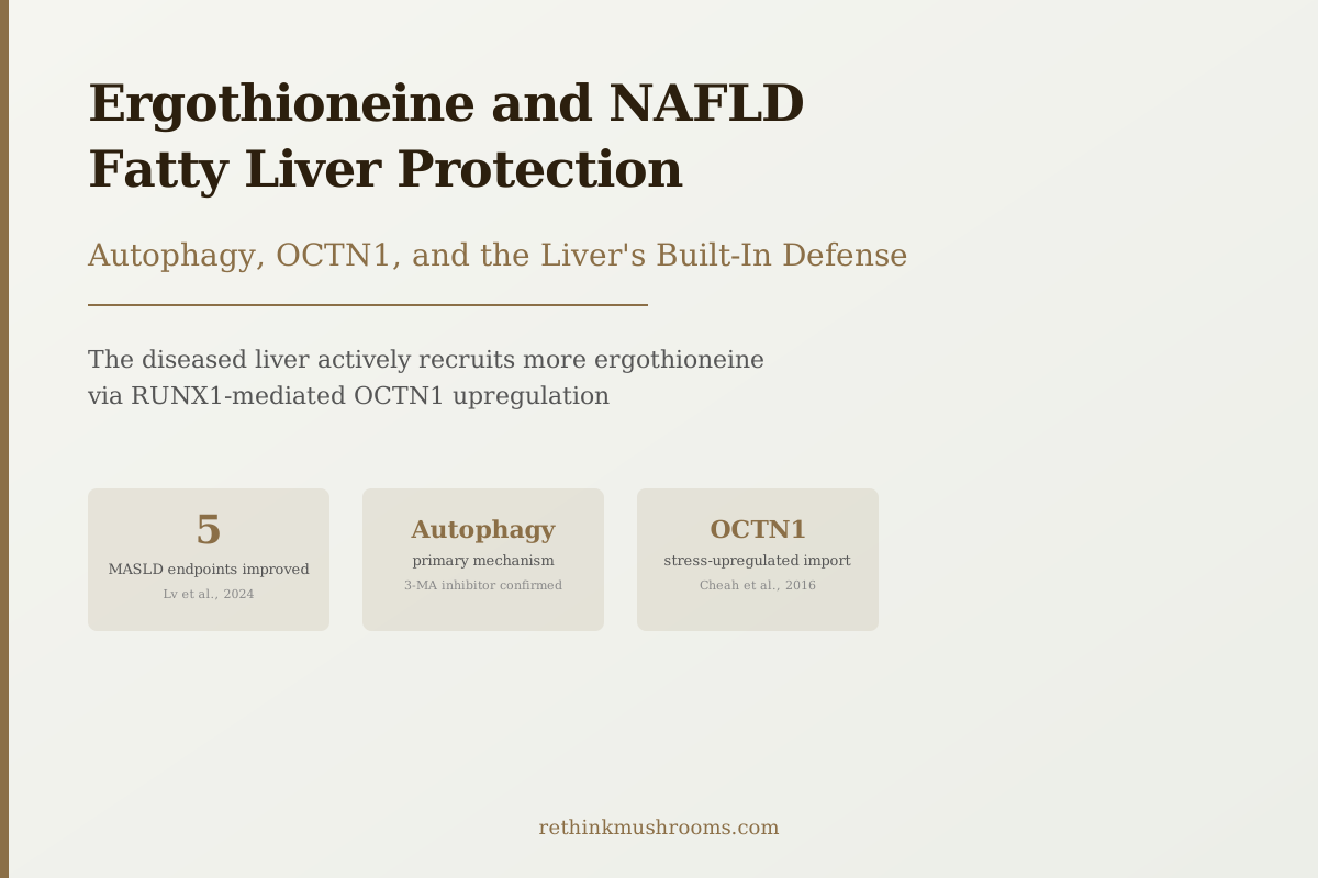
5 MASLD endpoints improved
In high-fat diet mice, ergothioneine improved body weight, blood lipids, insulin resistance, liver fat deposits, and glycogen storage, with autophagy confirmed as the primary mechanism.
Lv et al., Lipids Health Dis, 2024
Autophagy
primary mechanism confirmed by inhibitor test
OCTN1 upregulated
NAFLD liver recruits more ergothioneine
No approved drug
for NAFLD/MASLD in 2026
Lv et al., Lipids Health Dis, 2024
 View as image ↗
Here is what makes the ergothioneine-NAFLD story unusual: when your liver is developing fatty liver disease, it actively tries to acquire more ergothioneine. Cheah and colleagues documented this in guinea pigs in 2016, finding that NAFLD livers had significantly higher ergothioneine concentrations than healthy livers, driven by the liver upregulating the OCTN1 transporter via a transcription factor called RUNX1.[1] The accumulation correlated with disease severity. The damaged liver was importing more of the compound the more damaged it became.
This framing matters because it reframes the question from "does ergothioneine help fatty liver?" to "why has the liver evolved to import more ergothioneine when it develops fatty liver?" The research across the past decade, culminating in a 2024 mechanistic study that confirmed autophagy as the primary protective pathway, provides a clearer answer than most NAFLD nutrition research offers.
For background on ergothioneine's biology and its dedicated transporter, see What Is Ergothioneine?. For the full liver health evidence across all injury types, see Ergothioneine and Liver Health: Hepatoprotective Research.
Key Takeaways
- NAFLD/MASLD livers actively upregulate the OCTN1 ergothioneine transporter via RUNX1 as a stress response, importing more ergothioneine as disease severity increases (Cheah et al., 2016)
- In high-fat diet mice with MASLD, ergothioneine treatment improved body weight, body fat, blood lipids, insulin resistance, and liver fat and glycogen deposition (Lv et al., 2024)
- The autophagy mechanism was confirmed by blocking autophagy with 3-methyladenine, which abolished ergothioneine's hepatic benefits, establishing autophagy as the primary protective pathway, not a secondary effect
- Dare et al. (2021) found ergothioneine combined with metformin showed synergistic protection against liver injury in diabetic rats, reducing oxidative stress, inflammation, and hypertriglyceridemia beyond metformin alone
- Kawano et al. (1983) established that ergothioneine inhibits lipid peroxide formation in liver tissue, the foundational mechanism for all subsequent NAFLD research
- No peer-reviewed human RCT has published full results for ergothioneine specifically in NAFLD populations; the ErgMS trial (metabolic syndrome with liver outcomes) is ongoing
What NAFLD and MASLD Actually Are
Non-alcoholic fatty liver disease (NAFLD) is the accumulation of excess fat in the liver in people who drink little or no alcohol. The condition was renamed in 2023 to metabolic dysfunction-associated steatotic liver disease (MASLD), reflecting its origin in metabolic dysfunction rather than the mere absence of alcohol. The terminology shift acknowledges that MASLD is a disease of metabolism, not just behavior.
MASLD is now estimated to affect about 25 to 30 percent of adults globally, making it the most common chronic liver condition worldwide. The disease exists on a spectrum from simple fat accumulation (steatosis) to steatohepatitis (fat plus inflammation), to fibrosis (scar tissue), to cirrhosis. The progression from steatosis to steatohepatitis to fibrosis is the primary clinical concern; fibrosis is the strongest predictor of liver-related mortality.
As of 2026, no pharmaceutical treatment is approved specifically for NAFLD or MASLD. Several drugs are in late-stage trials targeting various aspects of the disease's pathology. This context is important for interpreting ergothioneine research: a preclinical finding in a condition with no approved treatment carries different weight than a preclinical finding in a condition with established pharmacological options.
The OCTN1 Story: What the Liver Tells Us It Needs
In NAFLD, the liver faces chronically elevated oxidative stress from several sources: fat metabolism generates reactive oxygen species, inflammatory cells recruited to the liver generate more, and impaired mitochondria fail to neutralize them efficiently. The Cheah 2016 study showed that under this oxidative burden, the liver specifically upregulates OCTN1, the transporter responsible for importing ergothioneine from the bloodstream into liver cells.[1]
The regulatory mechanism was identified: RUNX1, a transcription factor normally associated with immune cell development, drives OCTN1 expression in the liver under stress conditions. The accumulation of ergothioneine in NAFLD livers correlated with cholesterol levels and disease severity, suggesting the import mechanism is tightly linked to the degree of metabolic dysfunction.
What this finding implies: the liver has a calibrated response to oxidative stress that involves specifically acquiring more ergothioneine. This is not a generic antioxidant recruitment response. It is a targeted mechanism for a specific compound. The corollary question (what happens when dietary ergothioneine is low and the liver cannot fill this demand) is addressed by the population data on ergothioneine status and disease outcomes.
For a dedicated analysis of liver ergothioneine levels and what they predict, see Liver Ergothioneine Levels and Disease Risk.
The 2024 Autophagy Breakthrough: Lv et al.
Lv and colleagues published in Lipids in Health and Disease in 2024 the most mechanistically decisive animal study of ergothioneine in MASLD to date.[2] Mice were fed a high-fat diet to induce MASLD and then treated with ergothioneine. The treated animals showed improvements across multiple metabolic endpoints: reduced body weight and body fat, improved blood lipid profiles, better insulin sensitivity, and normalized liver fat deposition and glycogen storage under microscopic examination.
The mechanistic investigation was the study's most important contribution. The researchers measured autophagy flux in liver tissue and found it was significantly increased by ergothioneine treatment. Autophagy is the cellular process by which cells break down and recycle damaged or excess components, including lipid droplets, dysfunctional mitochondria, and oxidized proteins. In MASLD, autophagy is impaired; the liver loses its ability to clear excess fat and damaged cellular material, which accelerates disease progression.
To test whether autophagy was actually driving the protection (rather than just occurring alongside it), Lv's group administered the autophagy inhibitor 3-methyladenine to ergothioneine-treated mice. The inhibitor abolished ergothioneine's protective effects on liver fat, inflammation, and oxidative damage. This is the cleanest possible mechanistic confirmation: blocking autophagy removed the benefit, which proves autophagy was responsible for it.
Ergothioneine also reduced oxidative damage markers and suppressed inflammatory cytokines in these animals. But the autophagy finding places it in a mechanistic category that most antioxidants do not occupy. Ergothioneine does not just neutralize reactive species; it restores the liver's own cellular cleanup machinery.
Lipid Peroxidation: The 1983 Foundation
Before autophagy, before Nrf2, before OCTN1: there was lipid peroxidation. Kawano and colleagues showed in 1983 that ergothioneine inhibited lipid peroxide formation in mouse liver.[3] Lipid peroxidation is the chain reaction by which reactive oxygen species damage cell membranes; once initiated, it spreads through the lipid bilayer, causing membrane rupture and cell death. In NAFLD, excess liver fat provides abundant substrate for lipid peroxidation, making this mechanism directly relevant.
The 1983 finding has been confirmed by every subsequent mechanistic review. Fu and Shen's 2022 Frontiers in Pharmacology review specifically named lipid peroxidation inhibition as one of ergothioneine's core hepatoprotective mechanisms across NAFLD, drug-induced injury, and fibrosis contexts.[10] The consistency of this mechanism across four decades and multiple research groups gives it strong evidentiary weight.
The Metabolic Syndrome Connection: Dare 2021
NAFLD and type 2 diabetes co-exist at very high rates. Between 60 and 80 percent of people with type 2 diabetes have NAFLD; the metabolic dysfunction driving one condition typically drives both. Dare and colleagues studied this overlap in 2021, testing ergothioneine in diabetic rats with liver injury.[4]
The study design combined ergothioneine with metformin, the first-line diabetes medication. The ergothioneine-plus-metformin group showed greater reduction in oxidative stress, inflammation, and hypertriglyceridemia than metformin alone, suggesting the combination was synergistic rather than merely additive. Liver injury markers were also reduced. This was the first study to directly examine ergothioneine in a diabetes-NAFLD model, and the results align with the broader mechanistic picture: ergothioneine's antioxidant and anti-inflammatory properties address the oxidative substrate that links metabolic dysfunction to liver damage.
The mushroom-diabetes-liver connection is explored further in Mushroom Antioxidants and AGEs in Diabetes, where ergothioneine's role in the broader mushroom-metabolic health story is covered.
Why Dietary Deficiency May Be Contributing to NAFLD Rates
Ames, writing in PNAS in 2018, argued that ergothioneine should be classified as a longevity vitamin, placing it in a category of nutrients whose deficiency accelerates age-related diseases.[5] His argument rested partly on the evolutionary conservation of OCTN1: a dedicated transporter is only worth maintaining across millions of years of evolution if the compound it transports serves essential functions.
The dietary implication is pointed: ergothioneine enters the human body almost entirely through mushrooms. Western diets are characteristically low in mushroom consumption. If ergothioneine deficiency accelerates oxidative damage in metabolically active organs like the liver, and if the liver actively compensates for this deficiency by upregulating OCTN1 under stress, then chronic low ergothioneine intake may be contributing to NAFLD susceptibility or progression.
This is a hypothesis, not a proven causal chain. But the population-level data from Smith et al. (2020) is consistent with it: in 3,236 participants followed prospectively, ergothioneine was the single metabolite most strongly associated with lower all-cause mortality and reduced cardiovascular and metabolic disease risk.[6] Low ergothioneine status, in that data, tracked with worse metabolic outcomes across the board.
The Fibrosis Risk: What Mao 2025 Adds
NAFLD becomes more dangerous when it progresses to fibrosis. Mao and colleagues' 2025 study in a CCl4-induced fibrosis model found that ergothioneine at 5 to 10 mg per kilogram per day for six weeks reduced fibrosis markers including alpha-smooth muscle actin, collagen I and III, laminin, and hydroxyproline, alongside reducing ALT, AST, and bilirubin.[7] The mechanism was inhibition of PLA2G2A, which blocked the TGF-beta/Smads signaling cascade that drives hepatic stellate cell activation and scar tissue deposition.
For NAFLD specifically: the majority of NAFLD-related mortality comes from fibrosis progression. A compound that addresses both the upstream steatosis mechanisms (autophagy, lipid peroxidation) and the downstream fibrosis pathway (TGF-beta/Smads) would address the full disease arc. That is what the combined evidence suggests, from animal models.
The Lam-Sidun 2021 Synthesis
The most systematic preclinical review of ergothioneine's cardiometabolic effects was published by Lam-Sidun and colleagues in 2021, examining all available evidence for liver and cardiovascular protection.[9] Their conclusion: ergothioneine showed the most consistent hepatoprotective profile of any mushroom-derived compound studied, with consistent reduction in hepatic oxidative stress, lipid accumulation, and insulin resistance across animal models. They identified it as the most promising mushroom-derived compound for metabolic health.
The review predated the Lv 2024 autophagy study and the Mao 2025 fibrosis study. Those subsequent findings have strengthened the mechanistic case considerably.
Evidence Map
Ergothioneine & Fatty Liver — A Deep Mechanistic Base, No Human Trial Yet
The ergothioneine–NAFLD case is unusual: the human trial rung at the top is still empty, but the preclinical base is the most mechanistically specific of any dietary compound in NAFLD. Bar width reflects how close each tier sits to a proven human outcome — the widest, most solid bars are the autophagy inhibitor test and the liver’s own adaptive import response, while the dashed bars top and bottom are what is still missing.
Human NAFLD randomized controlled trial
gapNot yet published
No peer-reviewed RCT of ergothioneine in NAFLD has published results. The ErgMS trial (metabolic syndrome, liver-function endpoints) is ongoing, and a 30-subject company pilot reporting AST/ALT/GGT drops has not been peer-reviewed
Observational human association
evidenceSmith 2020
In 3,236 prospectively followed adults, ergothioneine was the single metabolite most strongly associated with lower all-cause mortality and reduced cardiovascular and metabolic disease risk — consistent with, but not proof of, a liver benefit
Autophagy proven by inhibitor test
evidenceLv 2024
In high-fat-diet MASLD mice, ergothioneine improved weight, lipids, insulin resistance, and liver fat; blocking autophagy with 3-methyladenine abolished the benefit — the cleanest possible proof that autophagy is the mechanism, not a bystander
Adaptive transporter response (the liver asks for it)
evidenceCheah 2016
NAFLD livers upregulate the OCTN1 ergothioneine transporter via RUNX1, importing more of the compound as disease severity rises — an endogenous signal that the liver treats ergothioneine as protective
Anti-fibrotic pathway (blocks progression)
evidenceMao 2025
In a CCl4 fibrosis model, ergothioneine (5–10 mg/kg/day) reduced alpha-SMA, collagen I/III, laminin, and hydroxyproline by inhibiting PLA2G2A and the TGF-beta/Smads cascade that drives hepatic stellate cell scarring
Metabolic-syndrome synergy
evidenceDare 2021
In diabetic rats with liver injury, ergothioneine plus metformin cut oxidative stress, inflammation, and hypertriglyceridemia beyond metformin alone — the first test in a diabetes-NAFLD overlap model
Lipid-peroxidation foundation (four decades consistent)
evidenceKawano 1983 · Fu & Shen 2022
Ergothioneine inhibited lipid peroxide formation in liver tissue in 1983, and every subsequent mechanistic review has confirmed it — the substrate-level mechanism directly relevant to fat-laden NAFLD membranes
Large Phase 2/3 human NAFLD trials, dose-response & subgroups
gapDoes not yet exist
No adequately powered human trial has established dose, duration, or which patients benefit. The mechanistic case is specific and convergent, but it has not been confirmed in human NAFLD
How to read it: read this ladder from the middle out. The two widest, most solid bars are the strongest evidence — an autophagy inhibitor test that proves the mechanism, and the liver’s own upregulation of the ergothioneine transporter as it gets sicker. Below them, three independent labs add fibrosis, metabolic-synergy, and lipid-peroxidation mechanisms that converge on the same conclusion. Only observational human data (Smith 2020) sits above the mechanisms, and the human NAFLD trial rung at the very top is still empty. The strength of the mechanism does not substitute for the human trials that have not yet been run; nothing here is medical advice.
Sources: Cheah 2016 [1]; Lv 2024 [2]; Kawano 1983 [3]; Dare 2021 [4]; Smith 2020 [6]; Mao 2025 [7]; Tian 2023 [8]; Fu & Shen 2022 [10]. Bar widths are illustrative of proximity to a proven human outcome, not quantitative effect sizes.
What Human Evidence Exists
The Tian 2023 review in the British Journal of Nutrition argued for reclassifying ergothioneine as an essential dietary micronutrient and highlighted the ErgMS trial as the first human intervention study targeting metabolic syndrome with liver function outcomes as primary endpoints.[8] Full peer-reviewed results from ErgMS have not yet been published.
A company-reported pilot study involving 30 subjects with abnormal liver function given 30 mg of ergothioneine daily for 30 days showed reductions in AST, ALT, and GGT. That finding has not undergone peer review. It is the closest available signal for direct human liver enzyme reduction with ergothioneine supplementation.
The honest picture: the animal mechanistic evidence is compelling and increasingly specific. The human RCT data for NAFLD specifically does not yet exist in peer-reviewed form. Observational population data (Smith 2020) is consistent with the preclinical direction. The ErgMS trial results, when published, will be the first peer-reviewed human test of ergothioneine in a metabolic syndrome population with liver function as a primary outcome.
Dietary ergothioneine comes almost entirely from mushrooms. Why Only Fungi Make Ergothioneine explains why plant foods contain negligible amounts and which mushroom species are the richest sources for people wanting to increase their intake.
The Bottom Line
The liver's adaptive upregulation of OCTN1 in NAFLD (Cheah 2016) suggests ergothioneine plays a defined role in the liver's stress response to metabolic disease. Lv et al. (2024) confirmed autophagy as the primary protective mechanism in an MASLD animal model, with lipid peroxidation inhibition (Kawano 1983), TGF-beta/Smads fibrosis pathway blockade (Mao 2025), and synergy with metformin (Dare 2021) filling out the mechanistic picture. Human RCT data in NAFLD populations remains unpublished. The preclinical case is the most mechanistically specific for any dietary compound in NAFLD.
Frequently Asked Questions
Sources & References
- 1RTHM-00771·Cheah, Irwin K et al. (2016). “Liver ergothioneine accumulation in a guinea pig model of non-alcoholic fatty liver disease. A possible mechanism of defence?.” Free radical research.Study breakdown →PubMed →↩
- 2RTHM-01942·Lv, Xiaoyu et al. (2024). “Ergothioneine ameliorates metabolic dysfunction-Associated Steatotic Liver Disease (MASLD) by enhancing autophagy, inhibiting oxidative damage and inflammation..” Lipids in health and disease.Study breakdown →PubMed →↩
- 3RTHM-00010·Kawano, H et al. (1983). “Studies on ergothioneine. XI. Inhibitory effect on lipid peroxide formation in mouse liver..” Chemical & pharmaceutical bulletin.Study breakdown →PubMed →↩
- 4RTHM-01342·Dare, Ayobami et al. (2021). “L-ergothioneine and metformin alleviates liver injury in experimental type-2 diabetic rats via reduction of oxidative stress, inflammation, and hypertriglyceridemia..” Canadian journal of physiology and pharmacology.Study breakdown →PubMed →↩
- 5RTHM-00936·Ames, Bruce N (2018). “Prolonging healthy aging: Longevity vitamins and proteins..” Proceedings of the National Academy of Sciences of the United States of America.Study breakdown →PubMed →↩
- 6RTHM-00655·Smith, Einar et al. (2020). “Ergothioneine is associated with reduced mortality and decreased risk of cardiovascular disease..” Heart (British Cardiac Society).Study breakdown →PubMed →↩
- 7RTHM-02138·Mao, Yaping et al. (2025). “Ergothioneine Ameliorates Liver Fibrosis by Inhibiting Glycerophospholipids Metabolism and TGF-β/Smads Signaling Pathway: Based on Metabonomics and Network Pharmacology..” Journal of applied toxicology : JAT.Study breakdown →PubMed →↩
- 8RTHM-01805·Tian, Xiaoying et al. (2023). “Ergothioneine: an underrecognised dietary micronutrient required for healthy ageing?.” The British journal of nutrition.Study breakdown →PubMed →↩
- 9RTHM-00696·Lam-Sidun, Daniel et al. (2021). “Mushroom-Derived Medicine? Preclinical Studies Suggest Potential Benefits of Ergothioneine for Cardiometabolic Health..” International journal of molecular sciences.Study breakdown →PubMed →↩
- 10RTHM-01517·Fu, Tong-Tong et al. (2022). “Ergothioneine as a Natural Antioxidant Against Oxidative Stress-Related Diseases..” Frontiers in pharmacology.Study breakdown →PubMed →↩