Mushroom Taxonomy
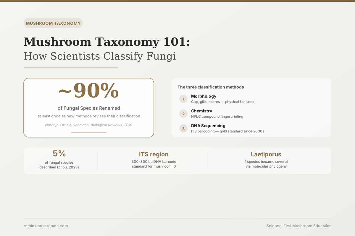
~5% of fungi described
Only about 5% of an estimated 2.2-3.8 million fungal species have been formally described and named. DNA sequencing is now the primary tool driving new classifications and correcting older ones that relied on physical appearance alone.
Zhou & May, Mycology, 2023
7 of 7
commercial reishi supplements sold as G. lucidum actually contained the distinct species G. lingzhi (Gunnels et al., 2020)
ITS barcoding
the 600-800 base pair DNA region now standard for mushroom species authentication
3 methods
morphology, chemistry, and DNA sequencing: the modern standard for classifying fungi
Zhou & May, Mycology, 2023
 View as image ↗
For most of the 20th century, mycologists thought chicken-of-the-woods was one species. The bright orange-yellow shelf mushroom fruiting on dead oaks was Laetiporus sulphureus, well described, well understood. In 2008, Lindner and Banik ran molecular analysis on Laetiporus specimens collected across North America and found what visual identification had completely missed: what appeared to be one organism was actually several distinct species, each occupying different geographic ranges and preferring different tree hosts.[5]
Nothing about the physical appearance of these specimens hinted at the division. The cap color, gill structure, spore morphology, and growth habit were nearly identical. Only DNA revealed the difference.
This is how scientists classify mushrooms today: not by appearance alone, but through a three-method approach that layers morphology, chemical profiling, and DNA sequencing. The Laetiporus story illustrates both why morphology dominated taxonomy for 200 years and why its limits became impossible to ignore once molecular tools arrived.
For a broader overview of how medicinal fungi fit into the taxonomic system, see Fruiting Body vs. Mycelium: The Great Mushroom Supplement Debate.
Key Takeaways
- Molecular phylogeny of Laetiporus (chicken-of-the-woods) revealed multiple cryptic species invisible to morphology, with distinct geographic distributions across North American tree types (Lindner & Banik, Mycologia, 2008)
- Modern mycologists use three methods: morphology (physical features), chemistry (compound profiling via HPLC), and DNA (ITS barcoding and multi-gene phylogenomics); DNA has been the primary reclassification driver since the early 2000s
- ITS (internal transcribed spacer) barcoding has been validated as a reliable DNA tool for identifying Ganoderma species in commercial supplements, distinguishing G. lucidum from G. lingzhi and other closely related species (Gunnels et al., PLoS ONE, 2020)
- Fungal taxonomy has a long, turbulent history of reclassification that genomics and phylogenomics are only now stabilizing; Naranjo-Ortiz and Gabaldón (2019) resolve the kingdom into nine phylum-level clades, including Basidiomycota (most culinary and medicinal mushrooms) and Ascomycota (cordyceps and truffles) (Naranjo-Ortiz & Gabaldón, Biological Reviews, 2019)
- Only an estimated 5% of 2.2-3.8 million fungal species have been described; phylogenomics and metagenomics are the tools expected to expand that figure significantly (Zhou & May, Mycology, 2023)
- Genetic analysis of Pleurotus (oyster mushroom) species showed that morphologically similar strains have significantly different genetic and chemical profiles, underscoring why strain-level classification beyond species matters for medicinal use (Lin et al., BioMed Research International, 2022)
Why Morphology Alone Kept Getting Things Wrong
Carolus Linnaeus formalized the binomial naming system for living organisms in the 18th century, and mycologists adopted it for fungi. Every species received a two-part Latin name: genus followed by species epithet. The first person to formally describe and name a specimen established the "type specimen," a physical reference sample kept in a herbarium or fungarium against which all later specimens could be compared. If a new collection matched the type, it was the same species. If it differed, it was new.
This system worked reasonably well for organisms with distinct, stable physical features. Fungi presented a persistent challenge. Many species look nearly identical in their mature, spore-bearing form. Others change appearance dramatically with age, light conditions, and host substrate. A young Ganoderma fruiting body looks quite different from a mature one. The same species can produce a flat or kidney-shaped cap depending on where it grows.
Thakur et al. (2024) reviewed Ganoderma lucidum specifically, documenting the range of morphological variation across cultivated strains and wild specimens.[6] A specimen might have a shiny red lacquered surface and a kidney-shaped cap, or a brown, woody, irregular growth, yet both could belong to the same species. Describing a species using morphology alone required specifying the range of variation, which was difficult without large sample collections.
Naranjo-Ortiz and Gabaldón (2019) reviewed the history of fungal classification and described it as a turbulent one, only now being settled by the arrival of genomics and phylogenomics.[1] Names accumulated along the way: a specimen originally classified under one name, then reclassified under another as more specimens became available, then reclassified again when a DNA tool revealed the earlier placement was wrong. Multiple names attached to the same species, and the same name was sometimes applied to multiple different organisms.
The Three Methods Scientists Use to Classify Fungi Today
Modern mycological classification uses a three-method standard. Each method addresses the limitations of the others, and the three together produce a species identification with substantially more confidence than any one alone.
Method 1: Morphology. Physical examination remains the first step. Mycologists document the macroscopic features (cap shape, color, texture, stem structure, pore or gill arrangement, color change when cut) and the microscopic features (spore shape and size under a microscope, hyphal structure, clamp connections). Morphological data is the starting point and provides context for interpreting the results of other methods. It is no longer sufficient alone for a reliable species identification of most fungi.
Method 2: Chemical profiling. High-performance liquid chromatography (HPLC) and mass spectrometry can generate a compound fingerprint for a mushroom sample, identifying the presence and relative quantities of specific molecules. Chemically similar species that look identical may produce different compound profiles. This was particularly useful in the period before DNA tools, when chemical analysis could confirm or complicate a morphological identification. It remains important for confirming that a specimen with the correct species DNA is also producing the expected bioactive compounds.
Method 3: DNA sequencing. This is now the primary classification tool. Scientists extract DNA from a small sample of mushroom tissue, amplify a target region via PCR (a process that makes millions of copies of a specific DNA sequence), sequence the amplified region, and compare it to a reference database to identify the organism. The most widely used target region is the ITS (internal transcribed spacer), discussed in detail below.
The interaction between these methods matters. A sample can match the morphological description of a species but fail the DNA check (suggesting it's a lookalike). A sample can have the right DNA but an anomalous compound profile (suggesting a variant or contaminant). The three-method standard protects against false identifications that a single method would miss.
ITS Barcoding: The 600-Base-Pair Key to Species Identity
The ITS region is a short section of ribosomal DNA, typically 600 to 800 base pairs in length. It sits between two coding regions of the ribosomal gene cluster, meaning it does not itself produce a protein. This position means it faces less evolutionary pressure to remain identical between generations, so it accumulates small mutations over time. Within a species, the ITS sequence is highly conserved; individuals of the same species have nearly identical ITS sequences. Between species, the ITS sequence differs in predictable ways. These properties make it ideal as a genetic barcode.
The standard workflow: extract DNA from a mushroom sample, amplify the ITS region using standard PCR primers, sequence the amplified product, and submit it to the UNITE or NCBI GenBank reference databases. The database returns the species whose ITS sequence most closely matches the query, along with a confidence percentage.
Gunnels et al. (2020) validated the ITS method specifically for identifying Ganoderma species in commercial herbal supplements.[3] This is not a trivial challenge: commercial supplement powders contain degraded DNA, mixed materials, and processing residues that can complicate amplification. Gunnels et al. confirmed that ITS barcoding reliably distinguishes between G. lingzhi and G. lucidum sensu stricto even from processed supplement powder, and that it detects species substitution that chemical testing alone would miss.
The practical relevance for anyone buying a reishi supplement is direct. When Gunnels et al. sequenced seven commercial supplements from independent suppliers, all of them sold as G. lucidum, every single one actually contained G. lingzhi rather than G. lucidum sensu stricto. Both belong to the same genus, but they are genetically distinct organisms with different geographic origins. Without ITS sequencing, that substitution is invisible.
Gunnels et al. (2020) · PLoS ONE
- Population
- Seven commercial reishi supplements (six powdered, one dried) from independent suppliers, all labeled Ganoderma lucidum
- Intervention
- Amplification and sequencing of the ITS region with genus-specific primers, followed by molecular phylogenetic analysis
- Finding
- All seven products contained G. lingzhi DNA rather than G. lucidum sensu stricto — a species substitution invisible to morphology or chemical testing
narrow-scope A small sample (seven products), but a clean, unanimous result that shows the method works even on processed supplement powder.
Gunnels T, Creswell M, McFerrin J, Whittall JB. The ITS region provides a reliable DNA barcode for identifying reishi/lingzhi (Ganoderma) from herbal supplements. PLoS ONE. 2020;15(11):e0236774.
For a detailed look at how DNA sequencing has been applied to reclassify major medicinal mushroom species, see How DNA Sequencing Is Rewriting Mushroom Classification.
What Molecular Analysis Found in Species Scientists Thought They Knew
The Laetiporus case introduced at the start of this article illustrates what molecular phylogeny does when applied to organisms previously classified on appearance alone.
Lindner and Banik (2008) analyzed collections of Laetiporus from across North America using multi-locus sequencing, meaning they amplified and compared multiple gene regions rather than just the ITS.[5] The results separated what had been treated as one species into a set of distinct taxa. L. cincinnatus favors hardwood stumps in the eastern US. L. conifericola grows on conifers in the west. The morphological differences between them are subtle enough that even experienced foragers routinely treat them as the same fungus. The genetic differences are not subtle at all.
The pattern extends to oyster mushrooms. Lin et al. (2022) examined genetic diversity across Pleurotus species and found that morphologically similar strains have significantly different genetic profiles.[4] Pleurotus ostreatus and Pleurotus pulmonarius, for example, look similar to casual observation and are sometimes sold interchangeably. Genetic analysis showed distinct compound profiles alongside the genetic distinction: the chemical composition, spanning sugars, amino acids, and lipid fractions, varied significantly across the species tested. Two products labeled "oyster mushroom" from different Pleurotus species are not equivalent from a compositional standpoint.
These two examples illustrate the general pattern: molecular tools consistently find more diversity within groups that morphology treated as uniform. For medicinal applications, this matters because the clinical research is species-specific. A trial conducted on G. lingzhi is not automatically applicable to a product labeled G. lucidum without genetic verification.
For the consumer implications of strain-level diversity, see Mushroom Genetics and Bioactive Variation: Why Strain Selection Matters.
The Frontier: Only 5% of the Fungal Kingdom Is Described
Zhou and May (2023) surveyed the current state of fungal taxonomy globally and documented a striking gap between what is known and what exists.[2] Current estimates place the total number of fungal species at between 2.2 and 3.8 million. Approximately 150,000 have been formally described and named. That is roughly 5% of the total.
The 95% that remain undescribed are not distributed randomly. Most are in understudied ecosystems: tropical soils, deep forest litter, marine sediments, and cryptic associations with other organisms. Some are certainly among the groups that already interest mycologists: there may be multiple undescribed species within genera that include known medicinal species, and their bioactive profiles are unknown.
Naranjo-Ortiz and Gabaldón (2019) place the major mushroom-bearing lineages, Basidiomycota (which includes most culinary and medicinal mushrooms) and Ascomycota (which includes cordyceps and truffles), among the nine phylum-level clades of fungi, lineages separated by deep evolutionary divergence.[1] That depth of divergence means even "closely related" fungal species can sit far apart on the tree of life, which helps account for the degree of chemical and genetic diversity that molecular tools keep revealing.
The tools Zhou and May identify for closing the 5% gap are phylogenomics (comparing entire genomes or large portions of them, rather than single gene regions) and metagenomics (sequencing all DNA in an environmental sample to detect organisms without isolating them individually). Both are now cost-effective enough to apply at scale. The result is likely to be a significant expansion of known fungal diversity over the next decade, including potential new species within medicinally important genera.
For context on how this genetic diversity within species affects medicinal applications, see The Genetic Diversity of Medicinal Mushroom Species.
Also see Basidiomycota vs. Ascomycota: The Two Major Mushroom Groups for the specific differences between the two major fungal divisions most relevant to medicinal mushroom research.
The Bottom Line
Scientists classify fungi using morphology, chemical profiling, and DNA sequencing, with ITS barcoding becoming the standard tool since the early 2000s. Molecular methods have consistently revealed hidden diversity in species that morphology treated as uniform, including multiple cryptic Laetiporus species across North America and significant genetic variation within Pleurotus. Fungal taxonomy has a long, turbulent history of reclassification that phylogenomics is only now stabilizing (Naranjo-Ortiz & Gabaldón, 2019), and Zhou and May (2023) found that only about 5% of an estimated 2.2-3.8 million fungal species have been formally described. For medicinal applications, accurate species identification matters because bioactive compound profiles are species-specific, and DNA verification is now commercially available for supplement authentication.
Frequently Asked Questions
Sources & References
- 1RTHM-01109·Naranjo-Ortiz, Miguel A et al. (2019). “Fungal evolution: diversity, taxonomy and phylogeny of the Fungi..” Biological reviews of the Cambridge Philosophical Society.Study breakdown →PubMed →↩
- 2RTHM-01839·Zhou, Li-Wei et al. (2023). “Fungal taxonomy: current status and research agendas for the interdisciplinary and globalisation era..” Mycology.Study breakdown →PubMed →↩
- 3RTHM-00615·Gunnels, Tess et al. (2020). “The ITS region provides a reliable DNA barcode for identifying reishi/lingzhi (Ganoderma) from herbal supplements..” PloS one.Study breakdown →PubMed →↩
- 4RTHM-01568·Lin, Pei et al. (2022). “Genetic and Chemical Diversity of Edible Mushroom Pleurotus Species..” BioMed research international.Study breakdown →PubMed →↩
- 5RTHM-00142·Lindner, Daniel L et al. (2008). “Molecular phylogeny of Laetiporus and other brown rot polypore genera in North America..” Mycologia.Study breakdown →PubMed →↩
- 6RTHM-02005·Thakur, Pratibha et al. (2024). “Exploring Ganoderma lucidum: morphology, cultivation and market potential..” World journal of microbiology & biotechnology.Study breakdown →PubMed →↩