Chaga & Inflammation
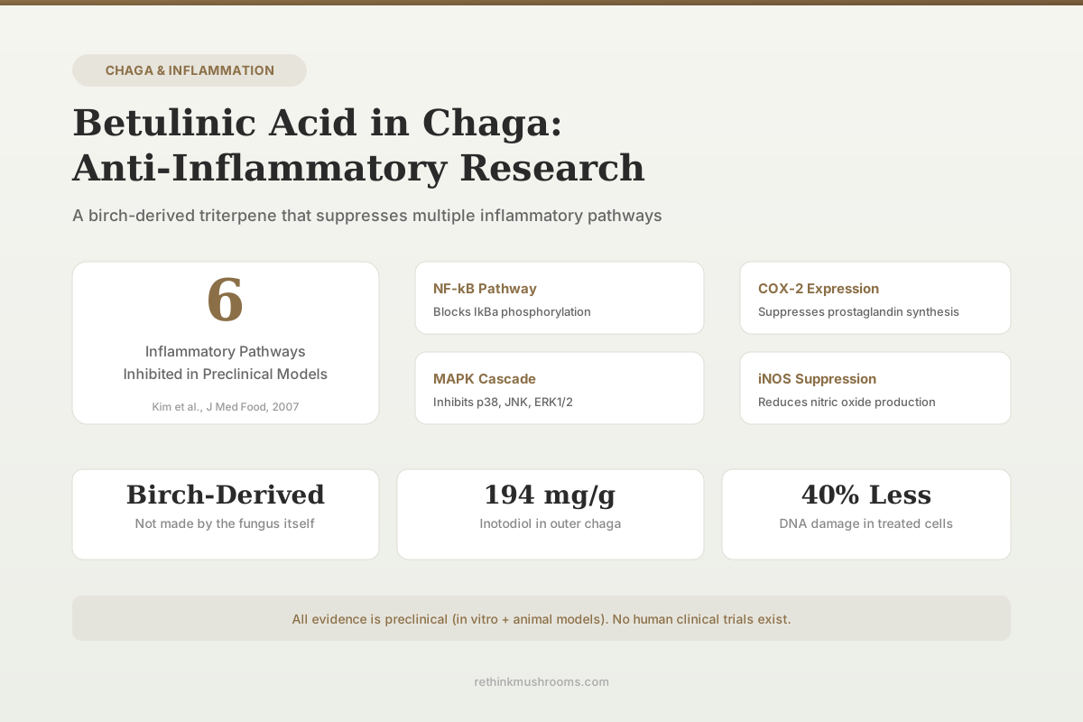
6 pathways inhibited
Chaga's betulinic acid suppresses NF-κB, COX-2, iNOS, TNF-α, IL-1β, and IL-6 in preclinical inflammatory models.
Kim et al., J Med Food, 2007
Birch-Derived
not made by the fungus itself
194 mg/g
inotodiol in outer chaga
40% Less
DNA damage in treated cells
Kim et al., J Med Food, 2007
 View as image ↗
Betulinic acid is a pentacyclic triterpene that chaga (Inonotus obliquus) does not produce on its own. It absorbs this compound directly from the birch trees it parasitizes.[1] This detail matters because it determines which chaga products actually contain betulinic acid and which do not. The compound has drawn attention from inflammation researchers for its capacity to shut down multiple pro-inflammatory signaling cascades simultaneously, from NF-κB nuclear translocation to COX-2 protein expression.[2] For a broader look at how chaga targets chronic inflammatory processes, see our overview of chaga for chronic inflammation.
The evidence is entirely preclinical. No human clinical trials have tested betulinic acid from chaga for any inflammatory condition. What exists is a body of in vitro and animal research identifying specific molecular targets and dose-response relationships that explain how this birch-derived compound interacts with inflammatory pathways.
Key Takeaways
- Chaga's betulinic acid originates from birch bark, not from the fungus itself, meaning lab-grown mycelium on grain contains zero or trace amounts (Windsor et al., 2025)
- A 70% ethanol extract of I. obliquus inhibited NF-κB activation by blocking IκBα phosphorylation and suppressed both iNOS and COX-2 protein expression in LPS-stimulated macrophages (Kim et al., 2007)
- Oral chaga extract at 100 mg/kg reduced TNF-α, IL-1β, IL-6, and IFN-γ mRNA expression in DSS-induced colitis in mice (Mishra et al., 2012)
- Inotodiol concentrations in the outer part of chaga reached 194.1 mg/g dry weight, with trametenolic acid at 106.3 mg/g (Kim et al., 2020)
- I. obliquus extract attenuated histamine-induced vasodilation in second-order arterioles, preserving endothelial junction integrity in live mice (Javed et al., 2019)
- I. obliquus polysaccharide inhibited phosphorylation of IKKα/β, IκBα, p65, p38, JNK, and ERK1/2 in infected macrophages, blocking both NF-κB and MAPK pathways simultaneously (Yan et al., 2021)
What Is Betulinic Acid and How Does Chaga Acquire It?
Betulinic acid (C30H48O3) belongs to the lupane family of pentacyclic triterpenes. Birch trees (Betula species) synthesize it in their bark as part of their own defense chemistry. When Inonotus obliquus colonizes a birch trunk, the fungus grows slowly through the bark over years, concentrating birch-derived compounds including betulin, betulinic acid, and related triterpenes in its sclerotium (the dark, hardened mass known as the chaga conk).[3]
The biosynthetic pathway runs through betulin as an intermediate: birch trees first produce betulin in large quantities (up to 30% of outer bark dry weight in some Betula species), and enzymatic oxidation converts a portion to betulinic acid. Chaga concentrates both compounds as it grows through the bark over 5 to 20 years, with relative proportions varying by geographic origin and host tree species.
This means chaga's triterpenoid profile depends heavily on its host tree. Kim et al. (2020) analyzed the composition of triterpenoids in I. obliquus using HPLC and LC-ESI-MS, finding that inotodiol and trametenolic acid were the dominant triterpenoids. Inotodiol reached 153.9 mg/g (dry basis) in the inner part and 194.1 mg/g in the outer part. Trametenolic acid measured 94.5 mg/g in the inner part and 106.3 mg/g in the outer part.[4] Betulinic acid itself is present at lower concentrations than these dominant triterpenoids but remains a pharmacologically significant component of the extract.
Windsor et al. (2025) demonstrated this host-dependency using complementary analytical techniques across multiple chaga dietary supplements. Wildcrafted chaga canker contained triterpenoids (inotodiol, trametenolic acid) and phenolics, while fermented grain products marketed as "chaga" lacked these compounds entirely. Iodine-starch assays confirmed starch-rich composition in fermented grains and its complete absence in authentic chaga canker.[5] For detailed analytical comparisons of wild versus cultivated chaga, see our article on betulinic acid content in wild vs. lab-grown chaga.
How Does Betulinic Acid Reduce Inflammation?
NF-κB and Akt Pathway Suppression
Kim et al. (2007) tested a 70% ethanol extract of I. obliquus (IOE70) in LPS-stimulated RAW 264.7 macrophage cells and mapped the specific signaling nodes it disrupts. IOE70 markedly inhibited the phosphorylation of IκBα, Akt, and MAPKs (including JNK) in dose-dependent patterns. The mechanism: IOE70 blocks NF-κB activation through the phosphatidylinositol 3-kinase/Akt/IκB pathway while simultaneously inhibiting JNK activation. This dual suppression reduced both iNOS and COX-2 protein expression, cutting off nitric oxide and prostaglandin production at their transcriptional source.[2]
This is consistent with the broader betulinic acid literature. Amiri et al. (2022), reviewing betulinic acid's anti-inflammatory activities across multiple experimental models in Frontiers in Pharmacology, documented that the compound blocks NF-κB by inhibiting IκB phosphorylation and degradation through the proteasome pathway, preventing NF-κB translocation from cytoplasm to nucleus and shutting down transcription of IL-1β, IL-6, IL-12, NO, and PGE2.
COX-2 and Prostaglandin Suppression
Debnath et al. (2013) compared the anti-inflammatory activity of I. obliquus extracts against germinated brown rice extracts in LPS-stimulated RAW 264.7 cells. The chaga-containing preparation suppressed nitric oxide production, PGE2 release, and protein expression of both iNOS and COX-2. It also reduced secretion of TNF-α, IL-1β, and IL-6.[6] The COX-2 suppression is relevant because this enzyme catalyzes the conversion of arachidonic acid to prostaglandin H2, the rate-limiting step in inflammatory prostaglandin synthesis. For a deeper examination of COX-2 inhibition mechanisms, see our article on Inonotus obliquus and COX-2 inhibition.
MAPK Signaling Cascade
The MAPK pathway (p38, JNK, ERK1/2) represents a parallel inflammatory signaling axis that operates alongside NF-κB. Yan et al. (2021) demonstrated that I. obliquus polysaccharide (IOP) inhibited phosphorylation of all three MAPK branches in Toxoplasma gondii-infected RAW264.7 macrophages. IOP blocked phosphorylation of IKKα/β, IκBα, and p65 in the NF-κB pathway, plus p38, JNK, and ERK1/2 in the MAPK pathway. It also prevented NF-κB p65 and c-Jun translocation from cytoplasm to nucleus and downregulated TLR2 and TLR4 protein expression.[7]
This dual-pathway inhibition (NF-κB plus MAPK) is a recurring finding across chaga research and appears to be a feature of multiple chaga bioactives, not betulinic acid alone. The compound works as part of a triterpenoid ensemble, and isolating its individual contribution from whole extracts remains a methodological challenge in this field.
Chaga Extracts and Intestinal Inflammation
Mishra et al. (2012) conducted the most directly relevant in vivo study. Female C57BL/6 mice received dextran sulfate sodium (DSS) at 2% in drinking water for 7 days to induce acute intestinal inflammation mimicking ulcerative colitis. Mice treated orally with an aqueous extract of I. obliquus (IOAE) at 50 and 100 mg/kg body weight showed significant improvements across multiple inflammation markers.[8]
Histological examination revealed that IOAE suppressed edema, mucosal damage, and crypt loss. It attenuated DSS-induced iNOS levels and myeloperoxidase accumulation in colon tissues (myeloperoxidase is a marker of neutrophil infiltration). IOAE also inhibited mRNA expression of TNF-α, IL-1β, IL-6, and IFN-γ in colon tissues. The co-suppression of TNF-α and iNOS together with IL-1β suggests interference at a transcriptional level upstream of individual cytokine production.
This study used an aqueous extract, which would contain both water-soluble polysaccharides and some triterpenes. Separating betulinic acid's contribution from other bioactive components was not the study's aim. For the full picture of chaga's preclinical IBD evidence, see our article on chaga and inflammatory bowel disease.
Microvascular Inflammation and Histamine Response
Javed et al. (2019) used intravital microscopy to observe chaga's effects on inflammation in real time in living tissue. In C57BL/6 mice, I. obliquus extract attenuated histamine-induced inflammatory conducted vasodilation in second-order arterioles of the gluteus maximus muscle.[9]
Histamine disrupts endothelial cell-to-cell junctions, impairing the conducted vasodilation that normally carries signals rapidly through vascular endothelium. This disruption is associated with microvascular dysfunction and, over time, cardiovascular disease progression. The chaga extract blocked this histamine-driven disruption, preserving junctional integrity.
The study also tested mast cell activation by compound 48/80, which similarly disrupted endothelial junctions and conducted vasodilation. The histamine antagonist pyrilamine blocked histamine's effects but not compound 48/80's, suggesting mast cells release additional mediators beyond histamine that also compromise vascular integrity. The authors identified mast cells as a putative future target for I. obliquus research, a direction that has not yet been followed up with betulinic acid-specific studies.
This study is notable for using intravital microscopy, allowing real-time observation of vascular responses in living tissue rather than measuring biomarkers in isolated cells. The arteriolar vasodilation response is a functional readout of endothelial communication, not just a molecular marker. When histamine disrupts gap junctions between endothelial cells, the conducted vasodilation signal fails to propagate along the vessel wall. Chaga extract preserved this propagation, suggesting protection of connexin-based gap junctions or the signaling molecules that pass through them.
Polysaccharides vs. Triterpenoids: Separating the Sources
A persistent challenge in chaga inflammation research is that whole extracts contain both polysaccharides (primarily beta-glucans) and triterpenoids (including betulinic acid, inotodiol, and trametenolic acid). These compound classes appear to act through overlapping but distinct mechanisms.
Yan et al. (2021) worked specifically with I. obliquus polysaccharide (IOP), demonstrating that the polysaccharide fraction alone could suppress NF-κB and MAPK pathways and downregulate TLR2/TLR4.[7] Kim et al. (2007) used a 70% ethanol extract, which preferentially extracts triterpenoids and smaller molecules over high-molecular-weight polysaccharides, yet found similar NF-κB and MAPK suppression.[2]
This convergence suggests that both compound classes target the same downstream pathways but through different upstream receptors and binding events. Polysaccharides interact with pattern recognition receptors like TLR2 and Dectin-1 on the macrophage surface, triggering intracellular signaling cascades from outside the cell. Triterpenoids like betulinic acid, being lipophilic small molecules, appear to cross cell membranes and modulate intracellular kinase cascades more directly without requiring receptor-mediated entry.
The extraction method determines which compound class dominates a given preparation. Hot water extracts (traditional chaga tea) favor polysaccharides. Ethanol or methanol extracts concentrate triterpenoids. Dual-extraction products claim to capture both fractions, but standardization across commercial products is inconsistent. No study has yet tested purified betulinic acid isolated from chaga against chaga polysaccharide in a head-to-head anti-inflammatory comparison, which makes it difficult to determine relative potency or whether the two classes produce synergistic effects. For a broader look at NF-κB inhibition by chaga, see our article on chaga and NF-κB inhibition research.
Why Chaga Source Determines Anti-Inflammatory Potential
The distinction between wildcrafted chaga canker and commercially produced mycelium-on-grain products is not academic. It directly determines whether betulinic acid is present at all.
Ern et al. (2024), reviewing the therapeutic properties of I. obliquus, confirmed that bioactive triterpenoids, polysaccharides, polyphenols, and lignin metabolites are responsible for health-related effects, but their concentrations vary dramatically depending on growth substrate and harvesting conditions.[10] Fordjour et al. (2023) noted that secondary metabolites including steroids, terpenoids, and other compounds exist in chaga, with demonstrated antioxidant, anti-inflammatory, antiviral, and antitumor activities, but these findings apply specifically to properly sourced material from birch hosts.[3]
Park et al. (2004) demonstrated that chaga extract at concentrations of 10 to 500 micrograms/mL provided over 40% reduction in H2O2-induced DNA fragmentation in human lymphocytes, measured by comet assay.[11] While this study measured antioxidant rather than anti-inflammatory activity directly, oxidative damage and inflammation are mechanistically linked through shared NF-κB activation pathways. For related research on betulinic acid's anticancer activity from chaga, see our article on betulinic acid from chaga: anti-cancer research.
Where the Evidence Stands
Every study cited here is preclinical. The in vivo work uses mouse models. The in vitro work uses isolated macrophage cell lines. No human trial has tested betulinic acid from chaga for any inflammatory condition.
Several methodological gaps constrain interpretation:
Extract complexity. Most studies use crude extracts containing dozens of bioactive compounds. Attributing observed anti-inflammatory effects specifically to betulinic acid requires isolation studies that largely do not exist for chaga-sourced material. The broader betulinic acid literature (using purified compound from birch bark or synthetic sources) fills some of this gap, but chaga-specific attribution remains weak.
Bioavailability. Betulinic acid has poor water solubility and limited oral bioavailability in its native form. A 2014 review in the European Journal of Pharmaceutical Sciences estimated oral bioavailability of betulinic acid below 1% in rat models. This raises questions about how much reaches systemic circulation after oral consumption of chaga tea, tinctures, or capsules. Formulation strategies like nanoencapsulation and cyclodextrin complexation have improved bioavailability in laboratory settings but are not standard in commercial chaga products.
Dose translation. Mouse studies used 50-100 mg/kg body weight of crude extract, not purified betulinic acid. Converting these doses to human equivalents requires accounting for species differences in metabolism, the fraction of betulinic acid in the extract, and the unknown contribution of co-administered compounds.
Model relevance. DSS-induced colitis and LPS-stimulated macrophages are standard inflammation models, but they represent acute, chemically induced inflammation rather than the chronic, low-grade inflammatory states that drive most human inflammatory diseases.
The molecular pharmacology is internally consistent across multiple research groups and extraction methods. Betulinic acid and related chaga triterpenoids converge on NF-κB and MAPK suppression through well-characterized kinase cascades. Whether this translates to meaningful anti-inflammatory effects in humans consuming chaga products remains an open question that only clinical trials can answer.
The Bottom Line
Betulinic acid in chaga originates from birch bark, not from the fungus itself. Preclinical research shows it suppresses NF-κB activation, COX-2 expression, and multiple pro-inflammatory cytokines through overlapping signaling pathways. All evidence is from cell cultures and animal models. No human clinical trials exist, and bioavailability of betulinic acid from oral chaga consumption is poorly characterized.
Frequently Asked Questions
Sources & References
- 1RTHM-01694·Fordjour, Eric et al. (2023). “Chaga mushroom: a super-fungus with countless facets and untapped potential..” Frontiers in pharmacology.Study breakdown →PubMed →↩
- 2RTHM-00117·Kim, Ho-Gyoung et al. (2007). “Ethanol extract of Inonotus obliquus inhibits lipopolysaccharide-induced inflammation in RAW 264.7 macrophage cells..” Journal of medicinal food.Study breakdown →PubMed →↩
- 3RTHM-01694·Fordjour, Eric et al. (2023). “Chaga mushroom: a super-fungus with countless facets and untapped potential..” Frontiers in pharmacology.Study breakdown →PubMed →↩
- 4RTHM-01218·Kim, Jaecheol et al. (2020). “Composition of Triterpenoids in Inonotus obliquus and Their Anti-Proliferative Activity on Cancer Cell Lines..” Molecules (Basel.Study breakdown →PubMed →↩
- 5RTHM-02209·Windsor, Coleton et al. (2025). “What This 2025 Chaga Supplement Authentication Study Actually Found.” International journal of molecular sciences.Study breakdown →PubMed →↩
- 6RTHM-00283·Debnath, Trishna et al. (2013). “Anti-oxidant and anti-inflammatory activities of Inonotus obliquus and germinated brown rice extracts..” Molecules (Basel.Study breakdown →PubMed →↩
- 7RTHM-00730·Yan, Kexin et al. (2021). “Inhibitory Effects of Inonotus obliquus Polysaccharide on Inflammatory Response in Toxoplasma gondii-Infected RAW264.7 Macrophages..” Evidence-based complementary and alternative medicine : eCAM.Study breakdown →PubMed →↩
- 8RTHM-00258·Mishra, Siddhartha Kumar et al. (2012). “Orally administered aqueous extract of Inonotus obliquus ameliorates acute inflammation in dextran sulfate sodium (DSS)-induced colitis in mice..” Journal of ethnopharmacology.Study breakdown →PubMed →↩
- 9RTHM-00562·Javed, Sumreen et al. (2019). “Inonotus obliquus attenuates histamine-induced microvascular inflammation..” PloS one.Study breakdown →PubMed →↩
- 10RTHM-01886·Ern, Phoebe Tee Yon et al. (2024). “Chaga Therapeutic Properties: What This 2024 Review Actually Shows.” Mycology.Study breakdown →PubMed →↩
- 11RTHM-00076·Park, Yoo Kyoung et al. (2004). “Chaga mushroom extract inhibits oxidative DNA damage in human lymphocytes as assessed by comet assay..” BioFactors (Oxford.Study breakdown →PubMed →↩