Chaga and Inflammation
IκBα blocked
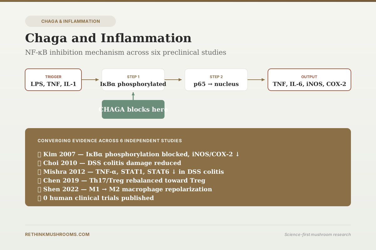
70% ethanol extract of chaga blocked IκBα phosphorylation and degradation in LPS-activated macrophages, preventing NF-κB p65 translocation to the nucleus and shutting down downstream inflammatory gene transcription.
Kim et al., Journal of Medicinal Food, 2007
5 Studies
converge on NF-κB pathway suppression across preparations and models
iNOS, COX-2, TNF-α ↓
three major inflammatory mediators reduced by chaga extracts
0 Human RCTs
no controlled trial has tested chaga for any inflammatory condition in humans
Kim et al., Journal of Medicinal Food, 2007
 View as image ↗
NF-κB is the transcription factor that most inflammatory pathways converge on. When toll-like receptors detect pathogens, when cytokine receptors sense IL-1 or TNF, when oxidative stress accumulates, NF-κB activates. Activated NF-κB enters the nucleus and drives transcription of inflammatory genes: TNF-α, IL-6, IL-1β, COX-2, iNOS, adhesion molecules, and dozens of others. Many pharmaceutical anti-inflammatory drugs act downstream of NF-κB (COX inhibitors block prostaglandin synthesis, TNF antibodies neutralize the cytokine). Compounds that act at NF-κB itself have the appeal of cutting off inflammation at its root, but also the risk of broad immune suppression if used systemically. Chaga (Inonotus obliquus) extracts have been shown across multiple independent studies to suppress NF-κB activation, and the mechanism has been characterized in detail. This article walks through what those studies measured. For the broader chaga for chronic inflammation context, see that separate article.
The consistent finding: chaga extracts block NF-κB activation upstream of nuclear translocation, specifically at the IκBα phosphorylation and degradation step. This means the p65/p50 dimer cannot enter the nucleus to drive inflammatory gene transcription. The effect has been replicated in LPS-stimulated macrophages, DSS-induced colitis, and broader immunomodulatory assays. Human trials have not been run.
Key Takeaways
- 70% ethanol extract of chaga (IOE70) inhibited LPS-induced iNOS and COX-2 expression in RAW 264.7 macrophages via suppression of IκBα phosphorylation and Akt/JNK signaling (Kim et al., Journal of Medicinal Food, 2007)
- Orally administered aqueous chaga extract decreased TNF-α, STAT1, IL-4, and STAT6 expression in DSS-induced colitis mice, ameliorating acute intestinal inflammation (Mishra et al., Journal of Ethnopharmacology, 2012)
- Chaga extract reduced colonic damage scores and suppressed NF-κB-driven inflammatory gene expression in DSS-induced colitis model (Choi et al., Journal of Biomedicine and Biotechnology, 2010)
- Inonotus obliquus polysaccharide modulated Th1/Th2 and Th17/Treg T-cell balance toward anti-inflammatory phenotypes in DSS-induced colitis mice (Chen et al., Artificial Cells, Nanomedicine, and Biotechnology, 2019)
- Chaga extracts at 25-200 μg/mL shifted macrophage polarization from pro-inflammatory M1 to anti-inflammatory M2 phenotype, increasing CD206 and Arg-1 while decreasing IL-1β and iNOS (Shen et al., eCAM, 2022)
- Chaga polysaccharide treatment downregulated TLR2, TLR4, and NF-κB p65 phosphorylation while upregulating Nrf2/HO-1 signaling in Toxoplasma gondii-induced liver injury (Xu et al., International Journal of Biological Macromolecules, 2019)
Why NF-κB Is the Central Inflammation Target
NF-κB (nuclear factor kappa B) exists in the resting cell as a p65/p50 heterodimer sequestered in the cytoplasm by IκBα (inhibitor of kappa B alpha). When an activating signal arrives (TLR4 detecting LPS, TNF receptor binding TNF-α, IL-1 receptor binding IL-1β), it triggers a kinase cascade. IKK (IκB kinase) phosphorylates IκBα. Phosphorylated IκBα is ubiquitinated and degraded by the proteasome. The freed p65/p50 dimer translocates to the nucleus, binds κB consensus sequences in gene promoters, and drives transcription of inflammatory genes.
Any compound that interferes with this sequence can suppress NF-κB-driven inflammation. The targets, in order of proximity to the final transcription step, include:
- TLR/receptor signaling at the membrane
- IKK kinase activity
- IκBα phosphorylation
- IκBα degradation
- p65 nuclear translocation
- p65 DNA binding
The chaga literature has characterized effects at multiple points in this chain, converging on IκBα stability and p65 translocation as the main interaction points.
Kim 2007: The Mechanistic Foundation
Kim and colleagues in 2007 published the foundational mechanism study of chaga's anti-inflammatory action.[1] They tested multiple solvent extracts (30%, 50%, 70%, 90% ethanol and water) for activity against LPS-stimulated RAW 264.7 macrophages. The 70% ethanol extract (IOE70) showed the strongest activity.
Findings at IOE70:
- Inhibited LPS-induced NO production (the hallmark macrophage inflammatory mediator)
- Inhibited iNOS protein expression (the enzyme that produces NO)
- Inhibited COX-2 protein expression (the enzyme that produces inflammatory prostaglandins)
- Suppressed IκBα phosphorylation
- Suppressed Akt and JNK phosphorylation
The authors concluded that IOE70's inhibition of iNOS and COX-2 was mediated by the Akt and JNK pathways feeding into NF-κB activation. The IκBα finding is particularly important: if IκBα is not phosphorylated, it cannot be degraded, and therefore the p65/p50 dimer remains trapped in the cytoplasm. Kim's data pinpointed the specific molecular step where chaga acts.
The choice of 70% ethanol rather than pure water matters. Different extraction solvents pull out different compound classes. 70% ethanol extracts triterpenoids, phenolic compounds, and some polysaccharides, while water extracts primarily pull polysaccharides and water-soluble phenolics. The strongest anti-inflammatory activity sitting in the 70% ethanol fraction suggests triterpenoids or mid-polarity phenolics as the active class, consistent with later work on betulin-derived triterpenoids covered in our betulinic acid in chaga article.
Choi 2010 and Mishra 2012: From Cell Culture to Colitis
In vitro activity against LPS-stimulated macrophages is a screening endpoint. In vivo activity in a disease model is more clinically relevant. Choi and colleagues in 2010 tested chaga in a dextran sulfate sodium (DSS)-induced colitis model in mice.[2] DSS colitis is the standard preclinical model for ulcerative colitis; it produces colonic epithelial damage, inflammatory infiltrate, weight loss, and diarrhea within days.
Chaga extract treatment reduced colonic damage scores, suppressed NF-κB-driven inflammatory gene expression, and preserved colonic tissue architecture. The effect maps the Kim 2007 cell culture finding onto an animal disease model.
Mishra and colleagues in 2012 extended the DSS colitis work using an orally administered aqueous chaga extract.[3] They found:
- Reduced TNF-α expression
- Reduced STAT1 expression (downstream of IFN-γ signaling, drives Th1 inflammation)
- Reduced IL-4 expression (drives Th2 inflammation)
- Reduced STAT6 expression (downstream of IL-4)
- Overall amelioration of acute inflammatory response
The dual reduction of Th1 (TNF-α, STAT1) and Th2 (IL-4, STAT6) markers indicates broader immune modulation than simple NF-κB suppression. This is relevant because inflammatory bowel disease involves a dysregulated mix of Th1, Th2, and Th17 responses, and a compound that damps the full range has a different profile than one that only hits one axis. For the condition-specific angle, see our chaga and inflammatory bowel disease article.
Chen 2019: T-Cell Balance Detail
Chen and colleagues in 2019 added mechanistic detail using Inonotus obliquus polysaccharide (IOP) in the same DSS colitis model.[4] They specifically measured T-cell phenotype distribution:
- Th1/Th2 ratio: modulated toward balance
- Th17/Treg ratio: shifted toward regulatory T-cell phenotype
- Amelioration of colitis severity
Th17 cells drive chronic inflammation in autoimmune and inflammatory diseases. Tregs (regulatory T cells) restrain Th17-driven inflammation. The Th17/Treg balance has become a central target in modern IBD drug development. Chen's data shows chaga polysaccharide pushes this balance toward the anti-inflammatory side in a disease model. Whether this would translate to a meaningful clinical effect in humans has not been tested.
Shen 2022: Macrophage Polarization Detail
Macrophages exist on a polarization spectrum. Classically activated M1 macrophages are pro-inflammatory: they produce TNF-α, IL-1β, iNOS, and reactive oxygen species. Alternatively activated M2 macrophages are anti-inflammatory and tissue-repair-oriented: they produce IL-10, arginase-1 (Arg-1), and CD206.
Shen and colleagues in 2022 tested chaga extracts against resting M0 macrophages and LPS-induced M1 macrophages.[5] At 25-200 μg/mL, chaga extracts:
- Significantly increased CD206 expression (M2 marker)
- Significantly increased Arg-1 expression (M2 marker)
- Decreased IL-1β expression (M1 cytokine)
- Decreased iNOS expression (M1 marker)
The net effect is a shift from M1 to M2 phenotype. This is different from simply blocking M1 activation; it is an active repolarization toward the resolution phenotype. The authors attributed the effect partly to chaga polysaccharides and chaga polyphenols, noting that different extracts had different potencies across the markers measured.
Xu 2019: The Liver Axis
Xu and colleagues' 2019 Toxoplasma gondii-induced liver injury study provides additional NF-κB mechanistic detail.[6] In that model, chaga polysaccharide:
- Reduced ALT, AST, MDA, and NO (injury markers)
- Decreased TNF-α, IL-6, IL-1β, IFN-γ, and IL-4 serum levels
- Downregulated TLR2 and TLR4 (upstream NF-κB activators)
- Reduced phosphorylation of NF-κB p65 and IκBα (the same molecular step as Kim 2007)
- Upregulated Nrf2 and HO-1 (antioxidant response)
The TLR/NF-κB axis reduction is the same chemistry as the macrophage and colitis findings, now in a liver infection context. The Nrf2/HO-1 activation adds an antioxidant arm, completing the classical anti-inflammatory protective profile. For the liver-specific angle, see our chaga and CCl4 liver damage article.
The Consistent Mechanism Across Studies
Six independent studies using different models (LPS-stimulated macrophages, DSS colitis, Toxoplasma liver injury, resting and activated M0/M1 macrophages, CCl4 liver injury from previous article) converge on the same NF-κB-related signaling profile. The pattern includes:
- IκBα phosphorylation suppressed → stable IκBα → sequestered NF-κB in cytoplasm
- p65/p50 nuclear translocation reduced → less inflammatory gene transcription
- Downstream mediators reduced: iNOS, COX-2, TNF-α, IL-6, IL-1β
- Upstream amplifiers reduced: TLR2, TLR4
- Parallel Nrf2/HO-1 antioxidant activation
- T-cell rebalancing: Th17 → Treg, Th1/Th2 normalization
- Macrophage repolarization: M1 → M2
No single study established every element of this profile. But the collection across six independent labs using four to five distinct models produces a coherent mechanistic picture. The active compound classes implicated include triterpenoids (especially the triterpenoid-rich 70% ethanol fractions), polysaccharides (in the colitis and liver injury work), and polyphenols (contributing in some extracts).
Compound-Specific Contributions: What Solvent Choice Reveals
The Kim 2007 extraction comparison is more informative than most single-preparation studies because it tested five different solvents (30%, 50%, 70%, 90% ethanol, and water) in parallel against the same inflammatory endpoint. The 70% ethanol extract outperformed the others. This narrows the candidate active compound class: highly water-soluble polysaccharides would have been strongest in the water extract, not the 70% ethanol extract. Purely hydrophobic triterpenoids would have been strongest in the 90% ethanol extract.
The 70% ethanol sweet spot captures mid-polarity compounds: smaller oligosaccharides, phenolic acids, flavonoids, and partially glycosylated triterpenoids. Betulinic acid and inotodiol, two of the best-characterized chaga triterpenoids, both sit in this polarity range. Chlorogenic acid, syringic acid, and gallic acid are also present in this fraction.
In the colitis studies (Choi 2010, Mishra 2012, Chen 2019), the active preparations were water or whole aqueous extracts rather than 70% ethanol. This suggests a different active class for the intestinal lumen compartment: chaga polysaccharides and water-soluble phenolics act directly on gut-localized immune cells without needing systemic absorption. Different compartments in the body may respond to different chaga compound classes, which complicates dose-finding for systemic versus intestinal conditions.
Why Human Data Is Still Missing
The preclinical record is stronger than for many natural products currently in early human trials. The absence of a chaga human anti-inflammatory trial has the same structural explanations as the rest of the chaga clinical gap: no pharmaceutical sponsorship (unpatentable), supplement regulation (no efficacy trial required), and chaga's documented oxalate safety signal constraining chronic-use trial design. For the safety counterweight, see our chaga long-term safety and combination safety articles.
For inflammatory bowel disease specifically, the existing preclinical chaga data would already be sufficient to justify a small Phase I/II trial in mild-to-moderate ulcerative colitis. A protocol would need: standardized polysaccharide-enriched extract with low oxalate content; 8-12 week duration to capture Mayo score change; standard IBD endpoints (clinical response, mucosal healing by endoscopy, fecal calprotectin); and kidney function monitoring given the oxalate signal. Such a trial has not been published or registered. The compound exists. The mechanism is characterized. The clinical translation has not happened.
An adjacent observation: COX-2 inhibition is the same mechanism that NSAIDs like celecoxib rely on for anti-inflammatory activity, and COX-2 suppression by chaga extracts has been documented in multiple studies. This does not mean chaga matches NSAIDs clinically. NSAID efficacy rests on decades of trial data across pain, arthritis, and other inflammatory conditions. Chaga has in vitro COX-2 suppression data and no clinical translation. The mechanistic overlap is real; the evidence-weight comparison is not close. For a focused piece on COX-2 specifically, see our Inonotus obliquus and COX-2 inhibition article.
The Bottom Line
Chaga (Inonotus obliquus) extracts suppress NF-κB-driven inflammation across at least six independent preclinical studies using different models and preparations. The mechanism blocks IκBα phosphorylation and degradation, preventing p65/p50 nuclear translocation and shutting down transcription of TNF-α, IL-6, IL-1β, iNOS, COX-2, and related inflammatory genes. Additional effects include Th17/Treg rebalancing, M1-to-M2 macrophage repolarization, and parallel Nrf2/HO-1 antioxidant activation. Active compound classes include triterpenoids (strongest in 70% ethanol extracts), polysaccharides (strongest in colitis and liver injury models), and polyphenols. No human clinical trial has tested chaga for any inflammatory condition. The clinical gap is not due to missing preclinical evidence; it is due to absent sponsorship and oxalate safety constraints on chronic-use trial design.
Frequently Asked Questions
Sources & References
- 1RTHM-00117·Kim, Ho-Gyoung et al. (2007). “Ethanol extract of Inonotus obliquus inhibits lipopolysaccharide-induced inflammation in RAW 264.7 macrophage cells..” Journal of medicinal food.Study breakdown →PubMed →↩
- 2RTHM-00177·Choi, Se Young et al. (2010). “Anti-inflammatory effects of Inonotus obliquus in colitis induced by dextran sodium sulfate..” Journal of biomedicine & biotechnology.Study breakdown →PubMed →↩
- 3RTHM-00258·Mishra, Siddhartha Kumar et al. (2012). “Orally administered aqueous extract of Inonotus obliquus ameliorates acute inflammation in dextran sulfate sodium (DSS)-induced colitis in mice..” Journal of ethnopharmacology.Study breakdown →PubMed →↩
- 4RTHM-00549·Chen, Yi-Fang et al. (2019). “Inonotus obliquus polysaccharide ameliorates dextran sulphate sodium induced colitis involving modulation of Th1/Th2 and Th17/Treg balance..” Artificial cells.Study breakdown →PubMed →↩
- 5RTHM-00796·Shen, Dayue et al. (2022). “In Vitro Immunomodulatory Effects of Inonotus obliquus Extracts on Resting M0 Macrophages and LPS-Induced M1 Macrophages..” Evidence-based complementary and alternative medicine : eCAM.Study breakdown →PubMed →↩
- 6RTHM-00601·Xu, Lu et al. (2019). “The polysaccharide from Inonotus obliquus protects mice from Toxoplasma gondii-induced liver injury..” International journal of biological macromolecules.Study breakdown →PubMed →↩