Psilocybin & Structural Neuroplasticity
~10% spine density increase
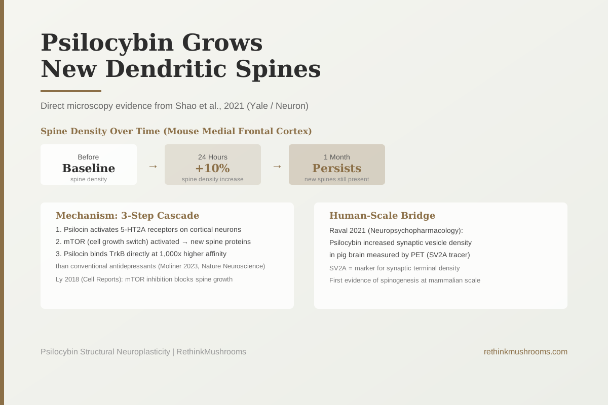
A 2021 Yale study used live brain imaging to watch psilocybin grow new dendritic spines in mouse frontal cortex by approximately 10% within 24 hours. The new spines were still present 1 month later.
Shao et al., Neuron, 2021
24 hours
time until new dendritic spines appeared after a single psilocybin dose in the Shao 2021 study
1 month
persistence of new dendritic spines in mouse frontal cortex after a single psilocybin dose
1,000x
higher affinity with which psilocin binds the TrkB receptor compared to conventional antidepressants (Moliner 2023, Nature Neuroscience)
Shao et al., Neuron, 2021
 View as image ↗
In 2021, Yale neuroscientists fitted a mouse with a small glass window over its skull and watched through a two-photon microscope, a device that uses infrared light to image individual neurons in a living brain, as a single dose of psilocybin grew new connections in the frontal cortex within hours. What they saw was not a metaphor for brain change. It was an actual physical observation of new dendritic spines forming on individual neurons, captured in real time and verified repeatedly.
The result, published in Neuron, showed that psilocybin produced approximately a 10% increase in dendritic spine density in the mouse medial frontal cortex within 24 hours of a single dose. One month later, the new spines were still there.[1]
This is the most concrete and directly measurable finding in the psilocybin neuroscience literature, and it is almost entirely absent from consumer-facing content on psychedelics and brain health. Most coverage mentions that psilocybin "promotes neuroplasticity" without explaining what that phrase actually describes. This article explains what dendritic spines are, what the Yale study found and how they found it, why depression erodes this same structure, and how the mechanism behind the spine growth relates to a 2023 discovery that changes how researchers understand the entire plasticity story.
For the broader context of how psilocybin affects the brain, see Psilocybin and the Default Mode Network: How It Quiets the Ego. For how the functional brain changes during a psilocybin experience relate to these structural changes, see Functional Connectivity Changes Under Psilocybin: fMRI Evidence.
Key Takeaways
- The Shao 2021 study (Neuron, Yale) used in vivo two-photon microscopy to directly observe that a single psilocybin dose grew new dendritic spines in mouse medial frontal cortex by approximately 10% within 24 hours, with persistence confirmed at 1 month
- Dendritic spines are the tiny protrusions on neuron branches that form the receiving end of synaptic connections. More spines means more potential connections and a more flexible, adaptable neural network
- Depression and chronic stress are associated with loss of dendritic spines in the prefrontal cortex (synaptic pruning), and psilocybin reversed this in the same brain region
- The spine growth involves mTOR (the cell's master growth-regulation switch) and TrkB (the receptor for BDNF, the brain's primary growth factor) downstream of 5-HT2A activation
- A 2023 Nature Neuroscience discovery found that psilocin directly binds TrkB at 1,000x higher affinity than conventional antidepressants, providing a mechanism for why the structural changes persist long after the drug clears
- A pig brain PET imaging study (Raval 2021) independently confirmed synaptic density increases from a single psilocybin dose, providing a bridge between mouse microscopy data and human-scale evidence
What Dendritic Spines Are and Why They Matter
A neuron is not a simple wire. It is a branching tree of processes that reach toward thousands of other neurons. The branches are called dendrites (from the Greek word for tree), and they are covered in tiny protrusions called dendritic spines.
Each dendritic spine is the receiving end of one synapse, the gap between two neurons where chemical signals are transmitted. A single neuron in the cortex may have thousands of dendritic spines, each forming a connection with a different sending neuron. The total connectivity of your brain, the number and strength of specific neuron-to-neuron links, is largely determined by how many dendritic spines exist and how large they are.
More spines on a neuron means more inputs from other neurons. A neuron with more dendritic spines is more connected to its local network. A region of the brain with higher spine density is more capable of forming and strengthening the patterns of activity that represent memories, learned behaviors, and flexible thinking.
Spines are not static. They grow, retract, enlarge, and shrink in response to experience and brain chemistry. The ability to change spine number and size is one of the core mechanisms of synaptic plasticity, the brain's capacity to reshape itself in response to what happens to it.
What Depression Does to Dendritic Spines
The prefrontal cortex, the region at the front of the brain that handles executive function, emotional regulation, and planning, is particularly vulnerable to a process called synaptic pruning during chronic stress and depression.
Stress hormones including cortisol reduce the availability of BDNF (brain-derived neurotrophic factor), the brain's primary protein for maintaining and growing synaptic connections. With less BDNF, dendritic spines in the prefrontal cortex retract. The brain region that should be providing top-down regulation of emotional responses is left with fewer connections and operates less effectively.
This is one of the best-supported structural correlates of depression. Post-mortem studies of people who died with depression find reduced dendritic spine density in the prefrontal cortex. Animal models of depression show the same reduction. And treatments that work against depression, whether conventional antidepressants, exercise, or psychotherapy, are associated with restoration of spine density.
Psilocybin acts in the prefrontal cortex. The Shao 2021 study measured spine growth in the medial frontal cortex specifically, the same region where depression-related atrophy occurs.
The Yale Study: What They Did and What They Found
Shao and colleagues wanted to answer a specific question: does psilocybin produce actual, visible, structural changes in the brain, or just functional changes in neural activity patterns?
To answer this, they used a technique called in vivo two-photon microscopy. A small transparent window was surgically fitted over a section of mouse skull. A two-photon microscope, which uses pulsed infrared light to image tissue at microscopic resolution without damaging it, was focused through this window onto individual neurons in the medial frontal cortex, just below the surface.
The researchers imaged the same neurons multiple times: before psilocybin, and at intervals after a single dose. They measured the number and size of individual dendritic spines on specific neurons, allowing them to track changes in real time in a living brain.
Within 24 hours of psilocybin administration, spine density had increased by approximately 10%.[1] The new spines were not just more numerous; they were also larger on average, which correlates with stronger synaptic connections. The changes persisted at the 1-month measurement point. Some individual spines that appeared after psilocybin were still present 30 days later.
The study also found behavioral correlates. Psilocybin ameliorated stress-induced behavioral deficits (modeled by chronic stress exposure before treatment) and increased excitatory neurotransmission in frontal cortex circuits. The behavioral improvement and the structural changes appeared together, though the study was not designed to prove a causal link between them in the mechanistic sense.
This is what "psilocybin promotes neuroplasticity" actually describes at the cellular level. Not an abstract change in brain activity, but new physical structures growing on individual neurons.
The Mechanism: Why Psilocybin Grows Spines
Understanding why psilocybin grows dendritic spines requires tracing what happens inside a neuron after psilocin (psilocybin's active metabolite) arrives.
Step 1: 5-HT2A activation. Psilocin primarily binds the 5-HT2A receptor, a serotonin receptor expressed at high density in the prefrontal cortex. When 5-HT2A is activated, it triggers a cascade inside the neuron via the Gq protein signaling pathway.
Step 2: mTOR activation. One downstream consequence of 5-HT2A activation is the stimulation of mTOR (mechanistic target of rapamycin), a protein complex inside cells that acts as a master switch for cellular growth and protein synthesis. mTOR activation is required for the formation of new dendritic spines, which require the synthesis of new structural proteins. Ly and colleagues established in their 2018 foundational study that mTOR inhibition blocks the spine-promoting effects of psychedelics.[3]
Step 3: TrkB activation and BDNF signaling. TrkB is the receptor for BDNF on neurons. When TrkB is activated by BDNF, it turns on a signaling cascade that promotes neuronal survival, growth, and structural change, including dendritic spine growth. This is the pathway that conventional antidepressants (SSRIs) are believed to eventually activate after weeks of daily use.
In 2023, Moliner and colleagues published a finding in Nature Neuroscience that significantly updated this picture.[4] They found that psilocin and LSD bind directly to TrkB, at a site in the transmembrane domain of TrkB receptor dimers, with affinities approximately 1,000 times higher than conventional antidepressants. This means psilocybin is not just indirectly activating TrkB through the BDNF pathway. It is directly engaging TrkB with extremely high potency.
The antidepressant and neuroplasticity effects in mice were shown to depend on this TrkB binding rather than solely on 5-HT2A activation. This is a mechanistic separation: the psychedelic experience (mediated by 5-HT2A) and the lasting structural plasticity (mediated by TrkB) appear to involve partially overlapping but distinct mechanisms.
This TrkB binding mechanism is a plausible explanation for why the new dendritic spines persist for a month. When TrkB is strongly activated, BDNF signaling sustains the maintenance machinery for new synaptic structures long after the drug has been metabolized and cleared from the brain.
For more on the 5-HT2A side of the mechanism, see Psilocybin and 5-HT2A Receptors: The Gateway to Psychedelic Effects.
The Pig Brain Evidence: Bridging Microscopy to Human Scale
The Shao 2021 study observed individual spines on individual neurons in mouse cortex. A separate 2021 study by Raval and colleagues provided evidence at a completely different scale: whole-brain changes in synaptic density measured via PET (positron emission tomography) imaging in pig brains.[2]
The PET method used in Raval 2021 measured SV2A, a protein found on synaptic vesicles (the small sacks inside neurons that carry neurotransmitters). SV2A density is a proxy for synaptic density: more synapses means more SV2A. This is the same PET methodology used in human brain imaging studies.
After a single psilocybin dose, pig brains showed increased SV2A signal, consistent with increased synaptic density, in multiple brain regions. At the same time, 5-HT2A receptor density decreased, consistent with the tolerance mechanism described in other research.
The pig brain result matters because pigs share more anatomical and neurochemical similarity with humans than mice do, and because the PET methodology is directly translatable to human PET studies. This is not direct human evidence for synaptogenesis, but it is a meaningful bridge between mouse microscopy data and human-scale measurements.
What Psilocybin's Structural Changes Mean for Depression Treatment
The convergence of the Shao 2021 spine data and the Moliner 2023 TrkB finding produces a plausible mechanistic account of how psilocybin might work as a rapid antidepressant.
Depression is associated with loss of dendritic spines in the prefrontal cortex, which reduces the functional capacity of the region most responsible for regulating emotional responses. Psilocybin, in a single dose, grows new dendritic spines in this same region within 24 hours via both mTOR-dependent spinogenesis and direct TrkB activation that sustains those spines for weeks. The result is a rapid restoration of synaptic connectivity in the brain region most damaged by depression, with effects that outlast the pharmacological action of the drug by a significant margin.
This is a biologically coherent account of a rapid antidepressant effect, and it is different from how SSRIs are understood to work (which primarily involves receptor desensitization and delayed neuroplasticity effects after weeks of use). It is also different from ketamine's antidepressant mechanism, though both ultimately involve mTOR-dependent spinogenesis.
What remains preclinical is the direct evidence: no human study has yet visualized psilocybin-induced dendritic spine growth using equivalent imaging methods. The pig brain PET data provides synaptic density changes at a translatable scale, but the specific resolution to see individual spines in a human frontal cortex, in a living person, is not yet technically achievable.
For the research on how the brain's structural plasticity relates to an enhanced period of learning and behavioral change after psilocybin, see The Plasticity Window: How Long Psilocybin Keeps the Brain Flexible. For the broader synaptogenesis research across the psychedelic class, see Psilocybin and Synaptogenesis: Creating New Neural Connections and Psilocybin and Neuroplasticity: Rewiring the Brain. For the detailed 24-hour timeline and what is known about when each structural change occurs, see Psilocybin and Dendritic Spine Growth: Structural Neuroplasticity in 24 Hours.
The Bottom Line
The Shao 2021 Yale study established, using direct in vivo imaging of individual neurons through a skull window, that a single psilocybin dose grows new dendritic spines in mouse frontal cortex by approximately 10% within 24 hours, with the new spines persisting at 1 month. The mechanism involves mTOR-dependent protein synthesis downstream of 5-HT2A activation and, as discovered in 2023, direct psilocin binding to TrkB at 1,000-fold higher affinity than conventional antidepressants. The pig brain PET imaging study by Raval 2021 independently confirmed increased synaptic density at a translatable scale. This converging evidence positions psilocybin as a compound that produces genuine structural brain changes rather than purely functional ones, with a biologically coherent mechanism that could explain rapid antidepressant effects.
Frequently Asked Questions
Sources & References
- 1RTHM-01434·Shao, Ling-Xiao et al. (2021). “Psilocybin induces rapid and persistent growth of dendritic spines in frontal cortex in vivo..” Neuron.Study breakdown →PubMed →↩
- 2. Source details for this reference are not yet available.
- 3RTHM-00983·Ly, Calvin et al. (2018). “Psychedelics Promote Structural and Functional Neural Plasticity..” Cell reports.Study breakdown →PubMed →↩
- 4RTHM-01756·Moliner, Rafael et al. (2023). “Psychedelics promote plasticity by directly binding to BDNF receptor TrkB..” Nature neuroscience.Study breakdown →PubMed →↩